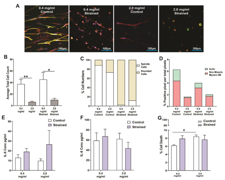
Figure 4.
Response of lung fibroblasts to mechanical strain within a 3D collagen-I microenvironment. Human fetal lung 1 (HFL1) fibroblasts were embedded in 3D collagen-1 gels of 0.4 mg/mL and 2.0 mg/mL concentrations and cultured for 24 h. The HFL1 fibroblast-seeded collagen gels were then left alone or mechanically strained for 48 h at a 1% amplitude and a frequency of 0.2 Hz. (A) Representative confocal images of HFL1 fibroblasts in 0.4 mg/mL and 2.0 mg/mL collagen-1 gels stained for non-muscle myosin IIB (red) and F-actin (green) after mechanical strain experiments. (B) Average total cell counts in 0.4 mg/mL and 2.0 mg/mL collagen-1 gels after strain or no strain for 48 h. (C) Percentage cell numbers of spindle and rounded shaped cells in 0.4 mg/mL and 2.0 mg/mL collagen-I gels after strain or no strain for 48 h. (D) Percentage positive pixel count per total number of pixels in images of 0.4 mg/mL and 2.0 mg/mL collagen-I gels after strain or no strain for 48 h. (E) Percentage positive pixel count per total number of pixels in images of 0.4 mg/mL and 2.0 mg/mL collagen-I gels after strain or no strain for 48 h. The concentration of (F) IL-6 and (G) IL-8 cytokines released from HFL1 fibroblasts in 0.4 mg/mL and 2.0 mg/mL collagen-1 gels after strain or no strain for 48 h. (G) The percentage of cell viability of HFL fibroblasts in 0.4 mg/mL and 2.0 mg/mL collagen-I gels after strain or no strain for 48 h. Mean ± SEM indicated for 3 technical replicates, n = 6. * p < 0.05, and ** p < 0.01.

