PLOS ONE logoLink to PLOS ONE
. 2022 Jan 20;17(1):e0262170. doi: 10.1371/journal.pone.0262170

Genomic surveillance of SARS-CoV-2 Spike gene by sanger sequencing

Tiago Souza Salles 1,#, Andrea Cony Cavalcanti 1,2,#, Fábio Burack da Costa 1, Vanessa Zaquieu Dias 1, Leandro Magalhães de Souza 2, Marcelo Damião Ferreira de Meneses 1, José Antônio Suzano da Silva 1,3, Cinthya Domingues Amaral 1, Jhonatan Ramos Felix 1,4, Duleide Alves Pereira 1, Stefanella Boatto 3, Maria Angélica Arpon Marandino Guimarães 5, Davis Fernandes Ferreira 1, Renata Campos Azevedo 1,*
Editor: Etsuro Ito6
PMCID: PMC8775319  PMID: 35051202

Abstract

The SARS-CoV-2 responsible for the ongoing COVID pandemic reveals particular evolutionary dynamics and an extensive polymorphism, mainly in Spike gene. Monitoring the S gene mutations is crucial for successful controlling measures and detecting variants that can evade vaccine immunity. Even after the costs reduction resulting from the pandemic, the new generation sequencing methodologies remain unavailable to a large number of scientific groups. Therefore, to support the urgent surveillance of SARS-CoV-2 S gene, this work describes a new feasible protocol for complete nucleotide sequencing of the S gene using the Sanger technique. Such a methodology could be easily adopted by any laboratory with experience in sequencing, adding to effective surveillance of SARS-CoV-2 spreading and evolution.

Introduction

The SARS-CoV-2 responsible for atypical pneumonia, evidenced in China by the end of 2019, was classified into the severe acute respiratory syndrome-related coronaviruses, member of Betacoronavirus genus, Coronaviridae family, been denominated Severe Acute Respiratory Syndrome Coronavirus 2 (SARS-CoV-2).

Coronaviruses are enveloped positive single-strand RNA viruses, with 30,000 bases in length, being the largest RNA genome identified up to date [1]. The SARS-CoV-2 genome has several ORFs; the first ORF1a/b stands at the RNA 5’ end and translates the non-structural proteins (nsP1 –nsP16). The RNA 3’ end holds the genes of the four structural (E, M, N e S) and accessories proteins. In the mature virus particle, protein S, a homo-trimeric type I fusion glycoprotein, is located on the surface of the virus particle and is responsible for binding to the cell receptor. In humans, the angiotensin-converting molecule (ACE-2) was assigned as the primary receptor for SARS-CoV2.

Several research groups have solved the complete structure of the SARS-CoV-2 S protein attached or not to the receptor ACE-2 [2]. This protein has approximately 1,273 amino acids, and its domains are delimited. Due to the relevance for virus attachment and entrance at susceptible cells, mutations in the receptor-binding domain (RDB) receive greater attention. In addition, mutations at other domains, like the amino (N) -terminal domain (NTD), can also lead to conformational changes in S protein structure and impact their function [3].

SARS-CoV-2 has particular evolutionary dynamics, and an extensive polymorphism is observed. However, the frequency of mutation across the SARS-CoV-2 genome is not uniform. Polymorphism (SNP) is mainly observed in protein S, RNA polymerase, RNA primase, and nucleoprotein [4]. According to the World Health Organization (WHO), isolates those present changes in amino acids that lead to suspected or confirmed cases with a phenotypic impact are considered variants of interest (VOI). Furthermore, these variants are classified as a concern (VOC) when they are associated with increased transmissibility, virulence, changes in the clinical presentation of COVID-19, and reduced containment measures, such as escaping diagnostic tools decreasing the effectiveness of vaccines and therapies [5].

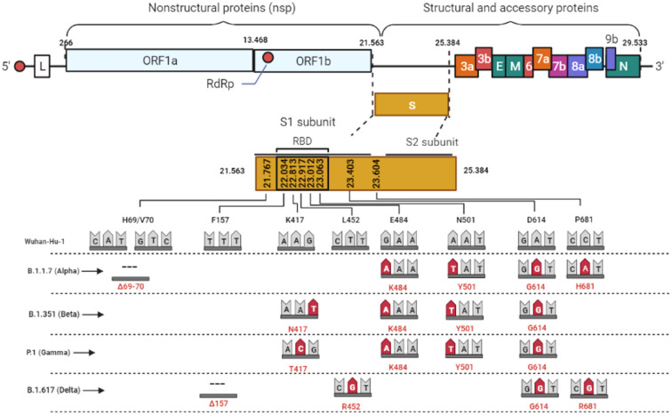
Since the S protein is the primary target of neutralizing antibodies, monitoring insertions, deletions, or substitutions of amino acids can reveal variants with the potential to evade vaccine immunity. In this context, genomic information is quickly shared through initiatives like the GISAID platform, and variants are counted and georeferenced [6]. Up to Jun 2021, five variants were classified as a concern (VOC), named; B.1.1.7 (Alpha), B.1.351(Beta), P.1(Gamma), B.1.617+ (Delta), first detected in the United Kingdom, South Africa, Brazil, and India, respectively (Fig 1).

Fig 1. Graphical representation of the SARS-CoV-2 genome.

Fig 1

Highlighted are the S gene and the main mutations described in the variants of concern. As a measure of comparison, the length of the S gene is already equivalent to the one of the whole Dengue virus genome.

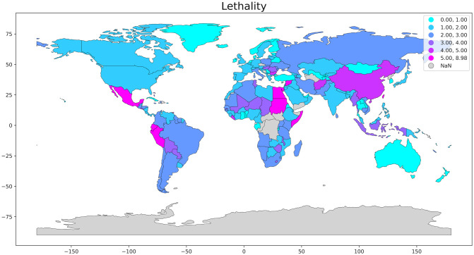
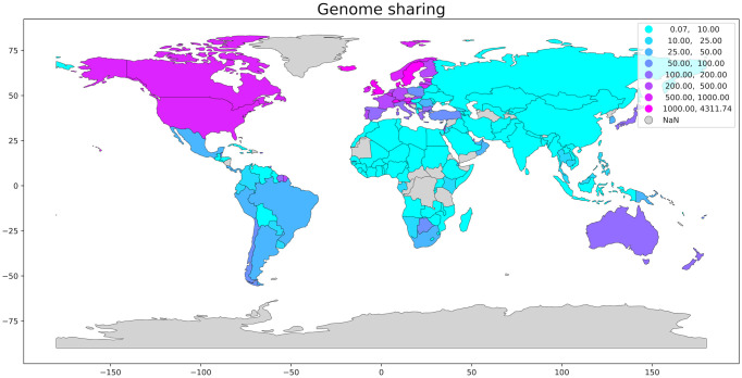
Early identification of the variants of concern (VOC) could provide excellent auxiliary information to decision making, allowing an earlier action towards measures to refrain the spreading of the virus such as reinforcement of mobility restriction or relaxation of such measures in areas where the variants are no present. Fig 2 shows a great variety in COVID-19 lethality in the different countries around the world. Unfortunately, due to the imposing genome size of the SARS-CoV-2, economic and laboratory challenges are manifest when monitoring the evolution of this virus. Fig 3 exhibits the significant disparity in the genome shared distribution per country.

Fig 2. Lethality [(Reported COVID-19 deaths)/(Reported COVID-19 cases)] per country.

Fig 2

We made use of the GISAID platform’s data to estimate COVID lethality and genome sharing per 105 inhabitants. We obtained the geospatial data for plotting the map in the open-source software library written for the Python programming language, Geopandas. The areas in grey are without reported data.

Fig 3. Normalized distribution of the genome shared per 105 inhabitants per country.

Fig 3

The areas in grey are without reported data. We have normalized the data to compare countries of different population sizes. The geospatial data for plotting the map was obtained in the open-source software library written for the Python programming language, Geopandas (source: GISAID platform).

Despite the reduction in the costs of new generation sequencing (NGS), the implementation of this system still requires a significant financial contribution, and the price per sample remains high for developing countries. The discrepancy in the number of sequences deposited in databases between countries reflects the difficulties of sequencing, as also shown in Table 1.

Table 1. Genome shared and lethality per country.

Country Genome shared Genome shared per 105 inhabitants§§ Reported COVID-19 cases Reported COVID-19 deaths Lethality§
United States of America 1,732,690 523 47,802,459 771,529 1.61%
United Kingdom 1,282,315 1,889 10,021,501 144,433 1.44%
Germany 260,519 311 5,650,170 100,476 1.78%
Denmark 218,679 3,775 466,817 2.841 0.61%
Canada 161,403 428 1,774,946 29,580 1,67%
France 121,009 185 7,285,128 116,314 1.60%
India 74,279 5 34,555,431 467,468 1.35%
Italy 71,623 118 4,968,341 133,486 2.69%
Brazil 75,292 35 22,043,112 613,339 2.78%
Mexico 38,365 30 3,872,263 293,186 7.57%
South Africa 23,634 40 2,952,500 89,771 3.04%
Russia 9,982 7 9,502,879 270,292 2.84%
China 1,203 0,1 127,631 5,697 4.46%
Iceland 9,812 2,875 17,446 35 0.20%

§ [(Reported COVID-19 deaths)/(Reported COVID-19 cases)].

§§Genome sharing data were normalized per 105 habitants to allow comparison between countries of rather different population sizes (source: GISAID platform).

Data collected up to November 28th 2021.

Unlike NGS methodologies, nucleotide sequencing based on the Sanger technique is widespread worldwide. In addition, the costs for sequencing small fragments are affordable. Therefore, to support the urgent surveillance of changes in SARS-CoV-2 S gene, this work describes a feasible protocol for complete nucleotide sequencing of the S gene using the Sanger technique. Thus, any laboratory with experience in sequencing can adopt this protocol.

Materials & methods

Ethics and study population

This work was previously approved by the Ethics Committee of Clementino Fraga Filho University Hospital (HUCFF/UFRJ) (number: 4.546.307). To evaluate this study, three samples from patients of confirmed COVID-19 presenting high viral load (Ct value < 20) were randomly selected. Patients were admitted to different hospitals in Rio de Janeiro, and a nasopharyngeal swab was collected to confirm clinical diagnosis by Rio de Janeiro Public Health Reference Laboratory—LACEN-RJ. Human samples were used after the conclusion of the diagnostic investigation. All patients’ personal information was anonymized, only the municipalities of residence were disclosed. Therefore, the ethics committee waived the requirement for informed consent from patients.

RNA extraction

According to the manufacturer’s instructions, the commercial kit MagMax Viral Pathogen (Thermo fisher, EUA) was used in the automated equipment King Fisher Apex (Thermo Fisher, EUA) to obtain the viral RNA from 200uL of respiratory secretion samples collected in nasopharyngeal swabs.

RT-qPCR for detection of SARS-CoV-2

The suspected samples of COVID-19 were tested in the diagnostic routine of the Noel Nutels Central Public Health Laboratory (LACEN-RJ) using the SARS-CoV-2 Duplex Kit (E/RP), Biomaguinhos (Fiocruz, Brasil). The reactions were performed using the QuantStudio 5 (Applied Biosystems, Thermo Fisher, EUA). The samples with ct values below 20 were selected for sequencing.

Amplification of the S protein gene

Six sets of primers targeting the S segment and two sets flanking it were designed based on the sequences deposited in GISAID until September 2020. 29 samples from 13 regions were aligned, and conserved regions were chosen using Clustal W program. An overlap of 100 nucleotides was programmed (Fig 4). Table 2 presents the sequence of primers used. Standard RT-PCR was performed using Superscript III one-step RT-PCR kit (Invitrogen, Carlsbad, CA, USA) according to the manufacturer’s instructions, with 0.7 μM primers and temperature conditions according to Table 3.

Fig 4. Agarose Gel Electrophoresis and schematic representation of the targeted fragments of each set of primers.

Fig 4

Amplification of S gene is visualized in agarose Gel Electrophoresis (A). Schematic representation of the targeted fragments of each set of primers is shown in (B). PR1 represents primer set 1, PR2 primer set 2, PR3 primer set 3, PR4 primer set 4, PR5 primer set 5, PR6 primer set 6, PR7 primer set 7 and PR8 primer set 8.

Table 2. List of primers used for the S gene amplification of SARS-CoV-2.

Primer
S segment
Sequence (5’–3’) Coding Position Product size (pb)
SP1 sense
ASP1antisense
GTTTGTTTTTCTTGTTTTATT
ACAGTGAAGGATTTCAACGTACAC
(21551–21574)
(22450–22474)
923pb
SP2 sense
ASP2 antisense
CGTGATCTCCCTCAGGGTTTT
TCAGCAATCTTTCCAGTTTGCC
(22190–22211)
(22810–22832)
620pb
SP3 sense
ASP3 antisense
GTAATTAGAGGTGATGAAGTCAGA
ACATAGTGTAGGCAATGATGGA
(22751–22775)
(23621–23643)
892pb
SP4 sense
ASP4 antisense
CTTGGCGTGTTTATTCTACAG
GCTTGTGCATTTTGGTTGACC
(23445–23466)
(24403–24424)
979pb
SP5 sense
ASP5 antisense
AGACTCACTTTCTTCCACAGCA
AGATGATAGCCCTTTCCACA
(24355–24377)
(24699–24719)
342pb
SP6 sense
ASP6 antisense
TTCTGCTAATCTTGCTGCTACT
GTTTATGTGTAATGTAATTTGACTCC
(24610–24632)
(25348–25372)
766pb
SP7 sense#
ASP7 antisense
TAGAGAAAACAACAGAGTT
TGAGGGAGATCACGCACTAA
(21492–21511)
(22184–22204)
712pb
SP8 sense#
ASP8 antisense
TTCTGCTAATCTTGCTGCTACT
CCTTGCTTCAAAGTTACAGTTCCA
(24610–24632)
(25409–25433)
825pb

# Set primers 7 and 8 flanks the S protein-coding region.

Nucleotide positions are according to the SARS-CoV-2 Wuhan (Genbank accession no.NC_ 045512-Wuhan-HU-1).

Table 3. RT-PCR cycle conditions.

Temperature Time
60°C 1 minute Reverse transcription and Transcriptase inactivation
50°C 45 minutes
94°C 2 minutes
95°C 15 seconds 40 cycles of amplification
53°C 30 seconds
68°C 1 minute
68°C 7 minutes Final extension

The amplification of the fragments was visualized by 1,5% Agarose Gel Electrophoresis. The samples were quantified with the nanodrop one (Thermo Scientific NanoDrop One Microvolume UV-Vis Spectrophotometers) for sequencing.

Nucleotide sequence determination and analysis

The nucleotide sequences were determined from 200 ng of the amplicon, using the Big Dye Terminator kit 3.1 (Applied Biosystems), following the manufacturer’s procedure. Amplicons were sequenced in the ABI 3730 genetic analyzer (Applied Biosystems, USA) following the manufacturer’s protocol. Raw sequence data were aligned, edited, assembled using the BioEdit Sequence Alignment Editor, Version 7.0.5.3.

The protocol described in this peer-reviewed article is published on protocols.io, https://dx.doi.org/10.17504/protocols.io.bx6kprcw and is included for printing as S1 File with this article.

Results and discussion

This methodology covered 100% of the S gene sequenced (3,822 pb). The sequences obtained were deposited at GISAID numbers EPI_ISL_4496739, EPI_ISL_4497141, EPI_ISL_4497286.

All the eight primers set produced single amplicons for the three samples used to evaluate this protocol (Fig 4A); therefore, sequencing reaction could be performed without extracting the bands from agarose gel. In addition, no mismatch in the primer regions that could lead to the escape of known VOCs was observed (S2 File).

The samples sequenced in this study originated from Rio de Janeiro City, Santo Antônio de Pádua and Seropédica, in Rio de Janeiro state. The obtained sequences were aligned with reference sequences of each VOC, in order to detect and compare mutations. Spike protein from Rio de Janeiro city sample displayed the same amino acid changes found in reference sequence of Gamma variant, suggesting that this sample is probably classified into P.1 lineage (Table 4). According to the literature, the P.1 lineage (gamma) emerged in Manaus, Amazonas, evolved from a B.1.1.28 clade in late November 2020 and replaced its parental lineage in less than two months[7]. We found a strain displaying similar spike protein with that of P.1 lineage circulating in Rio de Janeiro as early as February 2021.

Table 4. Protein S mutations of each VOC and studied sequences.

Sequences AA identity AA changes Mutations Accession number
Alpha (B.1.1.7) 99.214% 10 H69del, V70del, Y144del, N501Y, A570D, D614G, P681H, T716I, S982A, D1118H EPI_ISL_601443
Beta (B.1.351) 99.607% 5 D80A, E484K, N501Y, D614G, A701V EPI_ISL_660613
Gamma (P.1) 99.057% 12 L18F, T20N, P26S, D138Y, R190S, K417T, E484K, N501Y, D614G, H655Y, T1027I, V1176F |EPI_ISL_906071
Delta (B.1.617) 99.214% 10 T19R, E156G, F157del, R158del, A222V, L452R, T478K, D614G, P681R, D950N EPI_ISL_2047658
Rio de Janeiro 99.057% 12 L18F, T20N, P26S, D138Y, R190S, K417T, E484K, N501Y, D614G, H655Y, T1027I, V1176F EPI_ISL_4496739
Santo Antonio de Padua 99.764% 3 E484K, D614G, V1176F EPI_ISL_4497141
Seropedica 99.450% 7 E156D, E484K, D614G, D775V, T866P, M869K, V1176F EPI_ISL_4497286

Nucleotide positions are according to the SARS-CoV-2 Wuhan (GenBank accession no. NC_ 045512-Wuhan-HU-1).

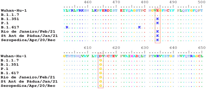
The samples from Santo Antônio de Pádua and Seropédica didn’t show similar mutation patterns with gamma VOC (Table 4), however, they presented some mutations of importance, like E484K and D614G (Fig 5). The change from glutamic acid to a lysine in the 484th amino acid position of the Spike protein (E484K) already occurred 228,871 times (4.27% of all samples with spike sequence) in 166 countries, according to GISAID Spike Glycoprotein Mutation Surveillance. This mutation has been reported in the literature to be related to enhanced host receptor binding [8] and antigenic drift [9] either alone or in association with other mutations [10]. The mutation D614G is widely spread and has already occurred 5,285,437 times (98.51% of all samples with Spike sequence) in 204 countries. It was reported to be related to the increase in infectivity of SARS-CoV-2, higher viral loads, increased replication fitness, and virulence [11,12].

Fig 5. Sequence alignment showing amino acid substitutions E484K and D614G.

Fig 5

Apart from the mutations of high importance, the sequence from Seropédica also presented some rare mutations. The amino acid substitutions D775V, T866P and M869K are present in less than three sequences in GISAID database. The effects of these mutations are still unknown.

Due to its essential role in establishing infection, as well as inducing immune response, the genomic surveillance of the S protein of SARS-CoV-2 is of paramount importance. Monitoring the emergence of new variants, and the interactions between their mutations, allow the scientific community to develop better strategies to control the pandemic.

The count of genomic sequences obtained in each country reveals a vast disproportion that becomes evident in surveillance platforms like GISAID. One of the reasons for this disparity is the limited access to NGS methodologies by most groups. Therefore, this work describes a protocol for complete nucleotide sequencing of the S gene using the Sanger technique, which could be helpful to keep tracking SARS-CoV-2 protein S evolution.

Supporting information

S1 File. The PDF of the protocol described in this peer-reviewed article published on protocols.io dx.doi.org/10.17504/protocols.io.bx6kprcw.

(PDF)

S2 File. Map of primer pairs in VOCs sequences.

(PDF)

S3 File. Flowchart describing the sequential steps of the protocol.

(PDF)

Acknowledgments

We want to thank all health professionals, especially LACEN-RJ staff, for their collaboration during the implementation of this protocol and for all efforts in facing the COVID-19 pandemic.

Data Availability

This methodology covered 100% of the S gene sequenced (3,822 pb). The sequences obtained were deposited at GISAID (numbers EPI_ISL_4496739, EPI_ISL_4497141, EPI_ISL_4497286) and GenBank (numbers OM064632, OM064633, OM064634).

Funding Statement

This work was material supported by Fundação Carlos Chagas Filho de Amparo à Pesquisa do Estado do Rio de Janeiro-FAPERJ [grant number E-26/201.840/2017] (RCA) and Coordenação de Aperfeiçoamento de Pessoal de Nível Superior - Brasil (CAPES) – Public Notice Number 09/2020 - Prevention and Combat against Outbreaks, Endemics, Epidemics and Pandemics. Process number 223038.014313/2020-19 (TSS and FBC). The funders had no role in study design, data collection and analysis, decision to publish, or preparation of the manuscript.

References

  • 1.Zhu N, Zhang D, Wang W, Li X, Yang B, Song J, et al. A Novel Coronavirus from Patients with Pneumonia in China, 2019. The New England journal of medicine. 2020;382: 727–733. doi: 10.1056/NEJMoa2001017 [DOI] [PMC free article] [PubMed] [Google Scholar]
  • 2.Wrapp D, Wang N, Corbett KS, Goldsmith JA, Hsieh C-L, Abiona O, et al. Cryo-EM structure of the 2019-nCoV spike in the prefusion conformation. Science (New York, NY). 2020;367: 1260–1263. doi: 10.1126/science.abb2507 [DOI] [PMC free article] [PubMed] [Google Scholar]
  • 3.Resende PC, Delatorre E, Gräf T, Mir D, Motta FC, Appolinario LR, et al. Evolutionary Dynamics and Dissemination Pattern of the SARS-CoV-2 Lineage B.1.1.33 During the Early Pandemic Phase in Brazil. Frontiers in Microbiology. 2021;11: 3565. doi: 10.3389/fmicb.2020.615280 [DOI] [PMC free article] [PubMed] [Google Scholar]
  • 4.Yin C. Genotyping coronavirus SARS-CoV-2: methods and implications. Genomics. 2020;112: 3588–3596. doi: 10.1016/j.ygeno.2020.04.016 [DOI] [PMC free article] [PubMed] [Google Scholar]
  • 5.WHO. World Health Organization: COVID-19 weekly epidemiological update, 25 February 2021. Special ed. 2021. https://apps.who.int/iris/handle/10665/339859.
  • 6.Elbe S, Buckland-Merrett G. Data, disease and diplomacy: GISAID’s innovative contribution to global health. Global challenges (Hoboken, NJ). 2017;1: 33–46. doi: 10.1002/gch2.1018 [DOI] [PMC free article] [PubMed] [Google Scholar]
  • 7.Naveca FG, Nascimento V, de Souza VC, Corado A de L, Nascimento F, Silva G, et al. COVID-19 in Amazonas, Brazil, was driven by the persistence of endemic lineages and P.1 emergence. Nature medicine. 2021;27: 1230–1238. doi: 10.1038/s41591-021-01378-7 [DOI] [PubMed] [Google Scholar]
  • 8.Starr TN, Greaney AJ, Hilton SK, Ellis D, Crawford KHD, Dingens AS, et al. Deep Mutational Scanning of SARS-CoV-2 Receptor Binding Domain Reveals Constraints on Folding and ACE2 Binding. Cell. 2020;182: 1295–1310.e20. doi: 10.1016/j.cell.2020.08.012 [DOI] [PMC free article] [PubMed] [Google Scholar]
  • 9.Ho D, Wang P, Liu L, Iketani S, Luo Y, Guo Y, et al. Increased Resistance of SARS-CoV-2 Variants B.1.351 and B.1.1.7 to Antibody Neutralization. Research square. 2021. doi: 10.21203/rs.3.rs-155394/v1 [DOI] [PMC free article] [PubMed] [Google Scholar]
  • 10.Wibmer CK, Ayres F, Hermanus T, Madzivhandila M, Kgagudi P, Oosthuysen B, et al. SARS-CoV-2 501Y.V2 escapes neutralization by South African COVID-19 donor plasma. bioRxiv: the preprint server for biology. 2021. [DOI] [PubMed] [Google Scholar]
  • 11.Korber B, Fischer WM, Gnanakaran S, Yoon H, Theiler J, Abfalterer W, et al. Tracking Changes in SARS-CoV-2 Spike: Evidence that D614G Increases Infectivity of the COVID-19 Virus. Cell. 2020;182: 812–827.e19. doi: 10.1016/j.cell.2020.06.043 [DOI] [PMC free article] [PubMed] [Google Scholar]
  • 12.Hou YJ, Chiba S, Halfmann P, Ehre C, Kuroda M, Dinnon KH 3rd, et al. SARS-CoV-2 D614G variant exhibits efficient replication ex vivo and transmission in vivo. Science (New York, NY). 2020;370: 1464–1468. doi: 10.1126/science.abe8499 [DOI] [PMC free article] [PubMed] [Google Scholar]

Decision Letter 0

Etsuro Ito

23 Nov 2021

PONE-D-21-34394Genomic surveillance of SARS-CoV-2 Spike protein by sanger sequencing.PLOS ONE

Dear Dr. Azevedo,

Thank you for submitting your manuscript to PLOS ONE. After careful consideration, we feel that it has merit but does not fully meet PLOS ONE’s publication criteria as it currently stands. Therefore, we invite you to submit a revised version of the manuscript that addresses the points raised during the review process.

The comments raised by the reviewers seem minor. Please provide your revised manuscript.

Please submit your revised manuscript by Jan 06 2022 11:59PM. If you will need more time than this to complete your revisions, please reply to this message or contact the journal office at plosone@plos.org. When you're ready to submit your revision, log on to https://www.editorialmanager.com/pone/ and select the 'Submissions Needing Revision' folder to locate your manuscript file.

Please include the following items when submitting your revised manuscript:

  • A rebuttal letter that responds to each point raised by the academic editor and reviewer(s). You should upload this letter as a separate file labeled 'Response to Reviewers'.

  • A marked-up copy of your manuscript that highlights changes made to the original version. You should upload this as a separate file labeled 'Revised Manuscript with Track Changes'.

  • An unmarked version of your revised paper without tracked changes. You should upload this as a separate file labeled 'Manuscript'.

If you would like to make changes to your financial disclosure, please include your updated statement in your cover letter. Guidelines for resubmitting your figure files are available below the reviewer comments at the end of this letter.

If applicable, we recommend that you deposit your laboratory protocols in protocols.io to enhance the reproducibility of your results. Protocols.io assigns your protocol its own identifier (DOI) so that it can be cited independently in the future. For instructions see: https://journals.plos.org/plosone/s/submission-guidelines#loc-laboratory-protocols. Additionally, PLOS ONE offers an option for publishing peer-reviewed Lab Protocol articles, which describe protocols hosted on protocols.io. Read more information on sharing protocols at https://plos.org/protocols?utm_medium=editorial-email&utm_source=authorletters&utm_campaign=protocols.

We look forward to receiving your revised manuscript.

Kind regards,

Etsuro Ito

Academic Editor

PLOS ONE

Journal Requirements:

1. Please ensure that your manuscript meets PLOS ONE's style requirements, including those for file naming. The PLOS ONE style templates can be found at 

https://journals.plos.org/plosone/s/file?id=wjVg/PLOSOne_formatting_sample_main_body.pdf and 

https://journals.plos.org/plosone/s/file?id=ba62/PLOSOne_formatting_sample_title_authors_affiliations.pdf

2. Thank you for providing the following Protocols.io DOI in your submission form [Protocols.io DOI]. In keeping with our submission requirements, please add the Protocols.io DOI to the Methods section of your manuscript as well using this format: “The protocol described in this peer-reviewed article is published on protocols.io, https://dx.doi.org/10.17504/protocols.io[........] and is included for printing as supporting information file 1 with this article.” For more information, please see our submission guidelines: https://journals.plos.org/plosone/s/submission-guidelines#loc-guidelines-for-specific-study-types

3. Please ensure that you have specified (1) whether consent was informed, (2) what type you obtained (for instance, written or verbal, and if verbal, how it was documented and witnessed). If your study included minors, state whether you obtained consent from parents or guardians. If the need for consent was waived by the ethics committee and (3) If you are reporting a retrospective study of medical records or archived samples, please ensure that you have discussed whether all data were fully anonymized before you accessed them and/or whether the IRB or ethics committee waived the requirement for informed consent. If patients provided informed written consent to have data from their medical records used in research, please include this information.

4. Thank you for stating the following financial disclosure: 

 (The funders had and will not have a role in study design, data collection and analysis, decision to publish, or preparation of the manuscript.)

At this time, please address the following queries:

a) Please clarify the sources of funding (financial or material support) for your study. List the grants or organizations that supported your study, including funding received from your institution. 

b) State what role the funders took in the study. If the funders had no role in your study, please state: “The funders had no role in study design, data collection and analysis, decision to publish, or preparation of the manuscript.”

c) If any authors received a salary from any of your funders, please state which authors and which funders.

d) If you did not receive any funding for this study, please state: “The authors received no specific funding for this work.

Please include your amended statements within your cover letter; we will change the online submission form on your behalf.

5. We note that the grant information you provided in the ‘Funding Information’ and ‘Financial Disclosure’ sections do not match. 

When you resubmit, please ensure that you provide the correct grant numbers for the awards you received for your study in the ‘Funding Information’ section.

6. Thank you for stating the following in the Acknowledgments Section of your manuscript: 

(This work was supported by Fundação de Amparo à Pesquisa do Estado do Rio de Janeiro-FAPERJ [grant number E-26/201.840/2017] and Coordenação de Aperfeiçoamento de Pessoal de Nível Superior - Brasil (CAPES) – Public Notice Number 09/2020 - Prevention and Combat against Outbreaks, Endemics, Epidemics and Pandemics. Process number 223038.014313/2020-19, "Digital Technologies for Monitoring, Mapping, and Control of Outbreaks, Endemics, Epidemics and Pandemics", held at the Federal University of Rio de Janeiro (TSS and FBC) )

We note that you have provided funding information that is not currently declared in your Funding Statement. However, funding information should not appear in the Acknowledgments section or other areas of your manuscript. We will only publish funding information present in the Funding Statement section of the online submission form. 

Please remove any funding-related text from the manuscript and let us know how you would like to update your Funding Statement. Currently, your Funding Statement reads as follows: 

 (The funders had and will not have a role in study design, data collection and analysis, decision to publish, or preparation of the manuscript.)

Additionally, because some of your funding information pertains to [commercial funding//patents], we ask you to provide an updated Competing Interests statement, declaring all sources of commercial funding. 

In your Competing Interests statement, please confirm that your commercial funding does not alter your adherence to PLOS ONE Editorial policies and criteria by including the following statement: "This does not alter our adherence to PLOS ONE policies on sharing data and materials.” as detailed online in our guide for authors  http://journals.plos.org/plosone/s/competing-interests.  If this statement is not true and your adherence to PLOS policies on sharing data and materials is altered, please explain how. 

Please include the updated Competing Interests Statement and Funding Statement in your cover letter. We will change the online submission form on your behalf.

7. We note that you have included the phrase “data not shown” in your manuscript. Unfortunately, this does not meet our data sharing requirements. PLOS does not permit references to inaccessible data. We require that authors provide all relevant data within the paper, Supporting Information files, or in an acceptable, public repository. Please add a citation to support this phrase or upload the data that corresponds with these findings to a stable repository (such as Figshare or Dryad) and provide and URLs, DOIs, or accession numbers that may be used to access these data. Or, if the data are not a core part of the research being presented in your study, we ask that you remove the phrase that refers to these data.

8. We note that Figures 2 and 3 in your submission contain [map/satellite] images which may be copyrighted. All PLOS content is published under the Creative Commons Attribution License (CC BY 4.0), which means that the manuscript, images, and Supporting Information files will be freely available online, and any third party is permitted to access, download, copy, distribute, and use these materials in any way, even commercially, with proper attribution. For these reasons, we cannot publish previously copyrighted maps or satellite images created using proprietary data, such as Google software (Google Maps, Street View, and Earth). For more information, see our copyright guidelines: http://journals.plos.org/plosone/s/licenses-and-copyright.

We require you to either (1) present written permission from the copyright holder to publish these figures specifically under the CC BY 4.0 license, or (2) remove the figures from your submission:

A. You may seek permission from the original copyright holder of Figures 2 and 3 to publish the content specifically under the CC BY 4.0 license.  

We recommend that you contact the original copyright holder with the Content Permission Form (http://journals.plos.org/plosone/s/file?id=7c09/content-permission-form.pdf) and the following text:

“I request permission for the open-access journal PLOS ONE to publish XXX under the Creative Commons Attribution License (CCAL) CC BY 4.0 (http://creativecommons.org/licenses/by/4.0/). Please be aware that this license allows unrestricted use and distribution, even commercially, by third parties. Please reply and provide explicit written permission to publish XXX under a CC BY license and complete the attached form.”

Please upload the completed Content Permission Form or other proof of granted permissions as an "Other" file with your submission.

In the figure caption of the copyrighted figure, please include the following text: “Reprinted from [ref] under a CC BY license, with permission from [name of publisher], original copyright [original copyright year].”

B. If you are unable to obtain permission from the original copyright holder to publish these figures under the CC BY 4.0 license or if the copyright holder’s requirements are incompatible with the CC BY 4.0 license, please either i) remove the figure or ii) supply a replacement figure that complies with the CC BY 4.0 license. Please check copyright information on all replacement figures and update the figure caption with source information. If applicable, please specify in the figure caption text when a figure is similar but not identical to the original image and is therefore for illustrative purposes only.

The following resources for replacing copyrighted map figures may be helpful:

USGS National Map Viewer (public domain): http://viewer.nationalmap.gov/viewer/

The Gateway to Astronaut Photography of Earth (public domain): http://eol.jsc.nasa.gov/sseop/clickmap/

Maps at the CIA (public domain): https://www.cia.gov/library/publications/the-world-factbook/index.html and https://www.cia.gov/library/publications/cia-maps-publications/index.html

NASA Earth Observatory (public domain): http://earthobservatory.nasa.gov/

Landsat: http://landsat.visibleearth.nasa.gov/

USGS EROS (Earth Resources Observatory and Science (EROS) Center) (public domain): http://eros.usgs.gov/#

Natural Earth (public domain): http://www.naturalearthdata.com/

9. Please review your reference list to ensure that it is complete and correct. If you have cited papers that have been retracted, please include the rationale for doing so in the manuscript text, or remove these references and replace them with relevant current references. Any changes to the reference list should be mentioned in the rebuttal letter that accompanies your revised manuscript. If you need to cite a retracted article, indicate the article’s retracted status in the References list and also include a citation and full reference for the retraction notice.

Reviewers' comments:

Reviewer's Responses to Questions

Comments to the Author

1. Does the manuscript report a protocol which is of utility to the research community and adds value to the published literature?

Reviewer #1: Yes

Reviewer #2: Yes

********** 

2. Has the protocol been described in sufficient detail?

Descriptions of methods and reagents contained in the step-by-step protocol should be reported in sufficient detail for another researcher to reproduce all experiments and analyses. The protocol should describe the appropriate controls, sample sizes and replication needed to ensure that the data are robust and reproducible.

Reviewer #1: Yes

Reviewer #2: Yes

********** 

3. Does the protocol describe a validated method?

The manuscript must demonstrate that the protocol achieves its intended purpose: either by containing appropriate validation data, or referencing at least one original research article in which the protocol was used to generate data.

Reviewer #1: Yes

Reviewer #2: No

********** 

4. If the manuscript contains new data, have the authors made this data fully available?

The PLOS Data policy requires authors to make all data underlying the findings described in their manuscript fully available without restriction, with rare exception (please refer to the Data Availability Statement in the manuscript PDF file). The data should be provided as part of the manuscript or its supporting information, or deposited to a public repository. For example, in addition to summary statistics, the data points behind means, medians and variance measures should be available. If there are restrictions on publicly sharing data—e.g. participant privacy or use of data from a third party—those must be specified.

Reviewer #1: Yes

Reviewer #2: Yes

********** 

5. Is the article presented in an intelligible fashion and written in standard English?

PLOS ONE does not copyedit accepted manuscripts, so the language in submitted articles must be clear, correct, and unambiguous. Any typographical or grammatical errors should be corrected at revision, so please highlight any specific errors that need correcting in the box below.

Reviewer #1: Yes

Reviewer #2: Yes

********** 

6. Review Comments to the Author

Please use the space provided to explain your answers to the questions above. You may also include additional comments for the author, including concerns about dual publication, research ethics, or publication ethics. (Please upload your review as an attachment if it exceeds 20,000 characters)

Reviewer #1: In the study of Salles and Cavalcanti et al., entitled “Genomic surveillance of SARS-CoV-2 Spike gene by sanger sequencing” the authors describe a new feasible protocol for complete nucleotide sequencing of the Spike (S) gene using the Sanger technique. Thus, the authors conclude that such a methodology could be easily adopted by any laboratory with experience in sequencing, adding to effective surveillance of SARS-CoV-2 spreading and evolution. The study is important and should be accepted after minor revisions.

Minor revisions:

1) Change title to “…SARS-CoV-2 Spike gene…” (and not protein);

2) Page 7, line 25: change to “…mainly in Spike (S) gene. Monitoring the S gene” (and not protein);

3) Page 7, line 29: change to “…SARS-CoV-2 S gene” (and not protein);

4) Page 7, line 30: change to “…S gene” (and not protein);

5) Page 7, line 33: change to “…Spike gene” (and not protein);

6) Page 7, line 34: change to “…SARS-CoV-2 Spike gene” (and not protein);

7) Page 8, line 41: Describe the acronym: “… been denominated severe acute respiratory syndrome coronavirus 2 (SARS‐CoV‐2).”;

8) Page 9, lines 86 and 87: change to: “Unlike NGS methodologies, nucleotide sequencing based on the Sanger technique is widespread worldwide.”;

9) Page 9, lines 88 and 89: change to “…SARS-CoV-2 S gene” and “S gene” (and not protein);

10) Page 10, line 133: change to “S gene” (and not protein);

11) Page 10, line 139: remove (data not shown);

12) Page 11, lines 152 and 156: update the number of times these mutations appear in GISAID;

13) Page 11, line 171: change to “S gene” (and not protein);

14) Page 14, line 250: update collected data;

15) Page 14, line 251 (Table 2): I suggest changing the primers’ names so they are not confused with the P1 (Gamma) variant and its sublineages;

16) Page 16, line 275: change to “…S gene” (and not protein);

17) Page 16, line 277: change to “…S gene” (and not protein).

Reviewer #2: The manuscript entitled "Genomic surveillance of SARS-CoV-2 Spike protein by Sanger sequencing" presents a protocol for complete sequencing of the S protein of SARS-CoV-2 using the Sanger technique, as an alternative to next-generation sequencing (NGS), in the investigation of mutations in the S protein of SARS-CoV-2 at lower cost.

The manuscript presents the steps of the protocol in a complete and detailed manner.

I recommend that a flowchart with the sequential steps of the proposed protocol be inserted.

lines 117 - 119 - I recommend that the data referring to cycling be presented in the form of a table, in order to facilitate understanding.

line 136 - I recommend replacing the term validate by evaluate

********** 

7. PLOS authors have the option to publish the peer review history of their article (what does this mean?). If published, this will include your full peer review and any attached files.

If you choose “no”, your identity will remain anonymous but your review may still be made public.

Do you want your identity to be public for this peer review? For information about this choice, including consent withdrawal, please see our Privacy Policy.

Reviewer #1: Yes: Fabrício Souza Campos

Reviewer #2: No

While revising your submission, please upload your figure files to the Preflight Analysis and Conversion Engine (PACE) digital diagnostic tool, https://pacev2.apexcovantage.com/. PACE helps ensure that figures meet PLOS requirements. To use PACE, you must first register as a user. Registration is free. Then, login and navigate to the UPLOAD tab, where you will find detailed instructions on how to use the tool. If you encounter any issues or have any questions when using PACE, please email PLOS at figures@plos.org. Please note that Supporting Information files do not need this step.

PLoS One. 2022 Jan 20;17(1):e0262170. doi: 10.1371/journal.pone.0262170.r002

Author response to Decision Letter 0


10 Dec 2021

Response to Reviewers

Dear Etsuro Ito,

We want to thank you for the comments. The manuscript was revised carefully, attending to the points raised during the review process. We hope that this version fully meets PLOS ONE’s publication criteria.

Kind regards,

Renata Campos Azevedo

Journal Requirements:

1. Please ensure that your manuscript meets PLOS ONE's style requirements, including those for file naming. The PLOS ONE style templates can be found at https://journals.plos.org/plosone/s/file?id=wjVg/PLOSOne_formatting_sample_main_body.pdf and https://journals.plos.org/plosone/s/file?id=ba62/PLOSOne_formatting_sample_title_authors_affiliations.pdf

R. The manuscript was revised according to style requirements.

2. Thank you for providing the following Protocols.io DOI in your submission form [Protocols.io DOI]. In keeping with our submission requirements, please add the Protocols.io DOI to the Methods section of your manuscript as well using this format: “The protocol described in this peer-reviewed article is published on protocols.io, https://dx.doi.org/10.17504/protocols.io[........] and is included for printing as supporting information file 1 with this article.” For more information, please see our submission guidelines: https://journals.plos.org/plosone/s/submission-guidelines#loc-guidelines-for-specific-study-types

R. The protocols.io DOI was added to the methods section using the format described (lines 227 up to 229 from the revised version). “The protocol described in this peer-reviewed article is published on protocols.io, https://dx.doi.org/10.17504/protocols.io.bx6kprcw and is included for printing as supporting information file 1 with this article.”

3. Please ensure that you have specified (1) whether consent was informed, (2) what type you obtained (for instance, written or verbal, and if verbal, how it was documented and witnessed). If the need for consent was waived by the ethics committee and (3) If you are reporting a retrospective study of medical records or archived samples, please ensure that you have discussed whether all data were fully anonymized before you accessed them and/or whether the IRB or ethics committee waived the requirement for informed consent.

R. The information regarding the waive of consent by the ethics committee was included (lines 134 up to 137). “Human samples were used after the conclusion of the diagnostic investigation. All patients' personal information was anonymized, only the municipalities of residence were disclosed. Therefore, the ethics committee waived the requirement for informed consent from patients.”

Financial disclosure:

a) Please clarify the sources of funding (financial or material support) for your study. List the grants or organizations that supported your study, including funding received from your institution.

b) b) State what role the funders took in the study. If the funders had no role in your study, please state: “The funders had no role in study design, data collection and analysis, decision to publish, or preparation of the manuscript.”

c) c) If any authors received a salary from any of your funders, please state which authors and which funders.

R. The financial disclosure was corrected.

This work was material supported by Fundação Carlos Chagas Filho de Amparo à Pesquisa do Estado do Rio de Janeiro-FAPERJ [grant number E-26/201.840/2017] (RCA) and Coordenação de Aperfeiçoamento de Pessoal de Nível Superior - Brasil (CAPES) – Public Notice Number 09/2020 - Prevention and Combat against Outbreaks, Endemics, Epidemics and Pandemics. Process number 223038.014313/2020-19 (TSS and FBC). The funders had no role in study design, data collection and analysis, decision to publish, or preparation of the manuscript.

Additionally, because some of your funding information pertains to [commercial funding//patents], we ask you to provide an updated Competing Interests statement, declaring all sources of commercial funding. In your Competing Interests statement, please confirm that your commercial funding does not alter your adherence to PLOS ONE Editorial policies and criteria by including the following statement: "This does not alter our adherence to PLOS ONE policies on sharing data and materials.” as detailed online in our guide for authors http://journals.plos.org/plosone/s/competing-interests. If this statement is not true and your adherence to PLOS policies on sharing data and materials is altered, please explain how.

R. Our research grant was from public organizations and not from commercial funding//patents. Therefore, the authors reaffirm that no competing interests exist.

7. We note that you have included the phrase “data not shown” in your manuscript. Unfortunately, this does not meet our data sharing requirements. PLOS does not permit references to inaccessible data. We require that authors provide all relevant data within the paper, Supporting Information files, or in an acceptable, public repository. Please add a citation to support this phrase or upload the data that corresponds with these findings to a stable repository (such as Figshare or Dryad) and provide and URLs, DOIs, or accession numbers that may be used to access these data. Or, if the data are not a core part of the research being presented in your study, we ask that you remove the phrase that refers to these data.

R. The data was included as supporting Information file 2.

8. We note that Figures 2 and 3 in your submission contain [map/satellite] images which may be copyrighted. All PLOS content is published under the Creative Commons Attribution License (CC BY 4.0), which means that the manuscript, images, and Supporting Information files will be freely available online, and any third party is permitted to access, download, copy, distribute, and use these materials in any way, even commercially, with proper attribution. For these reasons, we cannot publish previously copyrighted maps or satellite images created using proprietary data, such as Google software (Google Maps, Street View, and Earth). For more information, see our copyright guidelines: http://journals.plos.org/plosone/s/licenses-and-copyright.

R. We want to clarify that both Figures 2 and 3 were made originally for this article using the GISAID platform's data to estimate COVID Lethality and genome sharing per 105 inhabitants for various countries around the world, as cited in the figure caption. We obtained the geospatial data for plotting the map in the open-source software library written for the Python programming language, Geopandas. Like any open source project, Geopanda and its internal datasets are free for all to use and released under the liberal terms of the BSD-3-Clause license. We added Geopandas' reference to the bibliography to give its proper credit as a fundamental tool for plotting these maps.

9. Please review your reference list to ensure that it is complete and correct. If you have cited papers that have been retracted, please include the rationale for doing so in the manuscript text, or remove these references and replace them with relevant current references. Any changes to the reference list should be mentioned in the rebuttal letter that accompanies your revised manuscript. If you need to cite a retracted article, indicate the article’s retracted status in the References list and also include a citation and full reference for the retraction notice.

R. All references were checked, and no retracted article was cited.

Please find the answers to reviewers’ specific comments

Reviewer #1: In the study of Salles and Cavalcanti et al., entitled “Genomic surveillance of SARS-CoV-2 Spike gene by sanger sequencing” the authors describe a new feasible protocol for complete nucleotide sequencing of the Spike (S) gene using the Sanger technique. Thus, the authors conclude that such a methodology could be easily adopted by any laboratory with experience in sequencing, adding to effective surveillance of SARS-CoV-2 spreading and evolution. The study is important and should be accepted after minor revisions.

Minor revisions:

1) Change title to “…SARS-CoV-2 Spike gene…” (and not protein);

2) Page 7, line 25: change to “…mainly in Spike (S) gene. Monitoring the S gene” (and not protein);

3) Page 7, line 29: change to “…SARS-CoV-2 S gene” (and not protein);

4) Page 7, line 30: change to “…S gene” (and not protein);

5) Page 7, line 33: change to “…Spike gene” (and not protein);

6) Page 7, line 34: change to “…SARS-CoV-2 Spike gene” (and not protein);

9) Page 9, lines 88 and 89: change to “…SARS-CoV-2 S gene” and “S gene” (and not protein);

10) Page 10, line 133: change to “S gene” (and not protein);

13) Page 11, line 171: change to “S gene” (and not protein);

16) Page 16, line 275: change to “…S gene” (and not protein);

17) Page 16, line 277: change to “…S gene” (and not protein).

R: The manuscript was revised, and all phrases containing protein S written referring to gene S were correct.

7) Page 8, line 41: Describe the acronym: “… been denominated severe acute respiratory syndrome coronavirus 2 (SARS‐CoV‐2).”;

R: The acronym was described.

8) Page 9, lines 86 and 87: change to: “Unlike NGS methodologies, nucleotide sequencing based on the Sanger technique is widespread worldwide.”;

R: The term worldwide was used.

11) Page 10, line 139: remove (data not shown);

R: The statement was removed, and the data was included as supporting Information file 2.

12) Page 11, lines 152 and 156: update the number of times these mutations appear in GISAID;

R: The number of times the mutations appear was updated (lines 267 up to 277, revised version)

14) Page 14, line 250: update collected data;

R:The data was updated up to November 28th, 2021, from the table and maps.

15) Page 14, line 251 (Table 2): I suggest changing the primers’ names so they are not confused with the P1 (Gamma) variant and its sublineages;

R: Thank you for your suggestion. Sense primers are now named SP 1 up to 8 and antisense ASP 1 up to 8. Primers pairs was named as PR 1 up to 8

Reviewer #2: The manuscript entitled "Genomic surveillance of SARS-CoV-2 Spike protein by Sanger sequencing" presents a protocol for complete sequencing of the S protein of SARS-CoV-2 using the Sanger technique, as an alternative to next-generation sequencing (NGS), in the investigation of mutations in the S protein of SARS-CoV-2 at lower cost. The manuscript presents the steps of the protocol in a complete and detailed manner.

I recommend that a flowchart with the sequential steps of the proposed protocol be inserted.

R: The flowchart describing the sequential steps was included as supporting information file 3

lines 117 - 119 - I recommend that the data referring to cycling be presented in the form of a table, in order to facilitate understanding.

R: A table including the RT-PCR cycling was included in the Materials & Methods section.

line 136 - I recommend replacing the term validate by evaluate

R: The term validate was replaced as the suggestion (lines 130 and 237, revised version).

Decision Letter 1

Etsuro Ito

17 Dec 2021

Genomic surveillance of SARS-CoV-2 Spike gene by sanger sequencing.

PONE-D-21-34394R1

Dear Dr. Azevedo,

We’re pleased to inform you that your manuscript has been judged scientifically suitable for publication and will be formally accepted for publication once it meets all outstanding technical requirements.

Within one week, you’ll receive an e-mail detailing the required amendments. When these have been addressed, you’ll receive a formal acceptance letter and your manuscript will be scheduled for publication.

An invoice for payment will follow shortly after the formal acceptance. To ensure an efficient process, please log into Editorial Manager at http://www.editorialmanager.com/pone/, click the 'Update My Information' link at the top of the page, and double check that your user information is up-to-date. If you have any billing related questions, please contact our Author Billing department directly at authorbilling@plos.org.

If your institution or institutions have a press office, please notify them about your upcoming paper to help maximize its impact. If they’ll be preparing press materials, please inform our press team as soon as possible -- no later than 48 hours after receiving the formal acceptance. Your manuscript will remain under strict press embargo until 2 pm Eastern Time on the date of publication. For more information, please contact onepress@plos.org.

Kind regards,

Etsuro Ito

Academic Editor

PLOS ONE

Acceptance letter

Etsuro Ito

11 Jan 2022

PONE-D-21-34394R1

Genomic surveillance of SARS-CoV-2 Spike gene by sanger sequencing.

Dear Dr. Azevedo:

I'm pleased to inform you that your manuscript has been deemed suitable for publication in PLOS ONE. Congratulations! Your manuscript is now with our production department.

If your institution or institutions have a press office, please let them know about your upcoming paper now to help maximize its impact. If they'll be preparing press materials, please inform our press team within the next 48 hours. Your manuscript will remain under strict press embargo until 2 pm Eastern Time on the date of publication. For more information please contact onepress@plos.org.

If we can help with anything else, please email us at plosone@plos.org.

Thank you for submitting your work to PLOS ONE and supporting open access.

Kind regards,

PLOS ONE Editorial Office Staff

on behalf of

Prof. Etsuro Ito

Academic Editor

PLOS ONE

Associated Data

    This section collects any data citations, data availability statements, or supplementary materials included in this article.

    Supplementary Materials

    S1 File. The PDF of the protocol described in this peer-reviewed article published on protocols.io dx.doi.org/10.17504/protocols.io.bx6kprcw.

    (PDF)

    S2 File. Map of primer pairs in VOCs sequences.

    (PDF)

    S3 File. Flowchart describing the sequential steps of the protocol.

    (PDF)

    Data Availability Statement

    This methodology covered 100% of the S gene sequenced (3,822 pb). The sequences obtained were deposited at GISAID (numbers EPI_ISL_4496739, EPI_ISL_4497141, EPI_ISL_4497286) and GenBank (numbers OM064632, OM064633, OM064634).


    Articles from PLoS ONE are provided here courtesy of PLOS

    RESOURCES