Abstract
Cancer is one of the leading causes of morbidity and mortality worldwide. Colorectal cancer (CRC) is the third most frequently diagnosed cancer in men and the second in women. Standard patterns of antitumor therapy, including cisplatin, are ineffective due to their lack of specificity for tumor cells, development of drug resistance, and severe side effects. For this reason, new methods and strategies for CRC treatment are urgently needed. Current research includes novel platinum (Pt)- and other metal-based drugs such as gold (Au), silver (Ag), iridium (Ir), or ruthenium (Ru). Au(III) compounds are promising drug candidates for CRC treatment due to their structural similarity to Pt(II). Their advantage is their relatively good solubility in water, but their disadvantage is an unsatisfactory stability under physiological conditions. Due to these limitations, work is still underway to improve the formula of Au(III) complexes by combining with various types of ligands capable of stabilizing the Au(III) cation and preventing its reduction under physiological conditions. This review summarizes the achievements in the field of stable Au(III) complexes with potential cytotoxic activity restricted to cancer cells. Moreover, it has been shown that not nucleic acids but various protein structures such as thioredoxin reductase (TrxR) mediate the antitumor effects of Au derivatives. The state of the art of the in vivo studies so far conducted is also described.
Keywords: gold, Au(III) complex, colorectal cancer, anticancer drugs, organometallic, cancer therapy, cytotoxicity, metallodrugs
1. Introduction
Cancer is one of the leading causes of morbidity and mortality in the world, being responsible for approximately 9.6 million deaths in 2018 [1] and almost 10.0 million in 2020 [2]. According to global epidemiological data, colorectal cancer (CRC) is the third most frequently diagnosed cancer in men and the second in women. The worldwide burden of the disease is estimated to rise by 60% to over 2.2 million new cases and 1.1 million deaths by 2030. As reported in National Registry of Cancers, there has been a continuous increase in colon cancer incidence rate in Poland, with 4720 newly diagnosed cases [3,4].
Colon cancer develops as a result of the change of normal colonic epithelium including dysplasia and metaplasia to cancerous tumor, both polyposis and nonpolyposis, as a result of genetic alterations and the functional impact of these changes [5]. Colorectal tumors are driven by a wide range of spontaneous or induced mutations by mutagens, which is why they constitute a very heterogeneous group of cancers that are difficult to treat [6,7]. In the natural history of CRC development, a sequence of mutations accumulates, including antioncogene APC that becomes inactive and drives uncontrolled cell divisions. Further, K-Ras hyperactivity quickens cell divisions, and finally p53 becomes inactive. The accumulation of mutations in nonhereditary forms of CRC lasts about three decades [8].
It has been observed that median age at diagnosis with invasive cancer is about 70 years in developed countries [9]. The relationship between the aging and cancer is linked to the aging of lymphocytes (immunosenescence) [10] and with DNA defects that accumulate with age as well as with hormonal changes [11]. The development of CRC in a large number of cases begins decades before its detection through the adenoma–carcinoma sequence. By that time, it reaches a late stage, which complicates the treatment [12]. The risk for developing CRC is associated with personal features or habits [13] such as age [14], chronic diseases history including inflammatory bowel disease [15], Crohn’s disease [16] and sedentary lifestyle, obesity [17], unhealthy nutritional habits [18], smoking and alcohol consumption [19]. Therefore, a continuous increase in the incidence of CRC in developed countries can be attributed to an increasingly aging population, unfavorable modern eating habits and an increase in risk factors such as smoking, low physical activity and obesity [20].
In the case of early diagnosis, the basic treatment is surgery, but this is already ineffective in advanced cases with metastases, which constitute about 25% of diagnoses [21,22]. The effectiveness of a standard neoadjuvant cytotoxic therapy in these patients, based on oxaliplatin and other cisplatin analogues, has been drastically reduced by the lack of specificity towards cancer cells, rapid development of drug resistance and cancer recurrence [23].
Thus, current research has been focused on developing new metallodrugs based on other nonplatinum transition metals, such as gold (Au), silver (Ag), iridium (Ir) or ruthenium (Ru) [24]. Another approach is the substitution of a ligand and the modification of existing chemical structures that led to the synthesis of a wide range of metal-based compounds, some of which have shown improved cytotoxic and pharmacokinetic profiles [25]. Furthermore, nanoparticles, through their enhanced permeability and retention (EPR) effect, preferentially accumulate in tumors [26], which also makes them an attractive research topic. Despite an abundant literature on gold nanoparticles in experimental cancer biology, only a few of the gold-based nanodevices are currently being tested in clinical trials [27], and none of them are approved by health agencies. In recent years, the field of cytotoxicity of Au complexes has been rapidly developing, which is reflected in numerous reviews. Au(III) compounds seem to be a particularly promising and good alternative for platinum-based anticancer drugs due to their structural similarity to platinum (Pt)(II) [28]. However, most of the described compounds have an unclear mode of action and a lack of clinical relevance. That is why we are constantly looking for new and fully characterized Au(III) derivatives. In this review, we summarize the previous work and describe the most recent advances in the use of Au(III) derivatives in CRC treatment.
1.1. Colorectal Cancer Treatment
CRC can be divided into five stages: 0, I, II, III and IV, depending on the involvement of lymph nodes, formation of metastases and the grade of local invasion depth. The most advanced stage with the worst prognosis is stage IV. The therapeutic approach depends on the stage of the disease. Thus, tumors at stage 0 are surgically removed while patients with stage II and above (invasive cancer crossing the basement membrane) require more complex treatment methods that include surgery, chemotherapy and/or radiotherapy [12,29,30].
The standard therapy for CRC currently involves various medicines, either in combination or as single agents, such as: 5-fluorouracyl/leucovorin (5-FU/LV), capecitabine, irinotecan, oxaliplatin, bevacizumab, cetuximab, panitumumab, ziv-aflibercept, ramucirumab, regorafenib, trifluridine-tipiracil, pembrolizumab and nivolumab. The described mechanisms of action of cytostatics are varied and include interference in DNA replication and inhibition of the activities of vascular endothelial growth factor (VEGF) and epidermal growth factors (EGF) [31,32,33,34].
In surgical treatment of CRC that crosses the basement membrane, adjuvant and neoadjuvant therapy is proposed to be the gold standard. These terms (adjuvant and neoadjuvant) refer to the pairing of sequential steps of therapy, that is, chemotherapy followed by surgery, or surgery followed by chemotherapy, respectively.
In context of CRC therapy, the clinical prospective data on neoadjuvat therapy is limited—patients can benefit from these treatments [35], however the limitations are side effects such as neuropathy induced by oxaliplatin—that force for the invention of better and safer alternatives, such as other metallodrugs, including gold complexes [29].
The main reason for the low effectiveness of CRC treatment is the limited bioavailability and the lack of specificity towards cancer cells of conventional chemotherapeutic agents, which contributes to the destruction not only of cancerous cells but also of normal cells, and consequently leads to serious side effects. In addition, the development of drug resistance is observed [26,36,37,38,39].
In order to optimize anticancer therapy and reduce side effects, new alternative therapies in CRC are being investigated [13]. The most promising trends of research are the use of agarose tumor macrobeads [40,41,42], anti-inflammatory drugs [43,44,45], probiotics [46,47,48] and metal-based drugs [29,49,50,51,52].
1.2. Metallodrugs
Laboratories and scientific institutions around the world have researched many derivatives of metals such as Pt, Au and other metals for the treatment of tumors. Moreover, some of them have been patented or even implemented. A well-known drug is cisplatin, whose potent anticancer effect is derived from the interaction with DNA and impaired processes of replication, transcription and translation [53,54]. Unfortunately, the use of cisplatin may lead to neuro- and nephron-toxicity, along with evidence of either induced or intrinsic resistance to the treatment in some tumors [55,56]. Thus, development of both novel Pt- and other metal-based compounds is needed in order to obtain complexes with higher effectiveness, increased selectivity for tumor tissue, reduced toxicity, wider spectrum of activity, and ability to overcome tumor resistance often arising from cisplatin treatment [57].
Currently, the most abundant group of non-Pt, metal-based anticancer preparations are those incorporating Au. In fact, the group of reported compounds exhibiting anticancer activity based on Pt is less numerous than the group with same activity based on Au. Further down, medicinal preparations based on metallic Au (Au 0), also known as—depending on the size of particles—colloidal or nanoparticle Au, are by far the largest group among all therapeutic Au-based compounds [39,58,59].
Metallic Au particles are insoluble in blood and plasma, but the tumor cells exhibit the ability to greater accumulation of Au particles in relation to normal cells according to the EPR effect [60,61]. Despite the promising results of preclinical studies, low bioavailability and very rapid excretion from the body discourage further attempts to use metallic Au in the clinical setting [62,63]. Moreover, indicated accumulation of such compounds of Au in the liver and spleen is an important side effect [64,65,66,67].
The low bioavailability of Au(0)-based drugs has initiated research into Au complexes that are characterized by better bioavailability and solubility. During the last two decades, a large variety of Au(I) and Au(III) compounds are reported to possess relevant antiproliferative properties in vitro against selected human tumor cell lines, qualifying them as excellent candidates for further pharmacological evaluation. The unique chemical properties of the Au center confer very interesting and innovative pharmacological profiles to Au-based metallodrugs [68]. As previously described, Au(I) and Au(III) compounds are widely used in research as anticancer agents, however, most of them display limitations concerning solution stability under physiological conditions [24,25,29,69,70,71,72,73,74,75,76]. Au(III) compounds seem to be particularly promising and a good alternative for Pt-based anticancer drugs, due to their structural similarity [28]. Currently, researchers aim to develop different types of ligands able to stabilize the Au(III) cation and prevent its reduction under physiological conditions.
1.3. Perspectives for Gold-Based Compounds against Colorectal Cancer
There are several properties of Au that make it a potential anticancer agent in CRC. Since Au compounds have been used for centuries in the treatment of rheumatoid arthritis; their well-known anti-inflammatory and immunosuppressing properties made them promising drug candidates for CRC treatment [77].
Moreover, there is evidence that not nucleic acids, as in the case of cisplatin, but some selected protein targets, for example, thioredoxin reductase (TrxR), mediate the antitumor effects of Au derivatives [29,78,79,80]. This is particularly important as Trx-1 expression is upregulated in several human cancers, including CRC. The Trx/TrxR redox pathway is an attractive target for the development of new anticancer drugs, as elevated Trx-1 levels result in rapid tumor growth, inhibition of apoptosis, and reduced patient survival [81,82]. Gold complexes have multimodal mechanisms of action, and examples of them are listed below.
Direct DNA interaction—platin-derived compounds such as oxaliplatin are standard DNA-binding therapeutics applied in CRC—it was shown that some gold complexes cause DNA fragmentation rather than cross-linkage as a result of reversible and noncovalent DNA interaction [83,84,85,86].
Gold complexes can induce apoptosis by intrinsic mechanisms involving caspase 9 and 3 activation, cytochrome C release, and PARP cleavage [80,87,88,89].
Protein kinase C (PKC) is involved in cell proliferation, differentiation, migration, and survival. Hyperactivation of PKC signaling can be observed in cancers including CRC. Some gold complexes including aurothioglucose and aurothiomalate can inhibit PKC and consequently inhibit proliferation of cancer cells [90,91].
MEK/ERK (Ras) pathway—this pathway is necessary for proper cell divisions, and if hyperactivated it drives unhampered divisions of the cancer cell, its motility, mobility and insensitivity to induction of apoptosis. Ras hyperactivation is often involved in CRC development, but all signaling elements including growth receptors and downstream kinases (Raf, MEK, ERK) might participate in cancer progression [92].
The proteasome–ubiquitin pathway (UPS)—this cellular protein destination and degradation system was shown to be a good anticancer drug target, since proteasome inhibitors are applied in cancer treatment. Tumor cells with deregulated cell divisions are more sensitive to inhibition of UPS than normal cells that can enter the cell cycle, and block and escape death by proteasome inhibition. Particular gold complexes such as the Au(III) dithiocarbamate compound turned out to inhibit proteasome activity and induce accumulation of polyubiquitin complexes, both in vitro in tumor cell lines, as well as in xenografts resected from experimental animals [83,93].
Thus, the advantage of Au complexes is their multimodal mechanism of action targeting various elements crucial for cancer progression.
2. In Vitro Studies
Au(III) compounds, due to their similarity to cisplatin, were among the first metal complexes tested for anticancer activity. It was initially assumed that the mechanism of action of these compounds would be the same as Pt-based drugs, and based mainly on interactions with DNA [94,95]. However, subsequent studies revealed that the cytostatic effect of Au derivatives is rather multifaceted, and may include some selected proteins such as TrxR or deubiquitinases [78,79,80,96,97]. It is also known that these proteins are overexpressed in cancer cells, and inhibition of their activity is lethal to cancer cells, although with a much lower effect on noncancerous cells. Therefore, it is believed that the systemic toxicity of Au complexes will be significantly reduced compared to conventional therapy [29]. Notwithstanding, most of them display limitations concerning solution stability under physiological conditions and are easily reduced to Au(I) or Au(0), thus losing their activity. However, as the following examples show, it is possible to synthesize stable, resilient-to-reduction, organogold(III) complexes, which is the main goal in this field. To date, according to our knowledge, inorganic Au(III) complexes have not been described as cytostatic agents in CRC.
2.1. Organogold Derivatives
One of the first studies of Au(III) compounds on colon cancer cell lines was not satisfactory. In 1996, four analogues of the Au(III) complex [AuCl2(damp)] (damp = 2-[(dimethylamino)methyl]-phenyl) (2a–e) were evaluated for antitumor activity. The compounds have structural features in common with cisplatin, which was included as a comparison in the study (Figure 1). The derivatives have been tested on a panel of cell lines, among others made from human colorectal cancer such as: SW620, SW1116, SW403, HT29/219. The comparison of results for Au(III) compounds and cisplatin showed broadly similar growth-inhibiting properties and differential cytotoxicity, and the SW620 and SW1116 lines were the least sensitive to the compounds (Table 1). Therefore, these complexes might have the potential as an antitumor agent but in selected cancer types. In addition, although some compounds had some structural similarity to cisplatin, their mode of action seemed different [98]. Furthermore, the organogold compounds studied exhibit good stability within a physiological-like environment. Subsequent chemical and biological studies of 2a–e derivatives also confirmed the same properties [99].
Figure 1.
Structural comparison of organogold derivatives 2a–e, 4 with cisplatin (1).
Table 1.
Comparison of IC50 (µM) for complexes 2a–2e and cisplatin (1) against selected human cell lines [98].
| Symbol | Cell Line | |||||
|---|---|---|---|---|---|---|
| SW620 (Colon) |
SW1116 (Colon) |
HT29/219 (Rectum) |
ZR-75-1 (Breast) |
HT-1376 (Bladder) |
SK-OV-3 (Ovary) |
|
| 1 | 167 | 163 | 17 | 27 | 23 | 23 |
| 2a | 124 | 119 | 55 | 34 | 30 | 45 |
| 2b | 51 | 47 | 25 | 45 | 6,7 | 20 |
| 2c | 281 | 238 | 67 | 41 | 13 | 13 |
| 2d | 205 | 215 | 19 | 36 | 10 | 10 |
| 2e | 67 | 80 | 36 | 27 | 11 | 11 |
Calamai et al. designed, synthesized and evaluated for cytotoxicity four complexes (3a–3d, Figure 2) with a square-planar geometry, like cisplatin. The experiment was performed on a panel of five tumor cell lines, composed mainly of cell lines sensitive to cisplatin, e.g., HCT-8 with cisplatin and sodium tetrachloroaurate (NaAuCl3) as control. In a colon cancer cell line, all four investigated derivatives exhibited less cytotoxic effect (with IC50 ranging from 8 to 29 µΜ) than cisplatin (IC50 value 3.9 µΜ). On the contrary, their antitumor potency against other tumor cell lines was comparable to or even greater than cisplatin (Table 2) [84].
Figure 2.
Au complexes 3a–d with a square-planar geometry.
Table 2.
Characterization of the anticancer properties of new organogold compounds.
| Symbol | Proposed Mechanism of Action | Cell Line | IC50 Range (µM) |
|---|---|---|---|
| 3a–d | Bind to DNA | HCT8 | 8.0 ± 2.5 (3a) 11.6 ± 2.0 (3b) 29 (3c) 28.5 (3d) 3.9 ± 0.6 (cisplatin) |
| 4 | Inhibition of cathepsins B and K | DLD-1 HCT-116 HT-29 |
3.5 (DLD-1) 5.7 (HCT-116) 11.7 (HT-29) |
| 5a,b | Undetermined | Caco-2 HT-29 |
>120 (5a,b) 39.8 (5a) >120 (5b) |
| 6 | Intercalation of DNA, inhibition of topoisomerase I, II | SW620 | 15 |
| 7 | Undetermined | LS-174T | 74.0 |
| 8a–d | Undetermined | HT-29 | 5.2 ± 0.4 (8a) 18.1 ± 0.6 (8b) 17.7 ± 0.4 (8c) 33.7 ± 2.2 (8d) |
| 8e,g | Induction of apoptosis, G0/G1 cell cycle arrest |
HCT-116 | 47.0 ± 3.1 (8e) 67.0 ± 4.8 (8f) 14.9 ± 0.6 (8g) |
| 9a–d | Undetermined | LoVo | (2.40 ± 0.04) × 10−2 (9a) 3.8 ± 0.1 (9b) 7.6 ± 0.2 (9c) 7.9 ± 0.1 (9d) |
| 10a,b | Induction of ROS-dependent opening of the PTP. | HCT-116 | 15.8 ±2.1 (10a) 43.6 ± 5.4 (10b) |
| 11 | Modification of MtMP, release of cytochrome C to the cytoplasm, caspase-3 activation; inhibition of proteasome. |
Caco-2/TC7 | 1.00 ± 0.06 (11) 45.6 ± 8.08 (cisplatin) 2.1 ± 0.4 (auranorfin) |
| 12 | Distortion of DNA double helix | HCT-116 | |
| 13 | Inhibition of the zinc-finger protein PARP-1 | HCT116 p53+/+ HCT116 p53−/− |
2.1 ± 0.7 14.0 ± 1.1 |
| 14a,b | 14a proapoptotic activation, 14b pronecrotic actions |
HCT116 HCT116p53−/− |
0.48 ± 0.57 (14a) 0.33 ± 0.14 (14b) 0.23 ± 0.20 (14a) 0.27 ± 0.12 (14b) |
| 15 | Molecular target: sulfur-containing proteins |
WiDr | 9.8 ± 1.2 |
| 16 | Undetermined | HCT116 | >50 |
Cytostatic activity for square-planar cycloaurated Au(III) compounds on HCT-116 and HT29 cell lines was also studied. The most active thiosalicylate derivative 4 (Figure 1), with IC50 value 11.7 µM for HT29, was further tested in vivo. Additionally, it was proposed that the molecular targets of these compounds are thiol-containing biological molecules such as the cathepsin cysteine proteases, and it was found that they are able to inhibit both cathepsins B and K [100].
The cytotoxicity of two sterically different bithiazole Au(III) complexes, regular square-planar compound 5a and disordered square-pyramidal geometry in 5b (Figure 3), was also investigated. Of the three cancer cell lines studied, derivative 5a showed no cytotoxicity in Caco-2, but its toxicity in HT29 was similar to cisplatin (Table 2). Compound 5b had no anticancer value because it exhibited very little toxicity on the studied cell lines. This lack of activity might be due to steric construction [101].
Figure 3.
Au derivatives 5a,b and 6.
Wilson et al. reported that out of a series of four square-planar Au(III) chelates, only isoquinolylamidogold(III) chelate 6 (Figure 3) was sufficiently cytotoxic in the single-dose assay and promising for further studies. The data indicate that seven different colon cell lines were among the most susceptible to the Au(III) complex, with IC50 values below 20 μM. The lowest IC50 value was for the colon cancer cell line SW-620 (Table 2). The cytotoxicity of the investigated compound compares favorably with that of cisplatin and etoposide (a nonintercalating topoisomerase II inhibitor). Dual topoisomerase I and II inhibitors were given as the mechanism of action of the compounds. However, there is a need to improve the structure of the chelates so that they are less susceptible to precipitation from aqueous solutions, which may increase their cytotoxicity and thus the chances of further development [85].
Another approach was presented by researchers who synthesized a new bile acid cholylglycinato Au(III) complex 7 (Figure 4) based on the ability of bile acids for vectorializing the cytostatic activity of other agents. Cytostatic effect of the investigated compound was mild against human colon adenocarcinoma LS-174T in vitro, but 7 had a significantly higher IC50 value than cisplatin (Table 2). The appearance of colloidal Au during the process of hydrolysis under physiological conditions may explain the low cytostatic activity [102].
Figure 4.
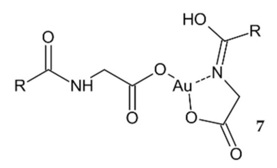
A new bile acid cholylglycinato Au(III) complex 7.
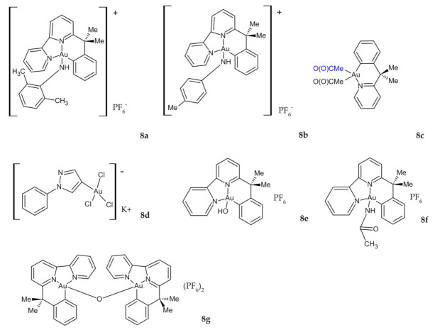
Investigations of the cytotoxicity scores of novel organogold (III) compounds 8a–d (Figure 5) revealed that all of these compounds, except for 8d, are generally stable under physiological conditions and exhibit significant cytotoxic properties on a limited panel of human tumor cell lines. However, negligible anticancer effects (Table 2) were generally measured on the HT29 line compared to those of cisplatin and oxaliplatin [103]. Massai et al. published further work on derivatives of these cyclometallated complexes 8e,g (Figure 5). All three compounds, especially 8g, cause moderate, but still significant. antiproliferative effects toward HCT-116 cancer cells, accompanied by a strong induction of apoptosis and a G0/G1 cell cycle arrest (Table 2). Given the fact that all these effects were greater on CRC cell line HCT-116 compared to the normal L-929 fibroblast cell line, there is a possibility to further investigate and characterize 8g as a potential anticancer agent on a larger and more complex panel of cancer cell lines, as well as to evaluate the mechanisms of its different toxicity effects between in vitro models of tumoral and healthy tissue [104].
Figure 5.
Novel organogold (III) compounds 8a–g.
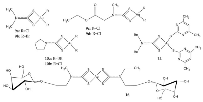
Considering the ability of dithiocarbamates to act as chelating ligands, many examples of Au(III) dithiocarbamate derivatives have been reported. The first more promising studies on the cytotoxic activity of Au(III) compounds in CRC were published by Ronconi et al., which described some of Au(I) and Au(III) complexes with dithiocarbamate ligands (DMDT = N,Ndimethyldithiocarbamate; DMDTM = S-methyl-N,N-dimethyldithiocarbamate; ESDT = thylsarcosinedithiocarbamate). Their preliminary studies have shown that the biological activity of the compounds should generally be attributed to the presence of the Au(III) metal center, and that the Au(I) compounds produce a less pronounced inhibition of cell growth compared to Au(III) analogues. Four complexes 9a–d (Figure 6) were selected for further in vitro cytotoxicity testing. Data regarding their in vitro antiproliferative activity against colon adenocarcinoma cell lines (LoVo), which are notoriously not very sensitive to cisplatin, are extremely interesting, because these new Au(III) complexes seem to also be cytotoxic against tumor cell lines resistant to cisplatin, overcoming their intrinsic resistance and supporting the hypothesis of a different mechanism of action. The IC50 values of investigated compounds ranged from 2.4 nM to 7.9 µΜ (Table 2), while cisplatin’s IC50 value was 56 µΜ [105,106].
Figure 6.
Au(III) dithiocarbamate derivatives 9a–d, 10a,b, 11, 16.
Treatment with the other two Au(III) compounds based on the pyrrolidinedithiocarbamates (PDT), 10a,b (Figure 6), showed a rapid (three-hour) dose-dependent decrease in cell viability in the HCT-116 colorectal carcinoma cells. It was found that the bromide derivative 10a was more effective than the chloride one 10b in inducing cell death and acting via elicited oxidative stress, with effects on the permeability transition pore, a mitochondrial channel whose opening leads to cell death (Table 2). Cisplatin did not show any cytotoxicity under the same experimental conditions [107]. Greater cytotoxicity of bromides is in agreement with the findings reported by Casini et al. [108] for other Au(III) anticancer agents.
Mixed thiolate–dithiocarbamate Au(III) complexes display high antiproliferative activity against colon cancer cell line Caco-2/TC7, without affecting differentiated enterocytes. The most promising derivative 11 (Figure 6) is characterized by a much higher cytotoxicity compared to cisplatin, and slightly higher than auranorfin (Table 2). Although it was assumed that TrxR was a potential target of the dithiocarbamate Au complexes, this was not supported, as reactive oxygen species (ROS) levels and TrxR activity remained unchanged during the experiment. Cell death studies showed that the complexes induced changes in mitochondrial membrane potential, cytochrome C release and caspase-3 activation. The complexes are characterized by high stability under physiological conditions, which gives the opportunity to develop new cytostatics in the treatment of colorectal cancer with the proteasome as a possible target [88].
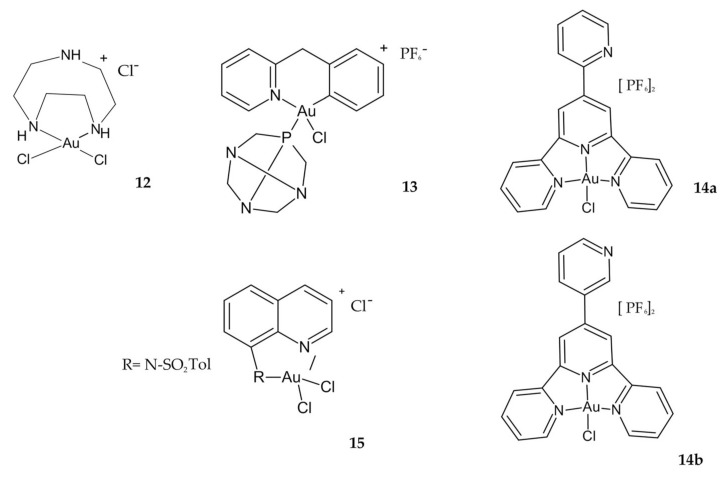
Another study showing high cytotoxicity against the colon cancer cell line was reported by Shi et al. Compared to cisplatin, Au(III) compound 12 (Figure 7) has demonstrated higher cytotoxicity for HCT-116 cell lines at all the concentrations used in the studies. At the concentration of 106 M, the compound showed 30% inhibition against the HCT-116 cell line, while at the same concentration, cisplatin shows 20% inhibition. Additionally, it has been shown that the compound 12 can induce DNA double helix distortion in its mechanism of action [86].
Figure 7.
Au(III) complexes 12, 13, 14a,b, 15.
The new Au(III) cyclometallated phosphine derivative 13, with PTA = 1,3,5-triazaphosphaadamantane ligand (Figure 7), which is not cytotoxic but is known in general to improve water solubility, has been proposed as a new antineoplastic agent in colorectal cancer. The derivative 13 was the most active of all investigated complexes in the study, twice as toxic as cisplatin against HCT-116 p53+/+ cells, and poorly effective on HCT-116 p53−/− (Table 2). This result suggests a similar dependence on p53 pathways for Au(III) complex as for cisplatin. Interestingly, compound 13 inhibited the zinc finger enzyme PARP-1 in nM concentrations, suggesting the possible design of selective inhibitors and the use of organometallic Au compounds in combination therapies with other anticancer drugs [89].
Both Au(III) complexes 14a and 14b incorporating 2,2′:6′,2′′-terpyridine ligand (Figure 7), showed excellent antiproliferative activities against HCT-116, higher than the free ligands and cisplatin. Additionally, compound 14a showed high selectivity against HCT-116 and HCT-116p53 −/− cells, confirmed by the selectivity index (SI). Most interestingly, the complex 14a exhibited proapoptotic activation, while 14b displayed pronecrotic actions [109].
Au(III) complexes containing quinoline ligands at position 8 with different groups arose due to the broad spectrum of medical applications of 8-hydroxyquinoline. It was found that compound 15 with an N-tosyl-8-aminoquinoline ligand (Figure 7) is the most active of the synthesized complexes in all cancer cell lines tested, including the cisplatin-resistant WiDr cell line, and acts by interacting with proteins. Moreover, this complex has proved to be the most stable compound in DMSO and saline solution, even after several hours [110].
The latest available work in this area describes Au(III) complexes with glycoconjugated dithiocarbamato ligands (among others 16, Figure 6). To improve the selective accumulation of an anticancer metal payload in malignant cells, carbohydrates (D-glucose, D-galactose, and D-mannose) were chosen as targeting agents exploiting the Warburg effect that accounts for the overexpression of glucose-transporter proteins (in particular GLUTs) in the phospholipid bilayer of most cancer cells. Unfortunately, the collected results indicate that the Au(III) complexes are not good substrates for GLUT and are inactive toward HCT-116 cells, with IC50 values higher than 50 µM (Table 2) [111].
2.2. Porphyrin Complexes
More comprehensive and promising results were presented by using porphyrin ligand, which can stabilize the Au(III) ion against demetallation and reduction by the biological reductant glutathione [112].
Preliminary studies of a series of Au(III) tetraarylporphyrin (TPP) derivatives confirmed their stability in the presence of glutathione and demonstrated a much greater potency than cisplatin in killing human cancer cells, including drug-resistant variants [113]. The 17 complex (Figure 8) was selected for further study of its antitumor activity and its mechanism against colon cancer. The investigated compound exhibited marked cytotoxicity against different colon cancer cell lines and IC50 values with 9-fold to 21-fold greater potency than that of cisplatin (Table 3). Furthermore, the 17 complex significantly induced apoptosis and cell cycle arrest and cleaved caspase 3, caspase 7, and poly(ADP-ribose) polymerase; released cytochrome C, and upregulated p53, p21, p27, and Bax. Furthermore, in vivo tests were also carried out [114]. The same complex was further studied by Altaf et al. with promising results. Compound 17 showed very high activity compared to Au(III) complexes of meso-1,2-di(1-naphthyl)-1,2-diaminoethane and cisplatin. It was about 7–8 times more potent than cisplatin against the HCT-15 cancer cell [115].
Figure 8.
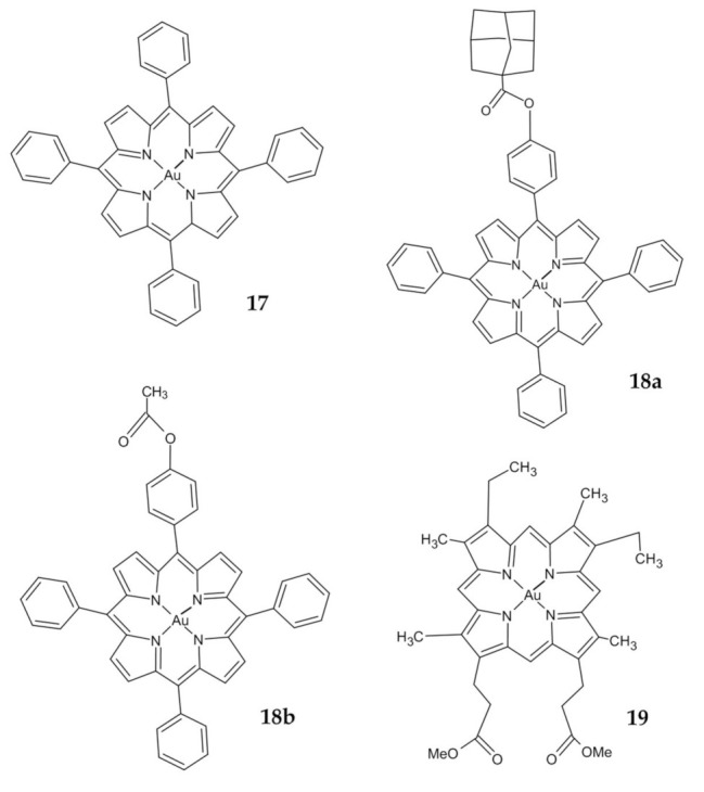
Au(III) porphyrin complexes 17, 18a,b, 19.
Table 3.
Characterization of the anticancer properties of porphyrin complexes.
| Symbol | Proposed Mechanism of Action | Cell Line | IC50 Range (µM) |
|---|---|---|---|
| 17a–e | Inducing apoptosis by a mitochondrial death pathway | SW1116 Colo 205 CRL-238 CCL-2134 HCT-15 HCT-15A2 |
0.20 ± 0.02 0.27 ± 0.02 1.41 ± 0.20 0.65 ± 0.13 0.86 ± 0.15 3.43 ± 0.46 |
| 18a,b | Inducing apoptosis by intrinsic pathway | HT-29 HCT-116 |
17.0 (18a) 3.5 (18b) 16.0 (18a) 3.0 (18b) |
| 19 | Inhibition the Trx, peroxiredoxin and deubiquitinases | HCT-116 NCM460 |
0.06 ± 0.01 1.5 ± 0.15 |
A novel Au(III) porphyrin analogs 18a and 18b (Figure 8) were prepared by modifying one of the peripheral phenyl groups of 17. Results revealed that 18b was more cytotoxic to the colon cancer line than 18a (Table 3). The investigated complexes reduced the survival of human CRC HT-29 and HCT-116 cell lines, caused cell cycle arrest in the G2/M phase, and decreased expression of cyclin B1 and cyclin-dependent kinase 1 (Cdk1) was observed with an increase in regulation of the active form of p53, p21, and Bcl-2 associated with X (Bax) [87]. Furthermore, they induced apoptosis by the intrinsic pathway, as previously described [114].
Tong et al. demonstrate an anticancer activity of Au(III) mesoporphyrin IX dimethyl ester (19) (Figure 8). This compound displayed a higher cytotoxicity in HCT-116 colon cancer cells compared to noncancerous colon epithelial cells (NCM460) with 25-fold differences in IC50 values (Table 3). Promising results from in vivo studies were also reported. The mechanism of action involves modification of the reactive cysteine residues and inhibiting the activity of thioredoxin, peroxiredoxin and deubiquitinases. Crucially, this study revealed that Au(III) induced ligand scaffold reactivity to target the thiol, which could be a useful tool in oncology [93].
2.3. N-Heterocyclic Carbenes (NHCs) Derivatives
A large part of the reviewed papers concerns research on N-heterocyclic carbenes (NHCs) Au complexes. It is widely accepted that the replacement of phosphine ligands by the isolobal NHC ligands frequently improves the properties of the new compounds for practical applications, which are them being water- and air-stable and easier to handle. Moreover, the imidazolium salts as the ligand precursor can be functionalized with almost any substituent, a rarely accessible feature in phosphines [116].
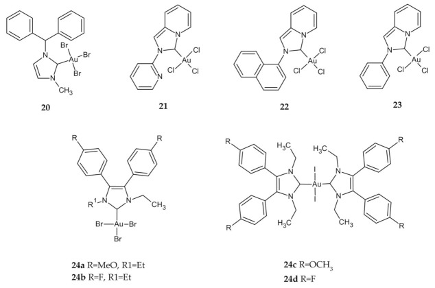
Lemke et al. described the synthesis and antiproliferative activity of a new halide, amino acid and dipeptide NHC Au(I) and NHC Au(III) complexes. In vitro cytostatic effect of compound 20 (Figure 9) was only mild against human colon adenocarcinoma HT-29, with an IC50 value higher than most active complexes and slightly higher compared to cisplatin (Table 4) [116].
Figure 9.
NHCs Au(III) derivatives 20–24.
Table 4.
Characterization of the anticancer properties of N-heterocyclic carbenes (NHCs).
| Symbol | Proposed Mechanism of Action | Cell Line | IC50 Range (µM) |
|---|---|---|---|
| 20 | Undetermined | HT-29 | 12.7 ± 1.2 |
| 21 | Undetermined | HCT-116 | 5.9 ± 3.6 |
| 22 | Undetermined | HCT-116 | 6.78 ± 2.01 |
| 23 | Undetermined | HCT-116 | 21.25 ± 1.37 |
| 24a,b | Inhibition of TrxR | HT-29 | 6.2 ± 1.0 (24a) 7.5 ± 2.9 (24b) |
| 24c,d | Undetermined | HT-29 | 0.26 ± 0.03 (24c) 0.30 ± 0.01 (24d) |
| 25a–g | Multiple molecular targets | HCT-116 | 4.40 ± 1.50 (25a) 1.10 ± 0.28 (25b) 0.49 ± 0.12 (25c) 0.23 ± 0.11 (25d) 0.25 ± 0.10 (25e) 0.52 ± 0.34 (25f) 0.20 ± 0.06 (25g) |
Other groups were evaluated in vitro for the cytotoxicities of Au and Ag NHCs complexes supported by a pyridine, annulated imidazole-2-ylidene (21) [117,118], imidazolium salt 1-naphthyl-2-pyridin-2-yl-2H-imidazo[1,5-a]pyridin-4-ylium hexafluorophosphate (22) [119], pyridyl[1,2-a]{2-acetylylphenylimidazol}-3-ylidene (23) [120] (Figure 9). All complexes of Au(III)–NHC exhibited lower cytotoxicity than Ag(I) and Au(I) complexes for nearly all cell lines tested. Despite that, compounds 21, 22, and 23 were more potent than cisplatin against the HCT-116 cell line (Table 4). Unfortunately, due to the lower cytotoxicity of Au(III)–NHC, studies involving the mechanism of action have not been performed for this complex.
Liu et al. have proposed NHC–Au halide complexes derived from 4,5-diarylimidazoles. The influence of the oxidation state of the metal (Au(I) or Au(III)) is relatively low in general, however, on the HT-29 cell line, the Au(III) complexes 24a and 24b (Figure 9) were less active than their Au(I) congeners, which coincides with earlier papers. All complexes inhibit TrxR with different IC50 values, nevertheless, other targets should be considered as part of the mode of action [121]. Researchers, encouraged by these promising results, have investigated the effect of halide exchange in Au–NHC complexes on their pharmacological properties. The growth-inhibitory effect against HT-29 cells was more than 10-fold higher for complexes 24c and 24d (Figure 9) than that of cisplatin or 5-FU. Moreover, this effect was independent of the oxidation state of Au and type of halides. Although the investigated complexes were successfully accumulated in the neoplastic tissue and localized in large amounts in the cell nucleus, their mode of action has not been clearly defined [122].
Fung et al. described the identification of multiple molecular targets for cyclometalated Au(III) complexes containing NHC ligands using photoaffinity groups. The IC50 values for complexes 25a–g (Figure 10) ranged from 4.4 µM to 0.2 µM in comparison to cisplatin with an IC50 value 11.8 µM (Table 4). Importantly, all the compounds showed much higher cytotoxicity to HCT-116 than to immortalized normal human hepatocyte (MIHA) cells, which may indicate their selectivity. Numerous tests showed the ability of complexes to bind to intracellular proteins such as mitochondrial heat shock protein 60 (HSP60), vimentin (VIM), nucleoside diphosphate kinase A (NDKA), nucleophosmin (NPM), nuclease-sensitive element binding protein (Y box binding protein, YB-1), and peroxiredoxin 1 (PRDX1). The fact that the complexes have multiple molecular targets can minimize the occurrence of drug resistance that is usually encountered with single-target anticancer agents due to naturally occurring genetic mutations [123].
Figure 10.
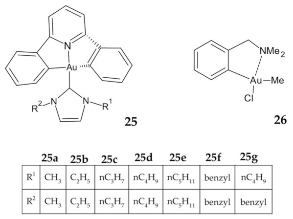
Cyclometalated Au(III) complexes 25, 26.
2.4. Chlorite–Cyanide Complex of Gold (III)
Recently we characterized the synthesis, in vitro safety as well as anticancer activity of a novel chlorite–cyanide complex of gold (III) named TGS121 (patent no: PL422125A1). The compound was prepared as described by Krajewska et al. The obtained Au(III) complex with the formula [Au(CN)4]2 (ClO2)Na is a sodium salt of chloride dioxide associated with Au(III)–cyanide group complex. This complex is water soluble, stable in a neutral pH and can be kept at room temperature. This complex turned out to be stable in cell culture medium and serum. Its relatively low molecular mass (692.5 g/mol) and the form of sodium salt makes it stable in physiological fluids and enables the passage through biological membranes such as the cell membrane. The novel Au(III) compound turned out to exert cytostatic/cytotoxic effects in cancer Ha-Ras transfected NIH3T3 fibroblasts selectively in comparison to the noncancer NIH3T3 cells (Table 5). Since Ras isoforms share 80% identity and share similar activity—one can conclude that in CRC with Ras hyperactivation (mostly K-Ras) (like HCT116), the compound TGS121 would also be effective [124,125].
Table 5.
Characterization of the anticancer properties of TGS121.
| Symbol | Proposed Mechanism of Action | Cell line | IC50 Range (µM) |
|---|---|---|---|
| TGS121 | Apoptosis induction, inhibition of Ras-mediated pathway, cell cycle arrest at G2/M |
Ras-3T3 | 0.231 ± 1.2 |
| NIH3T3 | 5.05 ± 2.6 |
3. In Vivo Studies
Promising cytotoxicity of many Au(III) complexes has been observed against tumor cells in vitro however, to our knowledge, there has been very little evaluation of these compounds on in vivo tumor models.
Thiosalicylate derivative of cycloaurated Au(III) 4 was tested in vivo against the colon HT29 tumor xenograft. Its cytotoxicity did not translate into in vivo pharmacological activity, with only modest inhibition of tumor growth. The lack of activity could in part be explained by the poor solubility of this complex, indicating that further work is necessary to improve both solubility and lipophilicity in order to improve biodistribution [100].
The second compound tested in vivo was an organogold(III) complex 26 (Figure 10), which exhibits promising in vitro cytotoxicity, but it failed to inhibit in vivo tumor growth in HT29 colon cancer xenografts [126].
Only two described Au(III) compounds exhibited favorable antitumor properties both in vitro and in vivo. The tetraarylporphyrin Au(III) complex 17, after intraperitoneal injection in mice at doses of 1.5 mg/kg and 3.0 mg/kg, significantly inhibited the proliferation of Colo205 tumor cells, induced apoptosis and inhibited colon cancer tumor growth [114]. The second one, Au(III) mesoporphyrin IX dimethyl ester 19, after intravenous injection twice per week for 21 days in nude mice bearing human colon cancer HCT-116 at doses 2 mg/kg, resulted in suppression of tumor growth by 72% compared to mice treated with vehicle control [93]. Acute toxicity studies have confirmed that the test compounds, in therapeutic concentrations, do not exhibit additional side effects in mice. Compound 19, after being transiently present in the liver and kidneys, was completely excreted in the urine. In contrast, complex 17 accumulated in the liver and kidneys with low urinary excretion [93]. The presented results suggest that these complexes may be a new potential therapeutic drug for colorectal cancer.
4. Scope and Limitations
This article is limited to gold compounds acting on human CRC cell lines, which might poorly represent the clinical disease. However, especially in CRC surgical treatment, neoadjuvant and adjuvant therapy is applied to reduce the micrometastatic area, and in this context, cell-based assays, especially clonogene assays, can mimic the in vivo situation [35]. We are observing a significant increase in publications on gold complexes. This indicates a great interest in this subject. Review work is also needed to systematize and summarize knowledge about this group of compounds.
5. Conclusions
Metallodrugs are very promising due to the fact that, depending on the choice of metal, its oxidation state, type, and number of coordinated ligands, we obtain a unique mechanism of drug action [72]. Therapies based on Au(III) compounds are particularly interesting and are currently being intensively developed due to their structural similarity to Pt(II) [28].
Colorectal cancer was shown to arise as a result of multiple genetic alternations in both oncogenes and tumor suppressor genes. If the initiated colon epithelium cells bearing mutations in APC or k-Ras can still control the DNA repair due to presence of active and functional p53, we talk about early and late adenoma—that is, nonmalignant and not-yet invasive. However, after genetic alternations that render the p53 pathway inactive, the initiated cells continue their unhampered divisions, despite DNA errors, and become malignant colon cancer cells [8]. That is why in colon cancer therapy, metal-based chemotherapeutics such as platin derivatives are used. Such drugs interfere with DNA, initiating cell-division catastrophe and subsequently cancer-cell death. The effectiveness of established metallodrugs is not always good due to resistance development in genetically instable cancer cells. In this context, novel drugs are needed.
The efficacy of Au(III) in the treatment of CRC has been proven in many in vitro assays using various colon cancer cell lines such as HT29, HT-116, COLO 205, and many others. Au(III) complexes are characterized by high efficiency, selectivity towards cancer cells, and compared to cisplatin display reduced toxicity, a broader spectrum of activity and the ability to overcome tumor resistance [57].
Given the structural and electronic similarity of Au(III) complexes to cisplatin and Pt-related anticancer drugs, it was assumed that their mechanism of action involves binding to DNA. However, it appears that DNA may not be the primary biological target of Au(III) complexes due to studies reporting low DNA binding affinity [115]. Although TrxR inhibition was found to be the main pathway of potent antitumor activity of many Au-NHC complexes [127], the reviewed studies on Au(III)–NHC complexes do not support this mode of action. Notwithstanding the fact that we observe a great deal of scientific interest in Au compounds as drug candidates, we still have insufficient data to describe modes of action and the mechanisms they involve [128].
So far, only two Au(III) derivatives are characterized by high cytotoxicity for colon cancer cells, confirmed by both in vitro and in vivo assays. More advances in metallodrug studies are expected to improve the therapeutic potential of Au(III) in colorectal cancer treatment.
Author Contributions
Conceptualization, A.G. and P.T.; writing—original draft preparation, A.G.; writing—review and editing, P.T., I.M.-B. and M.S.; supervision, M.B.-Z.; funding acquisition, P.T. and J.F. All authors have read and agreed to the published version of the manuscript.
Funding
This research was supported by the grant from the National Science Center (2017/25/B/NZ5/02848 to J.F.) and the Medical University of Lodz (#503/1-156-04/503-11-001-19 to J.F.). The research was funded by grant No. 1M15/3/M/MG/N/20 to P.T. The grant was supervised by I.M.-B. and was funded by a subsidy for science received by the Medical University of Warsaw.
Institutional Review Board Statement
Not applicable.
Informed Consent Statement
Not applicable.
Data Availability Statement
Data are contained within the article.
Conflicts of Interest
The authors declare no conflict of interest.
Footnotes
Publisher’s Note: MDPI stays neutral with regard to jurisdictional claims in published maps and institutional affiliations.
References
- 1.Bray F., Ferlay J., Soerjomataram I., Siegel R.L., Torre L.A., Jemal A. Global cancer statistics 2018: GLOBOCAN estimates of incidence and mortality worldwide for 36 cancers in 185 countries. CA Cancer J. Clin. 2018;68:394–424. doi: 10.3322/caac.21492. [DOI] [PubMed] [Google Scholar]
- 2.Sung H., Ferlay J., Siegel R.L., Laversanne M., Soerjomataram I., Jemal A., Bray F. Global Cancer Statistics 2020: GLOBOCAN Estimates of Incidence and Mortality Worldwide for 36 Cancers in 185 Countries. CA Cancer J. Clin. 2021;71:209–249. doi: 10.3322/caac.21660. [DOI] [PubMed] [Google Scholar]
- 3.Stewart B., Wild C.P. World Cancer Report 2014. WHO; Geneva, Switzerland: 2014. [Google Scholar]
- 4.Parkin D.M., Bray F., Ferlay J., Pisani P. Global Cancer Statistics, 2002. CA Cancer J. Clin. 2005;55:74–108. doi: 10.3322/canjclin.55.2.74. [DOI] [PubMed] [Google Scholar]
- 5.Ilyas M., Straub J., Tomlinson I.P.M., Bodmer W.F. Genetic pathways in colorectal and other cancers. Eur. J. Cancer. 1999;35:1986–2002. doi: 10.1016/S0959-8049(99)00298-1. [DOI] [PubMed] [Google Scholar]
- 6.Rawla P., Sunkara T., Barsouk A. Epidemiology of colorectal cancer: Incidence, mortality, survival, and risk factors. Prz. Gastroenterol. 2019;14:89–103. doi: 10.5114/pg.2018.81072. [DOI] [PMC free article] [PubMed] [Google Scholar]
- 7.Sideris M., Papagrigoriadis S. Molecular Biomarkers and Classification Models in the Evaluation of the Prognosis of Col-orectal Cancer. Anticancer Res. 2014;34:2061–2068. [PubMed] [Google Scholar]
- 8.Fearon E.R., Vogelstein B. A genetic model for colorectal tumorigenesis. Cell. 1990;61:759–767. doi: 10.1016/0092-8674(90)90186-I. [DOI] [PubMed] [Google Scholar]
- 9.Siegel R., DeSantis C., Virgo K., Stein K., Mariotto A., Smith T., Cooper D., Gansler T., Lerro C., Fedewa S., et al. Cancer treatment and survivorship statistics, 2012. CA: A Cancer J. Clin. 2012;62:220–241. doi: 10.3322/caac.21149. [DOI] [PubMed] [Google Scholar]
- 10.Pawelec G. Immunosenescence and cancer. Biogerontology. 2017;18:717–721. doi: 10.1007/s10522-017-9682-z. [DOI] [PubMed] [Google Scholar]
- 11.Anisimov V.N., Sikora E., Pawelec G. Relationships between cancer and aging: A multilevel approach. Biogerontology. 2009;10:323–338. doi: 10.1007/s10522-008-9209-8. [DOI] [PubMed] [Google Scholar]
- 12.Brenner H., Kloor M., Pox C.P. Colorectal cancer. Lancet. 2014;383:1490–1502. doi: 10.1016/S0140-6736(13)61649-9. [DOI] [PubMed] [Google Scholar]
- 13.Mármol I., Sánchez-De-Diego C., Pradilla Dieste A., Cerrada E., Rodriguez Yoldi M. Colorectal Carcinoma: A General Overview and Future Perspectives in Colorectal Cancer. Int. J. Mol. Sci. 2017;18:197. doi: 10.3390/ijms18010197. [DOI] [PMC free article] [PubMed] [Google Scholar]
- 14.Levin B., Lieberman D.A., McFarland B., Andrews K.S., Brooks D., Bond J., Dash C., Giardiello F.M., Glick S., Johnson D., et al. Screening and Surveillance for the Early Detection of Colorectal Cancer and Adenomatous Polyps, 2008: A Joint Guideline from the American Cancer Society, the US Multi-Society Task Force on Colorectal Cancer, and the American College of Radiology. CA Cancer J. Clin. 2008;58:130–160. doi: 10.3322/CA.2007.0018. [DOI] [PubMed] [Google Scholar]
- 15.Eaden J.A., Abrams K.R., Mayberry J.F. The risk of colorectal cancer in ulcerative colitis: A meta-analysis. Gut. 2001;48:526–535. doi: 10.1136/gut.48.4.526. [DOI] [PMC free article] [PubMed] [Google Scholar]
- 16.Canavan C., Abrams K., Mayberry J. Meta-analysis: Colorectal and small bowel cancer risk in patients with Crohn’s disease. Aliment. Pharmacol. Ther. 2006;23:1097–1104. doi: 10.1111/j.1365-2036.2006.02854.x. [DOI] [PubMed] [Google Scholar]
- 17.Martinez-Useros J., Garcia-Foncillas J. Obesity and colorectal cancer: Molecular features of adipose tissue. J. Transl. Med. 2016;14:1–12. doi: 10.1186/s12967-016-0772-5. [DOI] [PMC free article] [PubMed] [Google Scholar]
- 18.Willett W.C. Diet and Cancer: An Evolving Picture. JAMA. 2005;293:233–234. doi: 10.1001/jama.293.2.233. [DOI] [PubMed] [Google Scholar]
- 19.Pöschl G., Seitz H.K. Alcohol and Cancer. Alcohol. Alcohol. 2004;39:155–165. doi: 10.1093/alcalc/agh057. [DOI] [PubMed] [Google Scholar]
- 20.Kuipers E.J., Grady W.M., Lieberman D., Seufferlein T., Sung J.J., Boelens P.G., Van De Velde C.J.H., Watanabe T. Colorectal cancer. Nat. Rev. Dis. Primers. 2015;1:15065. doi: 10.1038/nrdp.2015.65. [DOI] [PMC free article] [PubMed] [Google Scholar]
- 21.Singhal S., Nie S., Wang M.D. Nanotechnology Applications in Surgical Oncology. Annu. Rev. Med. 2010;61:359–373. doi: 10.1146/annurev.med.60.052907.094936. [DOI] [PMC free article] [PubMed] [Google Scholar]
- 22.Kekelidze M., D’Errico L., Pansini M., Tyndall A., Hohmann J. Colorectal Cancer: Current Imaging Methods and Future Perspectives for the Diagnosis, Staging and Therapeutic Response Evaluation. World J. Gastroenterol. 2013;19:8502. doi: 10.3748/wjg.v19.i46.8502. [DOI] [PMC free article] [PubMed] [Google Scholar]
- 23.Colussi D., Brandi G., Bazzoli F., Ricciardiello L. Molecular Pathways Involved in Colorectal Cancer: Implications for Disease Behavior and Prevention. Int. J. Mol. Sci. 2013;14:16365–16385. doi: 10.3390/ijms140816365. [DOI] [PMC free article] [PubMed] [Google Scholar]
- 24.Markowska A., Kasprzak B., Jaszczyńska-Nowinka K., Lubin J., Markowska J. Noble Metals in Oncology. Contemp. Oncol. 2015;19:271. doi: 10.5114/wo.2015.54386. [DOI] [PMC free article] [PubMed] [Google Scholar]
- 25.Ndagi U., Mhlongo N., Soliman M.E. Metal Complexes in Cancer Therapy–an Update from Drug Design Perspective. Drug Des. Devel. Ther. 2017;11:599. doi: 10.2147/DDDT.S119488. [DOI] [PMC free article] [PubMed] [Google Scholar]
- 26.Wang A.Z., Langer R., Farokhzad O.C. Nanoparticle Delivery of Cancer Drugs. Annu. Rev. Med. 2012;63:185–198. doi: 10.1146/annurev-med-040210-162544. [DOI] [PubMed] [Google Scholar]
- 27.Nanospectra Biosciences, Inc. A Pilot Study of AuroLase(Tm) Therapy in Patients with Refractory and/or Recurrent Tumors of the Head and Neck. [(accessed on 15 December 2021)];2016 Available online: clinicaltrials.gov.
- 28.Shaw C.F. Gold-Based Therapeutic Agents. Chem. Rev. 1999;99:2589–2600. doi: 10.1021/cr980431o. [DOI] [PubMed] [Google Scholar]
- 29.Mármol I., Quero J., Rodríguez-Yoldi M.J., Cerrada E. Gold as a Possible Alternative to Platinum-Based Chemotherapy for Colon Cancer Treatment. Cancers. 2019;11:780. doi: 10.3390/cancers11060780. [DOI] [PMC free article] [PubMed] [Google Scholar]
- 30.Lee M.M., MacKinlay A., Semira C., Schieber C., Yepes A.J.J., Lee B., Wong R., Hettiarachchige C.K.H., Gunn N., Tie J., et al. Stage-based Variation in the Effect of Primary Tumor Side on All Stages of Colorectal Cancer Recurrence and Survival. Clin. Colorectal Cancer. 2018;17:e569–e577. doi: 10.1016/j.clcc.2018.05.008. [DOI] [PubMed] [Google Scholar]
- 31.Benson A.B., Venook A.P., Cederquist L., Chan E., Chen Y.-J., Cooper H.S., Deming D., Engstrom P.F., Enzinger P.C., Fichera A., et al. Colon Cancer, Version 1.2017, NCCN Clinical Practice Guidelines in Oncology. J. Natl. Compr. Cancer Netw. 2017;15:370–398. doi: 10.6004/jnccn.2017.0036. [DOI] [PubMed] [Google Scholar]
- 32.Van Cutsem E., Cervantes A., Nordlinger B., Arnold D., ESMO Guidelines Working Group Metastatic colorectal cancer: ESMO Clinical Practice Guidelines for diagnosis, treatment and follow-up. Ann. Oncol. 2014;25:iii1–iii9. doi: 10.1093/annonc/mdu260. [DOI] [PubMed] [Google Scholar]
- 33.Van Cutsem E., Nordlinger B., Cervantes A. Advanced colorectal cancer: ESMO Clinical Practice Guidelines for treatment. Ann. Oncol. 2010;21:v93–v97. doi: 10.1093/annonc/mdq222. [DOI] [PubMed] [Google Scholar]
- 34.Venook A. Critical Evaluation of Current Treatments in Metastatic Colorectal Cancer. Oncol. 2005;10:250–261. doi: 10.1634/theoncologist.10-4-250. [DOI] [PubMed] [Google Scholar]
- 35.Roth M.T., Zheng S. Neoadjuvant Chemotherapy for Colon Cancer. Cancers. 2020;12:2368. doi: 10.3390/cancers12092368. [DOI] [PMC free article] [PubMed] [Google Scholar]
- 36.Cho K., Wang X., Nie S., Chen Z., Shin D.M. Therapeutic Nanoparticles for Drug Delivery in Cancer. Clin. Cancer Res. 2008;14:1310–1316. doi: 10.1158/1078-0432.CCR-07-1441. [DOI] [PubMed] [Google Scholar]
- 37.Parveen S., Sahoo S.K. Polymeric nanoparticles for cancer therapy. J. Drug Target. 2008;16:108–123. doi: 10.1080/10611860701794353. [DOI] [PubMed] [Google Scholar]
- 38.Balducci L., Ades S. Faculty Opinions recommendation of Adjuvant chemotherapy for colon cancer in the elderly: Moving from evidence to practice. Oncology. 2009;23:162. doi: 10.3410/f.1158215.618336. [DOI] [PubMed] [Google Scholar]
- 39.Banerjee A., Pathak S., Subramanium V.D., Dharanivasan G., Murugesan R., Verma R.S. Strategies for targeted drug delivery in treatment of colon cancer: Current trends and future perspectives. Drug Discov. Today. 2017;22:1224–1232. doi: 10.1016/j.drudis.2017.05.006. [DOI] [PubMed] [Google Scholar]
- 40.Prehn R.T. The inhibition of tumor growth by tumor mass. Cancer Res. 1991;51:2–4. [PubMed] [Google Scholar]
- 41.Smith B.H., Gazda L.S., Conn B.L., Jain K., Asina S., Levine D.M., Parker T.S., Laramore M.A., Martis P.C., Vinerean H.V., et al. Hydrophilic Agarose Macrobead Cultures Select for Outgrowth of Carcinoma Cell Populations That Can Restrict Tumor Growth. Cancer Res. 2011;71:725–735. doi: 10.1158/0008-5472.CAN-10-2258. [DOI] [PubMed] [Google Scholar]
- 42.Ocean A.J., Parikh T., Berman N., Escalon J., Shah M.A., Andrada Z., Akahoho E., Pogoda J.M., Stoms G.B., Escobia V.B. Phase I/II Trial of Intraperitoneal Implantation of Agarose-Agarose Macrobeads (MB) Containing Mouse Renal Adenocar-cinoma Cells (RENCA) in Patients (Pts) with Advanced Colorectal Cancer (CRC) American Society of Clinical Oncology; Aleksandria, VA, USA: 2013. [Google Scholar]
- 43.Suh O., Mettlin C., Petrelli N.J. Aspirin use, cancer, and polyps of the large bowel. Cancer. 1993;72:1171–1177. doi: 10.1002/1097-0142(19930815)72:4<1171::AID-CNCR2820720407>3.0.CO;2-D. [DOI] [PubMed] [Google Scholar]
- 44.McDonald B.F., Quinn A.M., Devers T., Cullen A., Coulter I.S., Marison I.W., Loughran S. In-vitro characterisation of a novel celecoxib microbead formulation for the treatment and prevention of colorectal cancer. J. Pharm. Pharmacol. 2015;67:685–695. doi: 10.1111/jphp.12372. [DOI] [PubMed] [Google Scholar]
- 45.Lev-Ari S., Strier L., Kazanov D., Madar-Shapiro L., Dvory-Sobol H., Pinchuk I., Marian B., Lichtenberg D., Arber N. Celecoxib and Curcumin Synergistically Inhibit the Growth of Colorectal Cancer Cells. Clin. Cancer Res. 2005;11:6738–6744. doi: 10.1158/1078-0432.CCR-05-0171. [DOI] [PubMed] [Google Scholar]
- 46.Sah B., Vasiljevic T., McKechnie S., Donkor O. Effect of probiotics on antioxidant and antimutagenic activities of crude peptide extract from yogurt. Food Chem. 2014;156:264–270. doi: 10.1016/j.foodchem.2014.01.105. [DOI] [PubMed] [Google Scholar]
- 47.Choi S.S., Kim Y., Han K.S., You S., Oh S., Kim S.H. Effects of Lactobacillus Strains on Cancer Cell Proliferation and Oxi-dative Stress in Vitro. Lett. Appl. Microbiol. 2006;42:452–458. doi: 10.1111/j.1472-765X.2006.01913.x. [DOI] [PubMed] [Google Scholar]
- 48.Chong E.S.L. A potential role of probiotics in colorectal cancer prevention: Review of possible mechanisms of action. World J. Microbiol. Biotechnol. 2013;30:351–374. doi: 10.1007/s11274-013-1499-6. [DOI] [PubMed] [Google Scholar]
- 49.Dasari S., Tchounwou P.B. Cisplatin in cancer therapy: Molecular mechanisms of action. Eur. J. Pharmacol. 2014;740:364–378. doi: 10.1016/j.ejphar.2014.07.025. [DOI] [PMC free article] [PubMed] [Google Scholar]
- 50.Muhammad N., Guo Z. Metal-based anticancer chemotherapeutic agents. Curr. Opin. Chem. Biol. 2014;19:144–153. doi: 10.1016/j.cbpa.2014.02.003. [DOI] [PubMed] [Google Scholar]
- 51.Komeda S., Casini A. Next-Generation Anticancer Metallodrugs. Curr. Top. Med. Chem. 2012;12:219–235. doi: 10.2174/156802612799078964. [DOI] [PubMed] [Google Scholar]
- 52.Frezza M., Hindo S., Chen D., Davenport A., Schmitt S., Tomco D., Dou Q.P. Novel Metals and Metal Complexes as Platforms for Cancer Therapy. Curr. Pharm. Des. 2010;16:1813–1825. doi: 10.2174/138161210791209009. [DOI] [PMC free article] [PubMed] [Google Scholar]
- 53.Arnesano F., Natile G. Mechanistic insight into the cellular uptake and processing of cisplatin 30 years after its approval by FDA. Coord. Chem. Rev. 2009;253:2070–2081. doi: 10.1016/j.ccr.2009.01.028. [DOI] [Google Scholar]
- 54.Wheate N.J., Walker S., Craig G.E., Oun R. The status of platinum anticancer drugs in the clinic and in clinical trials. Dalton Trans. 2010;39:8113–8127. doi: 10.1039/c0dt00292e. [DOI] [PubMed] [Google Scholar]
- 55.Kelland L. The resurgence of platinum-based cancer chemotherapy. Nat. Rev. Cancer. 2007;7:573–584. doi: 10.1038/nrc2167. [DOI] [PubMed] [Google Scholar]
- 56.Shen D.-W., Pouliot L.M., Hall M.D., Gottesman M.M. Cisplatin Resistance: A Cellular Self-Defense Mechanism Resulting from Multiple Epigenetic and Genetic Changes. Pharmacol. Rev. 2012;64:706–721. doi: 10.1124/pr.111.005637. [DOI] [PMC free article] [PubMed] [Google Scholar]
- 57.Timerbaev A.R., Hartinger C.G., Aleksenko S.S., Keppler B.K. Interactions of Antitumor Metallodrugs with Serum Proteins: Advances in Characterization Using Modern Analytical Methodology. Chem. Rev. 2006;106:2224–2248. doi: 10.1021/cr040704h. [DOI] [PubMed] [Google Scholar]
- 58.De Araújo R.F., de Araújo A.A., Pessoa J.B., Neto F.P.F., da Silva G.R., Oliveira A.L.C.L., Carvalho T.G., Silva H.F.O., Eugênio M., Sant’Anna C., et al. Anti-inflammatory, analgesic and anti-tumor properties of gold nanoparticles. Pharmacol. Rep. 2017;69:119–129. doi: 10.1016/j.pharep.2016.09.017. [DOI] [PubMed] [Google Scholar]
- 59.Aminabad N.S., Farshbaf M., Akbarzadeh A. Recent Advances of Gold Nanoparticles in Biomedical Applications: State of the Art. Cell Biochem. Biophys. 2018;77:123–137. doi: 10.1007/s12013-018-0863-4. [DOI] [PubMed] [Google Scholar]
- 60.Upreti M., Jyoti A., Sethi P. Tumor microenvironment and nanotherapeutics. Transl. Cancer Res. 2013;2:309–319. doi: 10.3978/j.issn.2218-676X.2013.08.11. [DOI] [PMC free article] [PubMed] [Google Scholar]
- 61.Kalaydina R.-V., Bajwa K., Qorri B., DeCarlo A., Szewczuk M.R. Recent advances in “smart” delivery systems for extended drug release in cancer therapy. Int. J. Nanomed. 2018;ume 13:4727–4745. doi: 10.2147/IJN.S168053. [DOI] [PMC free article] [PubMed] [Google Scholar]
- 62.Pérez-Herrero E., Fernández-Medarde A. Advanced targeted therapies in cancer: Drug nanocarriers, the future of chemotherapy. Eur. J. Pharm. Biopharm. 2015;93:52–79. doi: 10.1016/j.ejpb.2015.03.018. [DOI] [PubMed] [Google Scholar]
- 63.Blanco E., Shen H., Ferrari M. Principles of nanoparticle design for overcoming biological barriers to drug delivery. Nat. Biotechnol. 2015;33:941–951. doi: 10.1038/nbt.3330. [DOI] [PMC free article] [PubMed] [Google Scholar]
- 64.Zaki A.A., Hui J.Z., Higbee E., Tsourkas A. Biodistribution, Clearance, and Toxicology of Polymeric Micelles Loaded with 0.9 or 5 Nm Gold Nanoparticles. J. Biomed. Nanotechnol. 2015;11:1836–1846. doi: 10.1166/jbn.2015.2142. [DOI] [PMC free article] [PubMed] [Google Scholar]
- 65.Balasubramanian S.K., Jittiwat J., Manikandan J., Ong C.N., Yu L., Ong W.-Y. Biodistribution of gold nanoparticles and gene expression changes in the liver and spleen after intravenous administration in rats. Biomaterials. 2010;31:2034–2042. doi: 10.1016/j.biomaterials.2009.11.079. [DOI] [PubMed] [Google Scholar]
- 66.Bednarski M., Dudek M., Knutelska J., Nowiński L., Sapa J., Zygmunt M., Nowak G., Luty-Błocho M., Wojnicki M., Fitzner K. The Influence of the Route of Administration of Gold Nanoparticles on Their Tissue Distribution and Basic Bio-chemical Parameters: In Vivo Studies. Pharmacol. Rep. 2015;67:405–409. doi: 10.1016/j.pharep.2014.10.019. [DOI] [PubMed] [Google Scholar]
- 67.Wojnicki M., Luty-Błocho M., Bednarski M., Dudek M., Knutelska J., Sapa J., Zygmunt M., Nowak G., Fitzner K. Tissue distribution of gold nanoparticles after single intravenous administration in mice. Pharmacol. Rep. 2013;65:1033–1038. doi: 10.1016/S1734-1140(13)71086-7. [DOI] [PubMed] [Google Scholar]
- 68.Nobili S., Mini E., Landini I., Gabbiani C., Casini A., Messori L. Gold compounds as anticancer agents: Chemistry, cellular pharmacology, and preclinical studies. Med. Res. Rev. 2009;30:550–580. doi: 10.1002/med.20168. [DOI] [PubMed] [Google Scholar]
- 69.Bertrand B., Williams M.R.M., Bochmann M. Gold(III) Complexes for Antitumor Applications: An Overview. Chem.-A Eur. J. 2018;24:11840–11851. doi: 10.1002/chem.201800981. [DOI] [PubMed] [Google Scholar]
- 70.Casini A., Sun R.W.-Y., Ott I. Medicinal Chemistry of Gold Anticancer Metallodrugs. Met. Ions Life Sci. 2018;18 doi: 10.1515/9783110470734-013. [DOI] [PubMed] [Google Scholar]
- 71.Jurgens S., Kuhn F.E., Casini A. Cyclometalated Complexes of Platinum and Gold with Biological Properties: State-of-the-Art and Future Perspectives. Curr. Med. Chem. 2018;25:437–461. doi: 10.2174/0929867324666170529125229. [DOI] [PubMed] [Google Scholar]
- 72.Lazarevic T., Rilak A., Bugarčić Ž.D. Platinum, palladium, gold and ruthenium complexes as anticancer agents: Current clinical uses, cytotoxicity studies and future perspectives. Eur. J. Med. Chem. 2017;142:8–31. doi: 10.1016/j.ejmech.2017.04.007. [DOI] [PubMed] [Google Scholar]
- 73.Parveen S., Arjmand F., Tabassum S. Development and future prospects of selective organometallic compounds as anticancer drug candidates exhibiting novel modes of action. Eur. J. Med. Chem. 2019;175:269–286. doi: 10.1016/j.ejmech.2019.04.062. [DOI] [PubMed] [Google Scholar]
- 74.Porchia M., Pellei M., Marinelli M., Tisato F., Del Bello F., Santini C. New insights in Au-NHCs complexes as anticancer agents. Eur. J. Med. Chem. 2018;146:709–746. doi: 10.1016/j.ejmech.2018.01.065. [DOI] [PubMed] [Google Scholar]
- 75.Yeo C., Ooi K., Tiekink E. Gold-Based Medicine: A Paradigm Shift in Anti-Cancer Therapy? Molecules. 2018;23:1410. doi: 10.3390/molecules23061410. [DOI] [PMC free article] [PubMed] [Google Scholar]
- 76.Zou T., Lum C.T., Lok C.-N., Zhang J.-J., Che C.-M. Chemical biology of anticancer gold(iii) and gold(i) complexes. Chem. Soc. Rev. 2015;44:8786–8801. doi: 10.1039/C5CS00132C. [DOI] [PubMed] [Google Scholar]
- 77.Ott I. On the medicinal chemistry of gold complexes as anticancer drugs. Coord. Chem. Rev. 2009;253:1670–1681. doi: 10.1016/j.ccr.2009.02.019. [DOI] [Google Scholar]
- 78.Casini A. Exploring the mechanisms of metalbased pharmacological agents via an integrated approach. J. Inorg. Biochem. 2012;109:97–106. doi: 10.1016/j.jinorgbio.2011.12.007. [DOI] [PubMed] [Google Scholar]
- 79.Chen X., Yang Q., Xiao L., Tang D., Dou Q.P., Liu J. Metal-Based Proteasomal Deubiquitinase Inhibitors as Potential An-ticancer Agents. Cancer Metastasis Rev. 2017;36:655–668. doi: 10.1007/s10555-017-9701-1. [DOI] [PMC free article] [PubMed] [Google Scholar]
- 80.Casini A., Messori L. Molecular Mechanisms and Proposed Targets for Selected Anticancer Gold Compounds. Curr. Top. Med. Chem. 2011;11:2647–2660. doi: 10.2174/156802611798040732. [DOI] [PubMed] [Google Scholar]
- 81.Powis G., Montfort W.R. Properties and Biological Activities of Thioredoxins. Annu. Rev. Pharmacol. Toxicol. 2001;41:261–295. doi: 10.1146/annurev.pharmtox.41.1.261. [DOI] [PubMed] [Google Scholar]
- 82.Raffel J., Bhattacharyya A.K., Gallegos A., Cui H., Einspahr J.G., Alberts D.S., Powis G. Increased expression of thioredoxin-1 in human colorectal cancer is associated with decreased patient survival. J. Lab. Clin. Med. 2003;142:46–51. doi: 10.1016/S0022-2143(03)00068-4. [DOI] [PubMed] [Google Scholar]
- 83.Arsenijevic M., Milovanovic M., Volarevic V., Djekovic A., Kanjevac T., Arsenijevic N., Dukic S., Bugarcic Z.D. Cytotoxicity of gold(III) Complexes on A549 Human Lung Carcinoma Epithelial Cell Line. Med. Chem. 2012;8:2–8. doi: 10.2174/157340612799278469. [DOI] [PubMed] [Google Scholar]
- 84.Calamai P., Carotti S., Guerri A., Mazzei T., Messori L., Mini E., Orioli P., Speroni G.P. Cytotoxic effects of gold(III) complexes on established human tumor cell lines sensitive and resistant to cisplatin. Anti-Cancer Drug Des. 1998;13:67–80. [PubMed] [Google Scholar]
- 85.Wilson C.R., Fagenson A.M., Ruangpradit W., Muller M.T., Munro O.Q. Gold (III) Complexes of Pyridyl-and Iso-quinolylamido Ligands: Structural, Spectroscopic, and Biological Studies of a New Class of Dual Topoisomerase I and II In-hibitors. Inorg. Chem. 2013;52:7889–7906. doi: 10.1021/ic400339z. [DOI] [PubMed] [Google Scholar]
- 86.Shi P., Jiang Q., Lin J., Zhao Y., Lin L., Guo Z. Gold(III) compounds of 1,4,7-triazacyclononane showing high cytotoxicity against A-549 and HCT-116 tumor cell lines. J. Inorg. Biochem. 2006;100:939–945. doi: 10.1016/j.jinorgbio.2005.12.020. [DOI] [PubMed] [Google Scholar]
- 87.Dandash F., Léger D.Y., Fidanzi-Dugas C., Nasri S., Brégier F., Granet R., Karam W., Diab-Assaf M., Sol V., Liagre B. In vitro anticancer activity of new gold(III) porphyrin complexes in colon cancer cells. J. Inorg. Biochem. 2017;177:27–38. doi: 10.1016/j.jinorgbio.2017.08.024. [DOI] [PubMed] [Google Scholar]
- 88.Quero J., Cabello S., Fuertes T., Mármol I., Laplaza R., Polo V., Gimeno M.C., Rodriguez-Yoldi M.J., Cerrada E. Proteasome versus Thioredoxin Reductase Competition as Possible Biological Targets in Antitumor Mixed Thiolate-Dithiocarbamate Gold(III) Complexes. Inorg. Chem. 2018;57:10832–10845. doi: 10.1021/acs.inorgchem.8b01464. [DOI] [PubMed] [Google Scholar]
- 89.Bertrand B., Spreckelmeyer S., Bodio E., Cocco F., Picquet M., Richard P., Le Gendre P., Orvig C., Cinellu M.A., Casini A. Exploring the potential of gold(iii) cyclometallated compounds as cytotoxic agents: Variations on the C^N theme. Dalton Trans. Camb. Engl. 2015;44:11911–11918. doi: 10.1039/C5DT01023C. [DOI] [PubMed] [Google Scholar]
- 90.Erdogan E., Lamark T., Stallings-Mann M., Jamieson L., Pellechia M., Thompson E.A., Johansen T., Fields A.P. Aurothiomalate Inhibits Transformed Growth by Targeting the PB1 Domain of Protein Kinase Cι. J. Biol. Chem. 2006;281:28450–28459. doi: 10.1074/jbc.M606054200. [DOI] [PubMed] [Google Scholar]
- 91.Islam S.M.A., Patel R., Acevedo-Duncan M. Protein Kinase C-ζ stimulates colorectal cancer cell carcinogenesis via PKC-ζ/Rac1/Pak1/β-Catenin signaling cascade. Biochim. Biophys. Acta Mol. Cell Res. 2018;1865:650–664. doi: 10.1016/j.bbamcr.2018.02.002. [DOI] [PubMed] [Google Scholar]
- 92.Wang Y., He Q.-Y., Che C.M., Tsao S.W., Sun R.W.-Y., Chiu J.-F. Modulation of gold(III) porphyrin 1a-induced apoptosis by mitogen-activated protein kinase signaling pathways. Biochem. Pharmacol. 2008;75:1282–1291. doi: 10.1016/j.bcp.2007.11.024. [DOI] [PubMed] [Google Scholar]
- 93.Tong K.-C., Lok C.-N., Wan P.-K., Hu D., Fung Y.M.E., Chang X.-Y., Huang S., Jiang H., Che C.-M. An anticancer gold(III)-activated porphyrin scaffold that covalently modifies protein cysteine thiols. Proc. Natl. Acad. Sci. USA. 2020;117:1321–1329. doi: 10.1073/pnas.1915202117. [DOI] [PMC free article] [PubMed] [Google Scholar]
- 94.Mirabelli C.K., Sung C.-M., Zimmerman J.P., Hill D.T., Mong S., Crooke S.T. Interactions of gold coordination complexes with DNA. Biochem. Pharmacol. 1986;35:1427–1433. doi: 10.1016/0006-2952(86)90106-1. [DOI] [PubMed] [Google Scholar]
- 95.Mirabelli C.K., Zimmerman J.P., Bartus H.R., Chiu-Mei S., Crooke S.T. Inter-strand cross-links and single-strand breaks produced by gold(I) and gold(III) coordination complexes. Biochem. Pharmacol. 1986;35:1435–1443. doi: 10.1016/0006-2952(86)90107-3. [DOI] [PubMed] [Google Scholar]
- 96.Bindoli A., Rigobello M.P., Scutari G., Gabbiani C., Casini A., Messori L. Thioredoxin reductase: A target for gold compounds acting as potential anticancer drugs. Coord. Chem. Rev. 2009;253:1692–1707. doi: 10.1016/j.ccr.2009.02.026. [DOI] [Google Scholar]
- 97.Nardon C., Boscutti G., Fregona D. Beyond platinums: Gold complexes as anticancer agents. Anticancer. Res. 2014;34:487–492. [PubMed] [Google Scholar]
- 98.Buckley R.G., Elsome A.M., Fricker S.P., Henderson G.R., Theobald B.R.C., Parish R.V., Howe A.B.P., Kelland L.R. Antitumor Properties of Some 2-[(Dimethylamino)methyl]phenylgold(III) Complexes. J. Med. Chem. 1996;39:5208–5214. doi: 10.1021/jm9601563. [DOI] [PubMed] [Google Scholar]
- 99.Parish R.V., Howe B.P., Wright J.P., Mack J., Pritchard R.G., Buckley R.G., Elsome A.M., Fricker S.P. Chemical and Bio-logical Studies of Dichloro (2-((Dimethylamino) Methyl) Phenyl) Gold (III) Inorg. Chem. 1996;35:1659–1666. doi: 10.1021/ic950343b. [DOI] [PubMed] [Google Scholar]
- 100.Zhu Y., Cameron B.R., Mosi R., Anastassov V., Cox J., Qin L., Santucci Z., Metz M., Skerlj R.T., Fricker S.P. Inhibition of the Cathepsin Cysteine Proteases B and K by Square-Planar Cycloaurated Gold (III) Compounds and Investigation of Their Anti-Cancer Activity. J. Inorg. Biochem. 2011;105:754–762. doi: 10.1016/j.jinorgbio.2011.01.012. [DOI] [PubMed] [Google Scholar]
- 101.Notash B., Amani V., Safari N., Ostad S.N., Abedi A., Dehnavi M.Z. The Influence of Steric Effects on Intramolecular Secondary Bonding Interactions; Cytotoxicity in Gold (III) Bithiazole Complexes. Dalton Trans. 2013;42:6852–6858. doi: 10.1039/c3dt00073g. [DOI] [PubMed] [Google Scholar]
- 102.Carrasco J., Criado J.J., Macias R., Manzano J.L., Marin J., Medarde M., Rodríguez E. Structural characterization and cytostatic activity of chlorobischolylglycinatogold(III) J. Inorg. Biochem. 2001;84:287–292. doi: 10.1016/S0162-0134(01)00172-6. [DOI] [PubMed] [Google Scholar]
- 103.Messori L., Marcon G., Cinellu M.A., Coronnello M., Mini E., Gabbiani O., Orioli P. Solution chemistry and cytotoxic properties of novel organogold(III) compounds. Bioorganic Med. Chem. 2004;12:6039–6043. doi: 10.1016/j.bmc.2004.09.014. [DOI] [PubMed] [Google Scholar]
- 104.Massai L., Cirri D., Michelucci E., Bartoli G., Guerri A., Cinellu M.A., Cocco F., Gabbiani C., Messori L. Organogold(III) compounds as experimental anticancer agents: Chemical and biological profiles. BioMetals. 2016;29:863–872. doi: 10.1007/s10534-016-9957-x. [DOI] [PubMed] [Google Scholar]
- 105.Ronconi L., Giovagnini L., Marzano C., Bettìo F., Graziani R., Pilloni G., Fregona D. Gold Dithiocarbamate Derivatives as Potential Antineoplastic Agents: Design, Spectroscopic Properties, and in Vitro Antitumor Activity. Inorg. Chem. 2005;44:1867–1881. doi: 10.1021/ic048260v. [DOI] [PubMed] [Google Scholar]
- 106.Ronconi L., Fregona D. The Midas touch in cancer chemotherapy: From platinum- to gold-dithiocarbamato complexes. Dalton Trans. 2009:10670–10680. doi: 10.1039/b913597a. [DOI] [PubMed] [Google Scholar]
- 107.Nardon C., Chiara F., Brustolin L., Gambalunga A., Ciscato F., Rasola A., Trevisan A., Fregona D. Gold(III)-pyrrolidinedithiocarbamato Derivatives as Antineoplastic Agents. ChemistryOpen. 2015;4:183–191. doi: 10.1002/open.201402091. [DOI] [PMC free article] [PubMed] [Google Scholar]
- 108.Casini A., Kelter G., Gabbiani C., Cinellu M.A., Minghetti G., Fregona D., Fiebig H.-H., Messori L. Chemistry, antiproliferative properties, tumor selectivity, and molecular mechanisms of novel gold(III) compounds for cancer treatment: A systematic study. JBIC J. Biol. Inorg. Chem. 2009;14:1139–1149. doi: 10.1007/s00775-009-0558-9. [DOI] [PubMed] [Google Scholar]
- 109.Czerwińska K., Golec M., Skonieczna M., Palion-Gazda J., Zygadło D., Szlapa-Kula A., Krompiec S., Machura B., Szurko A. Cytotoxic gold(iii) complexes incorporating a 2,2′:6′,2′′-terpyridine ligand framework—The impact of the substituent in the 4′-position of a terpy ring. Dalton Trans. 2017;46:3381–3392. doi: 10.1039/C6DT04584G. [DOI] [PubMed] [Google Scholar]
- 110.Casado-Sánchez A., Martín-Santos C., Padrón J.M., Mas-Ballesté R., Navarro-Ranninger C., Alemán J., Cabrera S. Effect of Electronic and Steric Properties of 8-Substituted Quinolines in Gold (III) Complexes: Synthesis, Electrochemistry, Stability, Interactions and Antiproliferative Studies. J. Inorg. Biochem. 2017;174:111–118. doi: 10.1016/j.jinorgbio.2017.06.004. [DOI] [PubMed] [Google Scholar]
- 111.Pettenuzzo N., Brustolin L., Coltri E., Gambalunga A., Chiara F., Trevisan A., Biondi B., Nardon C., Fregona D. CuIIand AuIIIComplexes with Glycoconjugated Dithiocarbamato Ligands for Potential Applications in Targeted Chemotherapy. ChemMedChem. 2019;14:1162–1172. doi: 10.1002/cmdc.201900226. [DOI] [PubMed] [Google Scholar]
- 112.Sun R.W.-Y., Li C.K.-L., Ma D.-L., Yan J.J., Lok C.-N., Leung C.-H., Zhu N., Che C.-M. Stable Anticancer Gold(III)-Porphyrin Complexes: Effects of Porphyrin Structure. Chem.-A Eur. J. 2010;16:3097–3113. doi: 10.1002/chem.200902741. [DOI] [PubMed] [Google Scholar]
- 113.Che C.-M., Sun R.W.-Y., Yu W.-Y., Ko C.-B., Zhu N., Sun H. Gold(iii) porphyrins as a new class of anticancer drugs: Cytotoxicity, DNA binding and induction of apoptosis in human cervix epitheloid cancer cellsElectronic supplementary information (ESI) available: Further experimental and crystallographic details. Chem. Commun. 2003:1718–1719. doi: 10.1039/b303294a. [DOI] [PubMed] [Google Scholar]
- 114.Tu S., Sun R.W.-Y., Lin M.C.M., Cui J.T., Zou B., Gu Q., Kung H.-F., Che C.M., Wong B.C.Y. Gold (III) porphyrin complexes induce apoptosis and cell cycle arrest and inhibit tumor growth in colon cancer. Cancer. 2009;115:4459–4469. doi: 10.1002/cncr.24514. [DOI] [PubMed] [Google Scholar]
- 115.Altaf M., Ahmad S., Kawde A.-N., Baig N., Alawad A., Altuwaijri S., Stoeckli-Evans H., Isab A.A. Synthesis, Structural Characterization, Electrochemical Behavior and Anticancer Activity of Gold (Iii) Complexes of Meso-1, 2-Di (1-Naphthyl)-1, 2-Diaminoethane and Tetraphenylporphyrin. New J. Chem. 2016;40:8288–8295. doi: 10.1039/C6NJ00692B. [DOI] [Google Scholar]
- 116.Lemke J., Pinto A., Niehoff P., Vasylyeva V., Metzler-Nolte N. Synthesis, structural characterisation and anti-proliferative activity of NHC gold amino acid and peptide conjugates. Dalton Trans. 2009:7063–7070. doi: 10.1039/b906140a. [DOI] [PubMed] [Google Scholar]
- 117.Dinda J., Samanta T., Nandy A., Saha K.D., Seth S.K., Chattopadhyay S.K., Bielawski C.W. N-Heterocyclic Carbene Sup-ported Au (I) and Au(III) Complexes: A Comparison of Cytotoxicities. New J. Chem. 2014;38:1218–1224. doi: 10.1039/C3NJ01463K. [DOI] [Google Scholar]
- 118.Rana B.K., Nandy A., Bertolasi V., Bielawski C.W., Saha K.D., Dinda J. Novel Gold(I)– and Gold(III)–N-Heterocyclic Carbene Complexes: Synthesis and Evaluation of Their Anticancer Properties. Organometallics. 2014;33:2544–2548. doi: 10.1021/om500118x. [DOI] [Google Scholar]
- 119.Samanta T., Munda R.N., Roymahapatra G., Nandy A., Saha K.D., Al-Deyab S.S., Dinda J. Silver (I), Gold (I) and Gold (III)-N-Heterocyclic Carbene Complexes of Naphthyl Substituted Annelated Ligand: Synthesis, Structure and Cytotoxicity. J. Organomet. Chem. 2015;791:183–191. doi: 10.1016/j.jorganchem.2015.05.049. [DOI] [Google Scholar]
- 120.Jhulki L., Dutta P., Santra M.K., Cardoso M.H., Oshiro K.G., Franco O.L., Bertolasi V., Isab A.A., Bielawski C.W., Dinda J. Synthesis and Cytotoxic Characteristics Displayed by a Series of Ag(i)-, Au(i)-and Au(Iii)-Complexes Supported by a Common N-Heterocyclic Carbene. New J. Chem. 2018;42:13948–13956. doi: 10.1039/C8NJ02008F. [DOI] [Google Scholar]
- 121.Liu W., Bensdorf K., Proetto M., Abram U., Hagenbach A., Gust R. NHC Gold Halide Complexes Derived from 4,5-Diarylimidazoles: Synthesis, Structural Analysis, and Pharmacological Investigations as Potential Antitumor Agents. J. Med. Chem. 2011;54:8605–8615. doi: 10.1021/jm201156x. [DOI] [PubMed] [Google Scholar]
- 122.Liu W., Bensdorf K., Proetto M., Hagenbach A., Abram U., Gust R. Synthesis, Characterization, and in Vitro Studies of Bis [1,3-Diethyl-4, 5-Diarylimidazol-2-Ylidene] Gold (I/III) Complexes. J. Med. Chem. 2012;55:3713–3724. doi: 10.1021/jm3000196. [DOI] [PubMed] [Google Scholar]
- 123.Fung S.K., Zou T., Cao B., Lee P.-Y., Fung Y.M.E., Hu D., Lok C.-N., Che C.-M. Cyclometalated Gold(III) Complexes Containing N-Heterocyclic Carbene Ligands Engage Multiple Anti-Cancer Molecular Targets. Angew. Chem. Int. Ed. 2017;56:3892–3896. doi: 10.1002/anie.201612583. [DOI] [PubMed] [Google Scholar]
- 124.Krajewska J., Włodarczyk J., Jacenik D., Kordek R., Taciak P., Szczepaniak R., Fichna J. New Class of Anti-Inflammatory Therapeutics Based on Gold (III) Complexes in Intestinal Inflammation–Proof of Concept Based on In Vitro and In Vivo Studies. Int. J. Mol. Sci. 2021;22:3121. doi: 10.3390/ijms22063121. [DOI] [PMC free article] [PubMed] [Google Scholar]
- 125.Lipiec S., Szymański P., Gurba A., Szeleszczuk Ł., Taciak P., Fichna J., Młynarczuk-Biały I. Innovative Gold Complexes with CN Group as Anticancer Agents—Possible Mechanisms of Action. In: Młynarczuk-Biały I., Biały Ł., editors. Advances in Biomedical Research—Cancer and Miscellaneous. Wydawnictwo Naukowe Tygiel Sp. z o. o.; Lubin, Poland: 2021. [(accessed on 15 December 2021)]. pp. 9–22. Available online: https://bc.wydawnictwo-tygiel.pl/publikacja/AA60CB34-1A06-4ECD-748B-83D6A9A5B19C. [Google Scholar]
- 126.Engman L., McNaughton M., Gajewska M., Kumar S., Birmingham A., Powis G. Thioredoxin reductase and cancer cell growth inhibition by organogold(III) compounds. Anti-Cancer Drugs. 2006;17:539–544. doi: 10.1097/00001813-200606000-00007. [DOI] [PubMed] [Google Scholar]
- 127.Karaaslan M.G., Aktaş A., Gürses C., Gök Y., Ateş B. Chemistry, structure, and biological roles of Au-NHC complexes as TrxR inhibitors. Bioorganic Chem. 2019;95:103552. doi: 10.1016/j.bioorg.2019.103552. [DOI] [PubMed] [Google Scholar]
- 128.Massai L., Grguric-Sipka S., Liu W., Bertrand B., Pratesi A. Editorial: The Golden Future in Medicinal Chemistry: Perspectives and Resources from Old and New Gold-Based Drug Candidates. Front. Chem. 2021;9 doi: 10.3389/fchem.2021.665244. [DOI] [PMC free article] [PubMed] [Google Scholar]
Associated Data
This section collects any data citations, data availability statements, or supplementary materials included in this article.
Data Availability Statement
Data are contained within the article.


