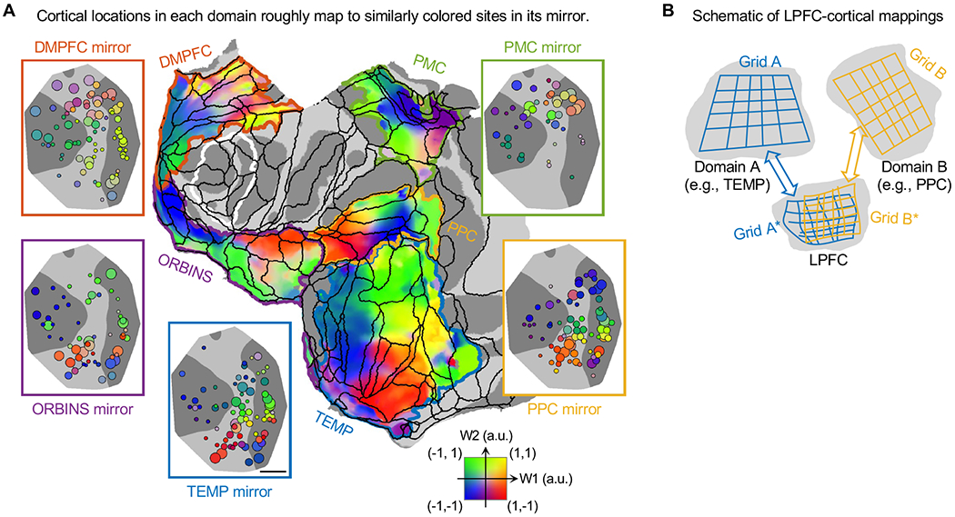
Figure 8. Summary of LPFC-Cortical Connectome: Overlapping Surface-to-Surface Mappings.

(A) Surface-to-surface mappings between LPFC and other cortical domains. Each non-LPFC domain (on flattened surface) and its mirror organization in LPFC (on MDS plane) are enclosed by curved and rectangle contours of the same color, respectively. The mirror organizations in LPFC are W1&2 maps of monkey J from Figures 5 and 6. The same 2D colormap indicates both W1&2 of LPFC sites (as circles) and W1&2 mapped to vertices (as overlay). On flattened surface, white contour encloses the MDS planes in Figures 1C and 1D, and black contours indicate borders of areas in CBCetal15 parcellation. Scale bar, 5 mm.
(B) Schematic of overlapping isomorphic mappings between LPFC and other cortical domains.
