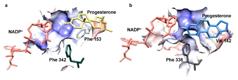
Figure 3.
Docking of progesterone into the active site of AtStR1 (a) and homology-modelled AtStR2 (b). The coulombic surface is displayed for important residues in the active site according to [32]. NADP+ (red), progesterone (yellow for AtStR1, blue for AtStR2) and the phenylalanine clamp residues (green for AtStR1, grey for AtStR2) are shown as stick models.

