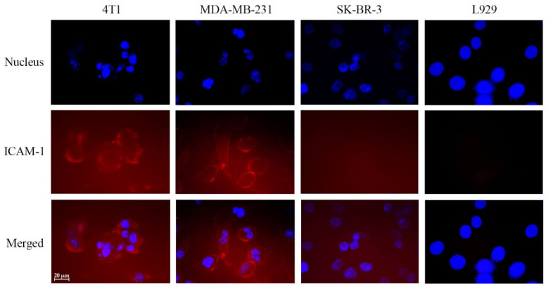
Figure 1.
Immunofluorescent imaging of intercellular adhesion molecule-1 (ICAM-1) expression in 4T1, MDA-MB-231, SK-BR-3, and L929 cells. The cells were seeded into chamber slides (30,000 cells/well) and incubated overnight. The cells were fixed with paraformaldehyde, quenched and stained for ICAM-1 detection (red). The cells nuclei were stained with DAPI (blue). The scale bar in the figure is 20 mm. Each picture was chosen as representative from a triplicated experiment.

