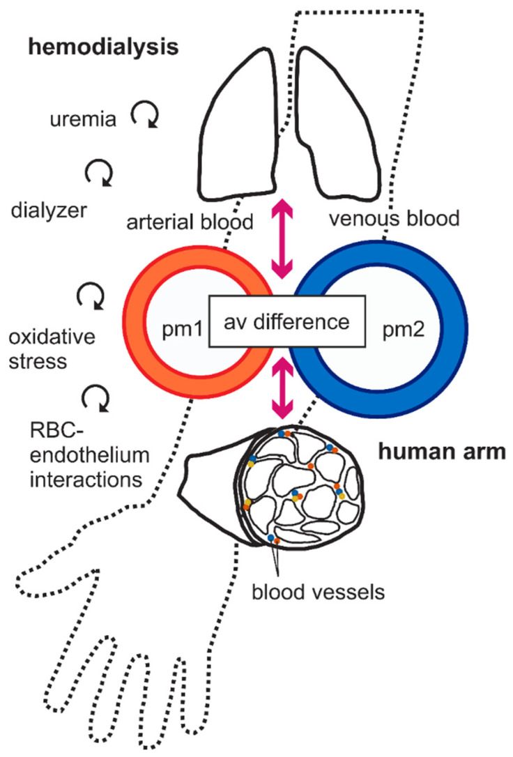
Figure 2.
A simplified scheme of the relationship among different compartments. Central and peripheral compartments are shown. Central compartment is consisting of plasma, pm1 and pm2. Peripheral compartments are consisting of organ tissues, especially arm muscle, with extracellular fluid, red blood cells (RBCs), etc. The continuity between arterial and venous systems via pulmonary (top) and peripheral (bottom) arm muscle is illustrated in the diagram. Arterial and venous blood samples were taken before (pre-HD) and after HD (post-HD) treatment. Plasma oxylipins were measured in pm1 and pm2, i.e., arterial shunt and subcutaneous vein, respectively. It is obvious that the arteriovenous (av) difference is caused by the circumstance that the peripheral tissues, especially the muscles, either produce, store, or degrade part of the oxylipins that pass through them. The curved vector graphs represent the hypothetical influence of CKD and hemodialysis in conjunction with shear stress, dialyzer, red blood cell (RBC)-endothelial interactions, and oxidative stress affecting plasma oxylipin biotransformation levels in peripheral tissues.

