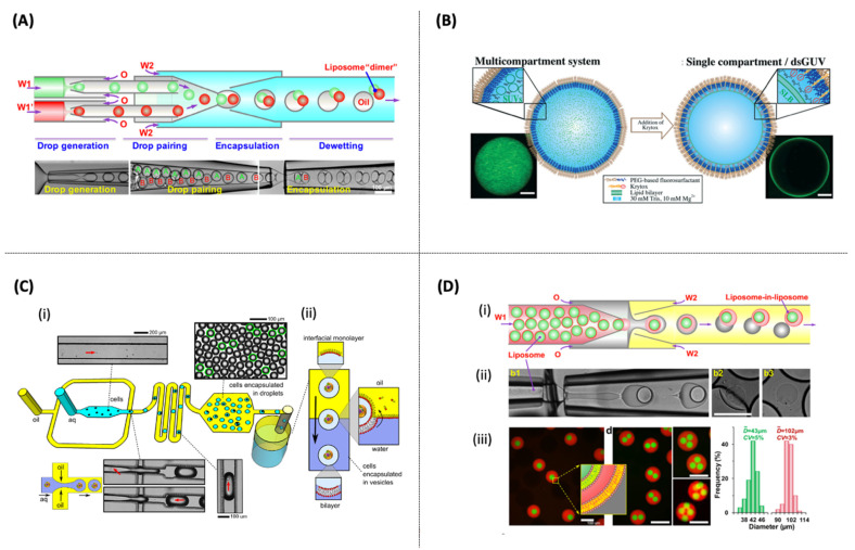
Figure 6.
(A) Schematic device and images of double emulsions production with two distinct drops [156] (B) Charge-controlled microfluidic for the formation of a multicompartmental vesicle. dsGUVs: droplet-stabilized GUVs. Scale bars: 10 μm [157] (C) (i) Microfluidic device used to encapsulate cells in w/o droplets encased in a lipid monolayer. (ii) Schematic depicting the transformation of cells-in-droplets to cells-in-vesicles [80] (D) (i,ii) Microfluidic preparation of double emulsions with an inner liposome and the assembly of vesosomes from emulsion dewetting. (iii) Confocal images of the monodisperse vesosomes with one, two, three, and four inner liposomes. Size distribution of the inner and outer liposomes of the vesosomes. Scale bars: 100 μm [159].

