Abstract
Background
Cleanliness of hospital surfaces helps prevent healthcare-associated infections, but comparative evaluations of various cleaning strategies during COVID-19 pandemic surges and worker shortages are scarce.
Purpose and methods
To evaluate the effectiveness of daily, enhanced terminal, and contingency-based cleaning strategies in an acute care hospital (ACH) and a long-term care facility (LTCF), using SARS-CoV-2 RT-PCR and adenosine triphosphate (ATP) assays. Daily cleaning involved light dusting and removal of visible debris while a patient is in the room. Enhanced terminal cleaning involved wet moping and surface wiping with disinfectants after a patient is permanently moved out of a room followed by ultraviolet light (UV-C), electrostatic spraying, or room fogging. Contingency-based strategies, performed only at the LTCF, involved cleaning by a commercial environmental remediation company with proprietary chemicals and room fogging. Ambient surface contamination was also assessed randomly, without regard to cleaning times. Near-patient or high-touch stationary and non-stationary environmental surfaces were sampled with pre-moistened swabs in viral transport media.
Results
At the ACH, SARS-CoV-2 RNA was detected on 66% of surfaces before cleaning and on 23% of those surfaces immediately after terminal cleaning, for a 65% post-cleaning reduction (p = 0.001). UV-C enhancement resulted in an 83% reduction (p = 0.023), while enhancement with electrostatic bleach application resulted in a 50% reduction (p = 0.010). ATP levels on RNA positive surfaces were not significantly different from those of RNA negative surfaces. LTCF contamination rates differed between the dementia, rehabilitation, and residential units (p = 0.005). 67% of surfaces had RNA after room fogging without terminal-style wiping. Fogging with wiping led to a -11% change in the proportion of positive surfaces. At the LTCF, mean ATP levels were lower after terminal cleaning (p = 0.016).
Conclusion
Ambient surface contamination varied by type of unit and outbreak conditions, but not facility type. Removal of SARS-CoV-2 RNA varied according to cleaning strategy.
Implications
Previous reports have shown time spent cleaning by hospital employed environmental services staff did not correlate with cleaning thoroughness. However, time spent cleaning by a commercial remediation company in this study was associated with cleaning effectiveness. These findings may be useful for optimizing allocation of cleaning resources during staffing shortages.
Introduction
Cleanliness of hospital surfaces helps prevent healthcare-associated infections [1,2]. Contamination of environmental surfaces with SARS-CoV-2 has been reported from various countries, under experimental conditions in the laboratory, or in facilities with bio-containment units [3–11]. However, larger, controlled, pre and post cleaning evaluations, particularly from U.S. health care facilities dealing with nosocomial outbreaks during the height of the pandemic are scarce.
Healthcare associated (HA) transmission of SARS-CoV-2 results from of a complex interplay of several factors, including patient census, nurse-to-patient ratio, adherence to isolation guidelines and policies for using personal protective equipment, patient acuity, and the prevalence of presymptomatic/asymptomatic carriers. However, these factors may not fully account for larger or sustained outbreaks. Environmental contamination and the quality of cleaning could also be an overlooked contributor to HA- transmission.
Furthermore, the shortage of environmental service (EVS) workers and their concerns regarding cleaning rooms of patients infected with SARS-CoV-2 further complicates cleaning and disinfection efforts [12–15]. This can force some healthcare facilities to use contingency-based approaches such as professional remediation companies. Moreover, long-term care facilities (LTCF) have experienced inordinately high infection and mortality rates [16–18]. The potential contribution of environmental contamination with SARS-CoV-2 to this is uncertain.
Adding to the challenge of investigating the role of environmental SARS-CoV-2 is that fact that the vast majority of studies, including this one, do not have the biosafety level necessary for the cultivation of live virus and must rely on viral RNA as a surrogate. Even though nucleic acid isolated from healthcare surfaces may not represent infectious or cultivable virus, and the infectious potential of environmental nucleic acid, including RNA, is not fully understood, we have shown that environmental nucleic acid from another common pathogen was correlated with nosocomial infections of that species [19].
Our objectives were to assess baseline environmental contamination with SAR-CoV-2 RNA at an acute-care hospital (ACH) and a LTCF and compare contamination rates between different wards or units and between outbreak and non-outbreak settings. Attempting to maintain cleaning standards and quality measures in the midst of critical environmental service worker shortages, the LTCF hired a commercial cleaning and remediation company as a contingency measure. Therefore, we also sought to evaluate the effectiveness of that approach.
Settings
The ACH is a 528-bed teaching hospital in the Finger Lakes Region of New York State. The baseline configuration pre-pandemic surge is such that 42% of the rooms are semi-private. Shared room configuration involves 2 beds in the same room separated by a curtain and having a single bathroom for 2 occupants. There are 48 intensive care beds and 14 special care nursery beds. It has the eleventh busiest emergency department and the second largest ventricle assist device program in the country. The LTCF has 201 beds and eight wards including a dementia unit. In the LTCF, 2 occupants share a single bathroom, but the room is larger and separated by a partial wall, curtain, or no physical barrier depending on the unit. There was an outbreak of nosocomial COVID-19 cases on one of the wards in the ACH and one of the buildings that contained several wards at the LTCF.
Methods
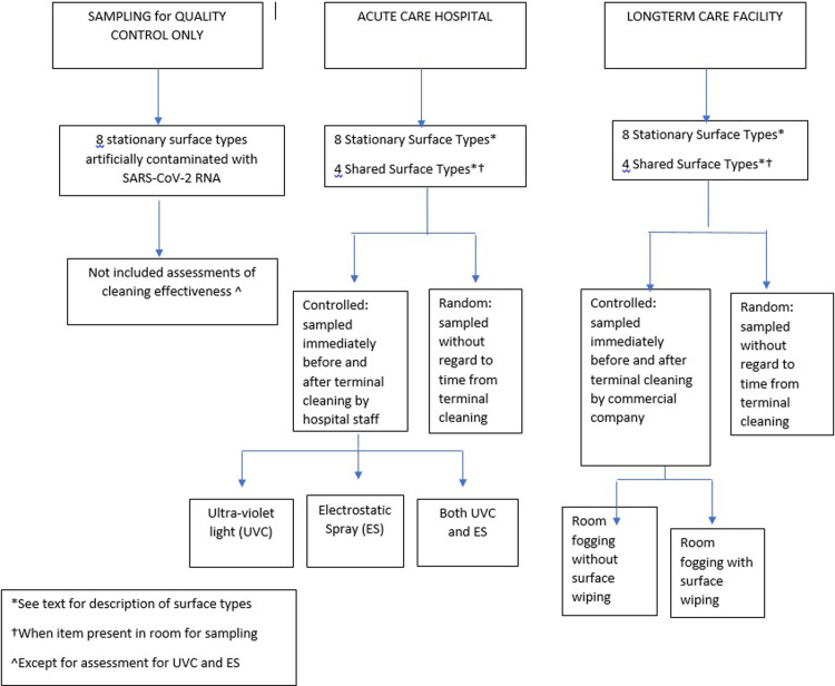
Three separate surface samplings were performed on eight of the same stationary surface types: 1) a test run using artificially contaminated (spiked) surfaces; 2) random sampling–meaning without regard to time of last terminal cleaning; and controlled sampling–meaning immediately before then immediately after terminal cleaning (Fig 1). Those eight stationary near-patient, high touch surfaces described below, were selected because these surfaces are conventionally most often used in studies of environmental contamination of healthcare facilities. Shared surface types (described below) were also sampled when available. Results from spiked surfaces were used primarily as quality control for sampling supplies and the diagnostic platform, and also to create an equal starting point for assessing enhancements to terminal cleaning.
Fig 1. Sampling approach.
To ensure the sampling method could reliably detect environmental RNA before formal sampling began, 100 cm2 test surfaces in vacated patient rooms were spotted with 1250 copies of SARS-CoV-2 genomic RNA (BEI Resources). After 10 minutes of drying time, these surfaces were sampled as positive controls. All such controls tested positive using the commercial assay described below. The mean cycle threshold of these pre-cleaned surfaces was 30. These results were for quality control only and were not included in the evaluation of cleaning effectiveness.
From 15th May 2020 to 15th June 2020, environmental surface sampling and assessments of cleaning thoroughness (adenosine triphosphate (ATP)). and effectiveness (reduction of viral RNA) were performed as previously described [19,20]. Briefly, swabs were streaked over targeted high-touch surfaces for a minimum of 20 seconds in a rolling motion to ensure contact with the entire swab surface. Dacron-tipped cobas® PCR Media Uni Swabs (Roche Molecular Systems, Inc. Branchburg, NJ) were pre-moistened with the transport media (sterile 40% guanidine hydrochloride Tris-HCl buffer) and analyzed on the cobas® 6800 System (Roche Molecular Systems, Inc. Branchburg, NJ). ATP testing of surfaces was performed using 3M™ Clean-Trace ATP monitoring system.) Sampling at both long-term and acute care facilities was performed using same equipment and in an identical manner by the same authors (DN, LR, EL).
At the ACH and the LTCF, for both random and controlled sampling, dedicated COVID-19 units were used as ‘positive control wards’, and wards which had zero COVID-19 patients identified were uses as ‘negative control wards’. Those that housed both COVID-19 and non-COVID patients were considered ‘mixed wards’. Inpatient dialysis units were considered separately. Outbreaks or nosocomial cases, by definition, occurred more than 7 days after admission (probable nosocomial) and 14 days definite nosocomial. Therefore “outbreaks” could only occur on mixed wards among initially COVID-19 negative patients or on all negative wards.
Random sampling: Assessment of the proportion of positive surfaces between cleanings (Ambient environmental contamination)
Eight stationary surfaces sampled in rooms on the outbreak and on the negative and positive ‘control’ wards. These included bed rails, call button/remote control, in-room telephone, over-bed tray table, sink and soap dispenser, chair, windowsill, and the floor. Walking computer workstations, hand-held glucometers, and vital sign machines represented shared surface samples and microwaves and refrigerator samples were taken from the staff common areas. Sampling occurred randomly throughout the daytime hours, without regard to the timing of daily or terminal cleaning. However, since most of these surfaces are cleaned daily, the time from last cleaning to sampling was most often less than 36 hrs. Sampling at various times of the day (i.e., randomly from last time of cleaning provides an estimate of the ambient level of contamination (proportion of positive surfaces) between daily or terminal cleanings.
Controlled sampling: Evaluation of cleaning effectiveness
At the ACH, immediately after a SARS-CoV-2 infected patient was discharged or transferred out of their room, nursing notified the authors who coordinated with EVS. The same eight stationary surface types sampled in the random assessment were sampled immediately before and after terminal cleaning involving surface wiping with a non-bleach sporicidal disinfectant containing hydrogen peroxide and peracetic acid (OxyCide ™). Additionally, at the ACH, three types of terminal cleaning enhancements were also evaluated: UV-C treatment at 60,000 mJ/cm2 (RD™ UVC Mobile System), adding electrostatic spraying (Clorox® Total 360®) following terminal cleaning, and adding both UV-C and electrostatic spraying. These enhancements were applied in addition to terminal cleaning when terminal cleaning was completed. To ensure each enhancement had an equal amount of RNA contamination present before cleaning, 100 cm2 surfaces in vacated patient rooms were spotted with 1250 copies of SARS-CoV-2 genomic RNA (BEI Resources) before terminal cleaning. During the post-cleaning sample collection, those spotted areas were included in the streaking of the target surface type (floor, bed rail, etc.).
At the LTCF, rooms were sampled before and after routine daily cleaning and also after a professional remediation service using three different proprietary room fogging agents. A room would have been sampled before and after daily or terminal cleaning by employed cleaning staff (2 times) or before and after cleaning by a commercial remediation company (2 times). Most rooms were sampled twice. A few could have been sampled on 4 separate occasions. Adenosine triphosphate (ATP) testing of surfaces was also performed using 3M™ Clean-Trace ATP monitoring system.
The proportions of sampled surfaces with detectible RNA were compared using the 2-sample percent defective tests or Chi squared tests. The mean cycle threshold values of SARS-CoV-2 genes were compared using 2-sample t-test of means.
Results
Randomized sampling: Ambient contamination on COVID-19, non-COVID-19, and inpatient dialysis units at the ACH and LTCF
ACH compared to LTCF
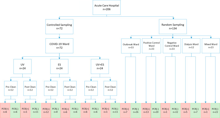
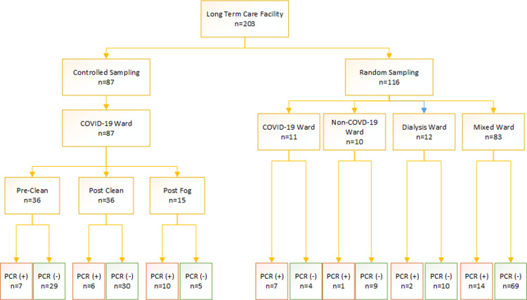
417 surface samples were collected: 206 from the ACH; and 203 from the LTCF and eight quality control samples not included in analysis. The sampling breakdown and number of positive surfaces is presented in Figs 2 and 3. Of surfaces swabbed, 29% (n = 59/206) at the ACH, and 23% (n = 47/203) at the LTCF had detectable RNA. Based on the proportion of positive surfaces, the difference between the overall contamination rates at the ACH and LTCF was not significant (p = 0.488). Similarly, surface contamination based on mean PCR mean cycle threshold values of the ORF1ab gene was also similar between the ACH and the LTCF. Mean cycle threshold value at the ACH was 35.4 compared to 35.9 at the LTCF (p = 0.179).
Fig 2. Sample breakdown at the acute care hospital.
Legend: ES = Electrostatic spray; UV light = Ultraviolet light.
Fig 3. Sample breakdown at the long-term care facility.
Legend: Fog = Chemical fogging of room.
ACH
At the ACH, 20% of 134 randomly sampled surfaces had detectible RNA (Fig 2). The proportion of surfaces with detectable RNA was not significantly different on the outbreak ward as compared to the dedicated COVID-19 ward (positive control): 7/33 versus 13/33 (p = 0.180). However, there was a significant difference (p = 0.001) when the outbreak ward was compared to the negative control ward, which only had 3% detectable surfaces (1/33).
LTCF
At the LTCF, 10% of the rooms in three units were sampled. 21% of 116 randomly sampled surfaces had viral RNA, which was not significantly different that that seen at the ACH (p = 1.000) (Fig 3). The COVID-19 positive resident rooms had a significantly higher proportion of surfaces with RNA than those of the mixed wards, 7/11 versus 14/83 (p = 0.002). The mixed ward had a higher proportion of surfaces with RNA (17%) than the target of non-COVID-19 ward (10%) (p = 0.035). Stratifying the units further, there was a significant difference between the dementia unit (11/28), and rehabilitation unit (2/28), or the residential unit (1/28) (p = 0.005).
Dialysis
Overall, for inpatient dialysis at the ACH combined with inpatient dialysis at the LTCF, 29% (7/24) of randomly sampled surfaces had viral RNA. A higher proportion of surfaces were positive at the ACH (5/12 or 42%) compared to the LTCF (2/12 or 17%).
Controlled sampling: Cleaning assessments with adenosine triphosphate measurements
ACH
At the ACH, 437 surfaces throughout the facility including on the outbreak wards, dedicated COVID-19 units, and negative control wards, underwent post cleaning ATP testing as part of routine protocols and quality assessments. The average ATP value was 32 (SD 16.8) relative light units (RLU). 35 surfaces had both adenosine triphosphate and PCR testing. 2 of 35 were positive for viral RNA. The mean ATP value of the two SARS-CoV-2 positive surfaces was 666 (SD 2.12) RLU. 33 of the 35 surfaces had no detectable SARS-CoV-2 RNA. The mean ATP of 33 negative surfaces was 1274 (SD 2216) RLU; 95% CI for difference -1394–178) p-value = 0.125.
On the dedicated COVID-19 wards at the ACH, RNA was detected on 24/36 (67%) of surfaces immediately before cleaning and on 8/36 (22%) of those surfaces immediately after terminal cleaning using a non-bleach sporicidal disinfectant containing hydrogen peroxide and peracetic acid (OxyCide ™) for a 65% post-cleaning reduction (p = 0.001).
UV-C combined with terminal cleaning resulted in an 83% reduction in the proportion of RNA- positive surfaces (50% positive pre-cleaning vs. 8.3% positive after cleaning, p = 0.025) (Fig 2). Terminal cleaning combined with electrostatic spraying (Clorox Pro Total 360 ®) resulted in a 50% reduction (100% vs. 50%, p = 0.006) in detectable target RNA. Spraying plus UV-C led to an 83% reduction (50% precleaned vs. 8.3% post cleaning, p = 0.028) When UV-C was used, there was 1 detectable RNA positive surface, and combining UVC with electrostatic did not result and any further reduction (1/12) compared to UVC alone.
LTCF
At the LTCF, RNA was detected on 10/15 (66%) surfaces after professional remediation company fogged patient rooms with a proprietary chlorine dioxide-based disinfectant. In this process, terminal-style surface wiping was not performed. In a separate assessment, detectable RNA was present on 19% of surfaces before cleaning versus 17% after cleaning that did involve terminal style surface wiping (17–19)/19 = -11% change in the proportion of positive surfaces). However, the amount of reduction depended on the time spent wiping and the spraying agent used (Fig 4). 16 surfaces underwent ATP testing before, and 16 were tested after cleaning. Their mean ATP value was 2308 RLU (SD 2782) (pre-cleaned), and 384 (SD 724; 95% CI for the mean difference in ATP = (408–3441) post cleaning, p = 0.016.
Fig 4. Effect of chemical type and cleaning time on percent of surfaces detected.
Limitations
Our study has several limitations. First, safety concerns and the lack of a Biosafety Containment Level 3 laboratory precluded the use of viral cultures to determine viability of virus, but nucleic acid of target pathogens can be consequential [19]. Furthermore, lack of culture-based assessments is not unique to our study [4–9]. Second, sponge sticks may be better at capturing pathogens from larger environmental surfaces such as floors and tray tables. These collection devices were unavailable and could not be used in automated PCR reaction tubes. Third, the commercial remediation service and EVS were not compared under identical conditions, but our main objective was not a direct head-to-head comparison of EVS vs contracted remediation service.
Discussion
At both the ACH and LTCF, viral RNA was detected more often on dedicated COVID-19 units, and on wards or units that were experiencing an outbreak or nosocomial transmission. Almost 30% of randomly sampled surfaces in dialysis units, including terminally cleaned dialysis machines, had detectible viral RNA. The overall facility level contamination was no different between an ACH and a corresponding LTCF. The room cleaning protocols used at the ACH significantly reduced environmental RNA, with UV-C light resulting in fewer positive surfaces than electrostatic spraying. Recognizing: 1) the challenges in cleaning the LTCF because rooms cannot be completely emptied of residents’ personal effects and small appliances for terminal styled-wiping as in a hospital; and 2) that the ATP values at the LTCF were on average higher than the ACH ATP values, our healthcare system invested in UV-C units for the LTCF.
Commercial cleaning services that spent more time per room cleaning resulted in a larger reduction in contamination, unlike previously published reports that showed time spent cleaning did not translate into more bioburden reduction [21]. Cleaning by this commercial remediation company that relied primarily on room fogging appeared to be less effective than environmental service employees of the hospital.
Given the scarcity of testing supplies, this study is noteworthy for its sample size. To our knowledge, it is the largest such U.S. study to date. It is also notable for the pragmatic, controlled assessments of cleaning practices under ‘real world’ conditions of staffing shortages during the height of the pandemic. Prior SARS-CoV-2 studies did not have a controlled assessment of cleaning, did not include shared equipment and ATP measurements [3–11].
Surface types that most often remained contaminated included flooring, hand-held glucometers, nursing call buttons, and walking computer workstations. However, none had Ct values lower than 29.
Conclusion
In summary, surface contamination with SARS-CoV-2 RNA significantly differed based on type of unit, disinfectant, and cleaning regimen. Although the same percent (10%) of rooms in each unit were sampled, the proportion of sample sites that were contaminated with SARS-COV-2 RNA appeared to be influenced by the occurrence of cases/ outbreaks on those units, and by the behavior of patients on the unit as in the case of the dementia ward where residents often wander and unable to understand or comply with masking guidelines. Analysis according to surface, unit, and cleaning regimen, was more informative than facility-level analysis. Additionally, none of the PCR-positive surfaces had Ct values less than 29. Just as patients who remain positive by PCR weeks after infection but with high CTs are not considered to be contagious, the environment viral load we encountered may also place the inanimate surfaces as unlikely source of contagion.
Although the environment may not be a major driver of SARS-CoV-2 transmission at least in community/public settings, the role that environmental contamination plays in nosocomial transmission in settings with heavy viral burdens and sick or physiologically vulnerable persons remain less understood. Perhaps another argument for meticulous cleaning of the environment of COVID-19 patients’ rooms is the company that SARS-CoV-2 may keep during the pandemic, e.g., Clostridioides difficle, MDR-Acinetobacter, or MDR-Candida that are known to persist in the environment longer than SARS-CoV-2.
Implications
This report is relevant to nursing homes and hospitals as the findings can assist in optimizing allocation of scarce cleaning and disinfection resources for maximum impact. For example, commercial remediation services can be expensive, and not a reasonable return on investment. On the other hand, UV-C machines also do “double-duty”, as most hospitals also use them for enhanced cleaning of environmentally hardier pathogens such as multi-drug resistant Acinetobacter baumannii, carbapenem resistant Enterobacteriaceae, Clostridioides difficile and Candida auris. Although not a reason to purchase an UV-C system, of interest was the psychology of UV-C awareness. Namely, that several cleaning staff who were initially reluctant to clean the rooms of discharged COVID-19 patients, had no hesitation if the rooms were first treated with UV-C. Furthermore, studies of the survivability of the virus on fomites have been criticized as lacking real-life generalizability [22,23]. Data in this report were obtained from a ‘typical’ general hospital and nursing home, representative of the type of settings (i.e., general/community/non-university-based hospitals) where much of health care is delivered. Studies for these settings may be underrepresented in research because most are carried out in large university-based hospitals.
Supporting information
(XLSX)
(XLSX)
(XLSX)
(XLSX)
Data Availability
All relevant data are within the manuscript and its Supporting Information files.
Funding Statement
The authors received no specific funding for this work.
References
- 1.Dancer SJ. Controlling hospital-acquired infection: focus on the role of the environment and new technologies for decontamination. Clin Microbiol Rev. 2014;27(4):665–90. doi: 10.1128/CMR.00020-14 [DOI] [PMC free article] [PubMed] [Google Scholar]
- 2.Carling PC, Parry MF, Von Beheren SM, et al. Identifying opportunities to enhance environmental cleaning in 23 acute care hospitals. Infect Control Hosp Epidemiol. 2008;29(1):1–7. doi: 10.1086/524329 [DOI] [PubMed] [Google Scholar]
- 3.Pastorino B, Touret F, Gilles M, de Lamballerie X, Charrel RN. Prolonged infectivity of SARS-CoV-2 in fomites. Emerg Infect Dis. 2020;26(9):2256–7. doi: 10.3201/eid2609.201788 [DOI] [PMC free article] [PubMed] [Google Scholar]
- 4.Feng B, Xu K, Gu S, et al. multi-route transmission potential of SARS-CoV-2 in healthcare facilities. J Hazard Mater. 2021; 402:123771. doi: 10.1016/j.jhazmat.2020.123771 [DOI] [PMC free article] [PubMed] [Google Scholar]
- 5.Wu S, Wang Y, Jin X, et al. Environmental contamination by SARS-CoV-2 in a designated hospital for coronavirus disease 2019. Am J Infect Control. 2020;48(8):910–4. doi: 10.1016/j.ajic.2020.05.003 [DOI] [PMC free article] [PubMed] [Google Scholar]
- 6.Guo ZD, Wang ZY, Zhang SF, et al. Aerosol and surface distribution of severe acute respiratory syndrome coronavirus 2 in hospital wards, Wuhan, China, 2020. Emerg Infect Dis. 2020;26(7):1583–91. doi: 10.3201/eid2607.200885 [DOI] [PMC free article] [PubMed] [Google Scholar]
- 7.Colaneri M, Seminari E, Piralla A, et al. Lack of SARS-CoV-2 RNA environmental contamination in a tertiary referral hospital for infectious diseases in Northern Italy. J Hosp Infect. 2020;105(3):474–6. [DOI] [PMC free article] [PubMed] [Google Scholar]
- 8.Ong SWX, Tan YK, Chia PY, et al. Air, surface environmental, and personal protective equipment contamination by Severe Acute Respiratory Syndrome Coronavirus 2 (SARS-CoV-2) from a symptomatic patient. JAMA. 2020;323(16):1610–2. doi: 10.1001/jama.2020.3227 [DOI] [PMC free article] [PubMed] [Google Scholar]
- 9.Ye G, Lin, Chen S, et al. Environmental contamination of SARS-CoV-2 in healthcare premises. J Infect. 2020;81(2): e1–e5. doi: 10.1016/j.jinf.2020.04.034 [DOI] [PMC free article] [PubMed] [Google Scholar]
- 10.Chin AWH, Chu JTS, Perera MRA, et al. Stability of SARS-CoV-2 in different environmental conditions. Lancet Microbe. 2020;1(1): e10. doi: 10.1016/S2666-5247(20)30003-3 [DOI] [PMC free article] [PubMed] [Google Scholar]
- 11.Ryu BH, Cho Y, Cho OH, et al. Environmental contamination of SARS-CoV-2 during the COVID-19 outbreak in South Korea. Am J Infect Control. 2020; 48(8): 875–879. doi: 10.1016/j.ajic.2020.05.027 [DOI] [PMC free article] [PubMed] [Google Scholar]
- 12.Memmott J. Keeping the hospital safe so people can heal. Available from: https://www.democratandchronicle.com/story/news/local/columnists/memmott/2020/09/13/keeping-hospital-safe-so-people-may-heal/5772158002/.
- 13.Ducharme J. ’No one mentions the people who clean it up’: What it’s like to clean professionally during the COVID-19 outbreak. Available from: https://time.com/5810911/covid-19-cleaners-janitors/.
- 14.Brown N, Cooke K. In fight for masks, hospital janitors sometimes come last. Available from: https://www.reuters.com/article/us-health-coronavirus-housekeepers/in-fight-for-masks-hospital-janitors-sometimes-come-last-idUSKBN21O2JF.
- 15.Litwin AS, Avgar AC, Becker ER. Superbugs versus outsourced cleaners: employment arrangements and the spread of health care–associated infections. ILR Review. 2017; 70:610–41. [Google Scholar]
- 16.Centers for Medicare and Medicaid Services. Nursing Home COVID-19 Data. Available from: https://www.cms.gov/files/document/6120-nursing-home-covid-19-data.pdf?_ga=2.220609944.1832926098.1598283932-535237757.1598283931. [PubMed]
- 17.Gaur S, Dumyati G, Nace DA, et al. Unprecedented solutions for extraordinary times: Helping long-term care settings deal with the COVID-19 pandemic. Infect Control Hosp Epidemiol. 2020;41(6):729–30. doi: 10.1017/ice.2020.98 [DOI] [PMC free article] [PubMed] [Google Scholar]
- 18.Kwiatkowski M, Nadolny TL, Priest J, et al. ‘A national disgrace’: 40,600 deaths tied to US nursing homes. Available from: https://www.usatoday.com/story/news/investigations/2020/06/01/coronavirus-nursing-home-deaths-top-40-600/5273075002/.
- 19.Lesho E, Carling P, Hosford E, et al. Relationships between cleaning, environmental DNA, and healthcare-associated infections in a new evidence-based design hospital. Infect Control Hosp Epidemiol. 2015;36(10):1130–8. doi: 10.1017/ice.2015.151 [DOI] [PubMed] [Google Scholar]
- 20.Clifford R, Sparks M, Hosford E, et al. Correlating cleaning thoroughness with effectiveness and briefly intervening to affect cleaning outcomes: How clean is cleaned? PLOS One. 2016;11(5): e0155779. doi: 10.1371/journal.pone.0155779 [DOI] [PMC free article] [PubMed] [Google Scholar]
- 21.Rupp ME, Adler A, Schellen M, et al. The time spent cleaning a hospital room does not correlate with the thoroughness of cleaning. Infect Control Hosp Epidemiol. 2013;34(1):100–2. doi: 10.1086/668779 [DOI] [PubMed] [Google Scholar]
- 22.Goldman E. Exaggerated risk of transmission of COVID-19 by fomites. Lancet Infect Dis. 2020;20(8):892–3. doi: 10.1016/S1473-3099(20)30561-2 [DOI] [PMC free article] [PubMed] [Google Scholar]
- 23.Lesho E, Laguio-Vila M, Walsh E. Stability and viability of SARS-CoV-2. New Eng J Med. 2020;382(20):1963–4. doi: 10.1056/NEJMc2007942 [DOI] [PubMed] [Google Scholar]
Associated Data
This section collects any data citations, data availability statements, or supplementary materials included in this article.
Supplementary Materials
(XLSX)
(XLSX)
(XLSX)
(XLSX)
Data Availability Statement
All relevant data are within the manuscript and its Supporting Information files.


