Abstract
Background
Salvage surgery following definitive radiotherapy or systemic treatment has become a feasible treatment option in selected patients with advanced initially unresectable non-small cell lung cancer. Recent clinical trials of neoadjuvant treatment have showed that surgery following immuno-chemotherapy is safely performed. Here, we present the first case of salvage surgery following immuno-chemotherapy with concurrent definitive radiotherapy for advanced lung large cell carcinoma.
Case presentation
A 44-year male was admitted to our hospital for salvage surgery. Ten months prior to this administration, he had been diagnosed with unresectable large cell carcinoma with malignant pericardial effusion (clinical stage IVA/T3N2M1A; no driver-gene alteration) originating from the right upper lobe (RUL). Due to rapid intrabronchial tumor growth causing severe dyspnea, emergency bronchial stenting in the right main bronchus using an expandable metallic stent had been performed. Thereafter, he had received immuno-chemotherapy with concurrent definitive radiotherapy. Despite dramatic radiographic response, he had suffered from persistent and refractory Pseudomonas aeruginosa lung infection associated with bronchial stent placement. As pericardial effusion had disappeared and no distant metastasis had developed, he was diagnosed with a potentially curable disease and was referred to our hospital. An extended sleeve resection was successfully performed, and pathological sections revealed that pathologic complete response was achieved with immuno-chemo-radiotherapy. The patient received no subsequent treatment, and is alive without tumor recurrence at 8 months after surgery.
Conclusions
Salvage surgery following immuno-chemotherapy with concurrent definitive radiotherapy for advanced non-small cell lung cancer may be feasible in selected patients, and may be considered as a treatment option to control local disease.
Supplementary Information
The online version contains supplementary material available at 10.1186/s40792-022-01371-3.
Keywords: Salvage surgery, Lung cancer, Immunotherapy, Chemotherapy, Radiotherapy
Background
Salvage surgery following definitive radiotherapy or systemic treatment has been increasingly adopted as a feasible treatment option in selected patients with advanced non-small cell lung cancer (NSCLC) which had been initially diagnosed as unresectable disease [1, 2]. Recent clinical trials of neoadjuvant treatment for resectable NSCLC have revealed that the addition of immuno-therapy using an antibody against programmed death 1 (PD-1) or programmed death-ligand 1 (PD-L1) to platinum-based chemotherapy (immuno-chemotherapy) may provide superior pathologic response and superior survival benefit over chemotherapy alone [3–6]. To further improve the therapeutic efficacy, immuno-chemotherapy with concurrent radiotherapy (cICRT) may be a promising treatment option. However, the feasibility of surgery following cICRT remains unknown. Here, we present the first successful case of salvage surgery following cICRT in which pathologic complete response (pCR) was achieved with cICRT for advanced NSCLC.
Case presentation
A 44-year male was admitted to our hospital for salvage surgery. The patient had quit smoking 4 years ago after a 20-pack-year history.
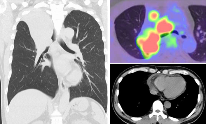
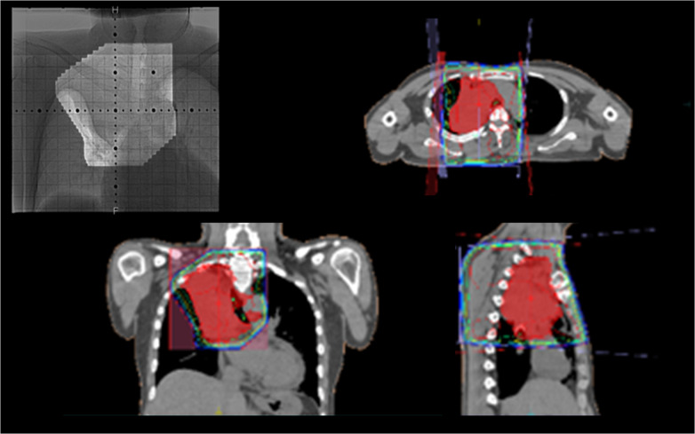
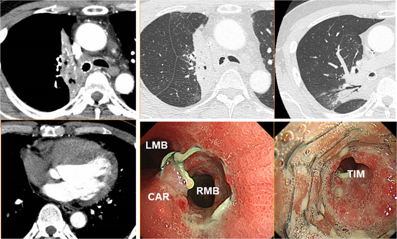
Ten months prior to the administration, he had been diagnosed with unresectable non-small cell lung cancer (clinical stage IVA/T3N2M1A) originating from the right upper lobe (RUL). Chest computed tomography (CT) had revealed a 4.8-cm tumor obstructing the right main bronchus, enlarged upper and lower paratracheal nodes, and pericardial effusion (Fig. 1). Transbronchial tumor biopsy had provided a pathological diagnosis of large cell carcinoma (no driver-gene alteration; proportion of tumor expressing PD-L1, 1%). Pathological evaluation of mediastinal nodes or pericardial effusion had not been performed. Due to rapid intrabronchial tumor growth causing severe dyspnea, emergency bronchial stenting in the right main bronchus using an expandable metallic stent (Ultraflex, Boston Scientific, Watertown, MA) had been performed. Thereafter, he had received immuno-chemotherapy (cisplatin plus pemetrexed with an anti-PD-1 antibody [pembrolizumab]) with concurrent definitive radiotherapy (60 Gy in 30 fractions, Fig. 2), and then had received maintenance pembrolizumab treatment. In total, he received 7 cycles of pembrolizumab treatment. Despite dramatic radiographic response, he had suffered from persistent and refractory Pseudomonas aeruginosa infection in the RUL and upper segment of the right lower lobe (S6) associated with bronchial stent placement (Fig. 3). As pericardial effusion had disappeared and no distant metastasis had developed, he was diagnosed with a potentially curable disease and was referred to our hospital.
Fig. 1.
Computed tomography and positron emission tomography at initial diagnosis revealed a large tumor obstructing the right main bronchus (left), enlarged upper and lower paratracheal nodes with strong uptake of fluorodeoxyglucose (right upper), and pericardial effusion (right lower)
Fig. 2.
A total dose of 60 Gy in 30 fractions were delivered to the primary tumor plus involved nodes. The endobronchial stent was included in the radiotherapy field
Fig. 3.
Computed tomography (CT) after bronchial stenting and immuno-chemo-radiotherapy showed dramatic tumor shrinkage and disappearance of pericardial effusion (left), and also showed persistent lung infection (right upper) associated with sent placement (right lower). Microbiological test of the “green–blue” sputum collected by deep coughing and by bronchoscopy revealed bacterial phagocytosis by neutrophils and growth of bacteria that was identified as Pseudomonas aeruginosa. CAR, carina; LMB, left main bronchus; RMB, right main bronchus; TIM, truncus intermedius
Whole-body CT and fluorodeoxyglucose-positron emission tomography (FDG-PET) revealed no nodal or distant metastasis, and the patients was diagnosed to have a curative disease after cICRT (ycIB/T2aN0M0). Preoperative pulmonary function tests revealed moderate lung airflow obstruction (forced vital capacity [FVC], 3600 mL; percentage of the predicted value of FVC [%FVC], 81%; forced expiratory volume in 1 s [FEV1], 2310 mL; percentage of the predicted value of FEV1 [%FEV1], 60%; FEV1/FVC, 64%) and mild reduction of diffusing capacity of the lung for carbon monoxide (DLCO, 58% of the predicted value). The blood oxygen saturation on room air at rest was normal (97%). The patient has not had any comorbidity other than obstructive pulmonary disease. Based on these results, we decided to perform a salvage surgery to control the persistent pulmonary infection and to achieve complete resection for the cure.
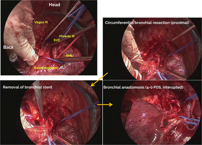
Through posterolateral thoracotomy, an extended sleeve resection of RUL plus S6 was performed due to severe fibrosis and persistent infection in S6 (Fig. 4). After severe adhesion was carefully dissected, pulmonary vessels were divided and standard nodal dissection was performed. As a simple suture ligation of the first branch of the right pulmonary artery (PA), the truncus anterior (A1 + 3), was difficult, a tangential resection was performed. After clamping the right main PA, the PA segment was excised and the defect was closed by a continuous 5–0 prolene (Ethicon Inc., Somerville, NJ) suture. Circumferential bronchial resection was performed, and the stent was removed. Frozen section examination confirmed that the proximal and distal bronchial stumps were pathologically tumor free. The bronchial ends were anastomosed end-to-end with interrupted 4–0 polydioxanone (PDS, Ethicon Inc., Somerville, NJ) sutures, and were covered with a pedicled flap of pericardial fat. To relief tension of the bronchial anastomosis site, division of pulmonary ligament and pericardial incision were performed (Additional file 1).
Fig. 4.
Through posterolateral thoracotomy, pulmonary vessels were divided and standard nodal dissection was completed. Then, right main bronchus and bronchus intermedius were exposed (left upper). Proximal transection of the main bronchus was performed (right upper), and the bronchial stent was removed (left lower). After distal transection of the bronchus intermedius and pathological confirmation of no malignant cells at the proximal and distal bronchial stumps, the bronchial ends were anastomosed end-to-end with interrupted 4–0 polydioxanone sutures (right lower)
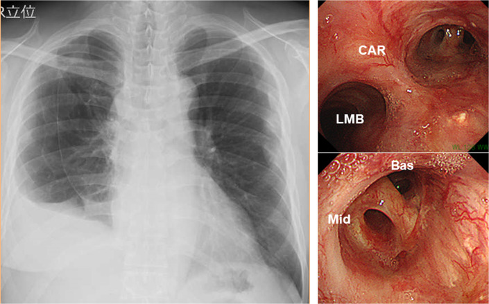
Pathological examination revealed no viable tumor cell in any resected specimen including pericardial effusion. Severe fibrosis with infiltration of a number of inflammatory cells was found in the resected lung including S6, which indicated the diagnosis of radiation pneumonitis and chronic infection. The postoperative course was uneventful, and bronchoscopy at 2 months after operation revealed excellent healing with good patency (Fig. 5). The patient received no subsequent treatment, and is alive without tumor recurrence at 9 months after surgery.
Fig. 5.
Chest roentgenogram (left) and bronchoscopy (right) at 4 months after surgery showed excellent lung expansion and bronchial healing. CAR carina, LMB left main bronchus, Mid right middle bronchus, Bas right basal bronchus
Discussion
Immuno-chemotherapy has become a standard treatment of care for unresectable NSCLC with no driver-gene alteration [7], and also may be useful as neoadjuvant treatment prior to surgery for resectable NSCLC [3–6]. A randomized controlled trial (CheckMate 816) comparing neoadjuvant immuno-chemotherapy with chemotherapy showed that the addition of immunotherapy did not increase incidence of postoperative adverse events. The pCR rate in the immuno-chemotherapy group was significantly higher (24.0% versus 2.2%) but still remains comparable with that achieved with neoadjuvant chemo-radiotherapy [8]. In the present case, pCR was achieved with cICRT even for initially unresectable disease, which may indicate that cICRT is more effective as neoadjuvant treatment for resectable and locally advanced NSCLC. Minegishi and coworkers recently reported a successful case of salvage surgery following maintenance treatment with an anti-PD-L1 antibody (durvalumab) following concurrent chemo-radiotherapy (cCRT) for initially unresectable locally advanced NSCLC [9]. In the present case, immunotherapy was concurrently prescribed with chemotherapy in addition to definitive dose of RT, and is the first case of salvage surgery following cICRT.
In the present case, definitive RT was performed in combination with systemic treatment. However, systemic treatment without RT is generally recommended as a standard treatment for clinical stage IVA disease with pericardial effusion. The patient had complained of severe dyspnea caused by rapid intrabronchial tumor growth, and had needed rapid symptom relief with effective local treatment. Accordingly, local treatment consisting RT following bronchial stenting was performed in the present case. Systemic immuno-chemotherapy without RT might provide a favorable pathologic response. The efficacy and safety of cICRT shall be examined in future clinical trials, such as the ESPADURVA trial [10, 11].
Safety concern exists regarding surgery following cICRT despite its potential advantages of superior anti-tumor effects. The present case indicates that surgery, even extended sleeve resection with bronchoplasty, after cICRT may be safely performed. In addition, definitive-dose radiotherapy was performed concurrently with immuno-chemotherapy in the present case. Recent clinical studies have shown that surgery following chemotherapy with concurrent definitive-dose of radiotherapy is safely performed [12, 13]. Again, the safety of surgery following “definitive” cICRT shall be extensively examined in future clinical trials [10].
Airway stents may be associated with significant complications, such as persistent infection as shown in the present case, and their removal is sometimes needed [14]. Some previous studies have reported that bronchoscopic removal is safe and feasible, but is sometimes associated with fetal complications, such as severe bleeding [15, 16]. In the present case, the metallic stent was easily removed after dissecting right main bronchus through thoracotomy. We have had no experience of removing a metallic bronchial stent either through thoracotomy or under bronchoscopy, and we dissected all vessels before removing the stent to prevent severe bleeding during surgery. In addition, we had planned to perform right pneumonectomy when the stent could not be safely removed.
Conclusions
Salvage surgery following “definitive” cICRT for advanced NSCLC may be feasible in selected patients, and may be considered as a treatment option to control local disease.
Supplementary Information
Additional file 1. Salvage surgery video.
Acknowledgements
The authors thank all colleagues in the Second Department of Surgery (Chest Surgery), Hospital of the University of Occupational and Environmental Health, Japan for helpful contribution to the perioperative care.
Abbreviations
- NSCLC
Non-small cell lung cancer
- PD-1
Programmed death 1
- PD-L1
Programmed death-ligand 1
- cICRT
Immuno-chemotherapy with concurrent radiotherapy
- pCR
Pathologic complete response
- RUL
Right upper lobe
- CT
Computed tomography
- FDG-PET
Fluorodeoxyglucose-positron emission tomography
- FVC
Forced vital capacity
- FEV1
Forced expiratory volume in 1 s
- PA
Pulmonary artery
Authors' contributions
AN was responsible for data collection and interpretation, and wrote the manuscript. FT revised the manuscript. AN, FT, TA, SS, MT, and KK performed the surgery and perioperative management of the patient. SS performed the pathological diagnosis. All the authors read and approved the final manuscript.
Funding
This work was not supported by any grants or funding.
Availability of data and materials
All data generated during this study are included in this published article.
Declarations
Ethics approval and consent to participate
Not applicable.
Consent for publication
Informed consent was obtained from the patient for the publication.
Competing interests
The authors have no competing interest.
Footnotes
Publisher's Note
Springer Nature remains neutral with regard to jurisdictional claims in published maps and institutional affiliations.
Contributor Information
Ayako Nawashiro, Email: nawa0407@med.uoeh-u.ac.jp.
Fumihiro Tanaka, Email: ftanaka@med.uoeh-u.ac.jp.
Akihiro Taira, Email: a-taira@med.uoeh-u.ac.jp.
Shinji Shinohara, Email: shinji5996@med.uoeh-u.ac.jp.
Masaru Takenaka, Email: m-takenaka@med.uoeh-u.ac.jp.
Koji Kuroda, Email: kuroda-k@med.uoeh-u.ac.jp.
Shohei Shimajiri, Email: shohei-s@med.uoeh-u.ac.jp.
References
- 1.Shimizu K, Ohtaki Y, Suzuki K, Date H, Yamashita M, Iizasa T, et al. Salvage surgery for non-small cell lung cancer after definitive radiotherapy. Ann Thorac Surg. 2021;11:862–873. doi: 10.1016/j.athoracsur.2020.10.035. [DOI] [PubMed] [Google Scholar]
- 2.Ohtaki Y, Shimizu K, Suzuki H, Suzuki K, Tsuboi M, Mitsudomi T, et al. Salvage surgery for non-small cell lung cancer after tyrosine kinase inhibitor treatment. Lung Cancer. 2021;153:108–116. doi: 10.1016/j.lungcan.2020.12.037. [DOI] [PubMed] [Google Scholar]
- 3.Provencio M, Nadal E, Insa A, García-Campelo MR, Casal-Rubio J, Dómine M, et al. Neoadjuvant chemotherapy and nivolumab in resectable non-small-cell lung cancer (NADIM): an open-label, multicentre single-arm, phase 2 trial. Lancet Oncol. 2020;21:1413–1422. doi: 10.1016/S1470-2045(20)30453-8. [DOI] [PubMed] [Google Scholar]
- 4.Shu CA, Gainor JF, Awad MM, Chiuzan C, Grigg CM, Pabani A, et al. Neoadjuvant atezolizumab and chemotherapy in patients with resectable non-small-cell lung cancer: an open-label, multicentre, single-arm, phase 2 trial. Lancet Oncol. 2020;21:786–795. doi: 10.1016/S1470-2045(20)30140-6. [DOI] [PubMed] [Google Scholar]
- 5.Rothschild SI, Zippelius A, Eboulet EI, Savic Prince S, Betticher D, Bettini A, et al. Durvalumab in addition to neoadjuvant chemotherapy in patients with stage IIIA(N2) non-small-cell lung cancer—a multicenter single-arm Phase II trial. J Clin Oncol. 2021 doi: 10.1200/JCO.21.00276. [DOI] [PubMed] [Google Scholar]
- 6.Spicer J, Wang C, Tanaka F, Saylors GB, Chen KN, Liberman M, et al. Surgical outcomes from the phase 3 CheckMate 816 trial: nivolumab (NIVO) + platinum-doublet chemotherapy (chemo) vs chemo alone as neoadjuvant treatment for patients with resectable non-small cell lung cancer (NSCLC) J Clin Oncol. 2021;39(15 suppl):8503. doi: 10.1200/JCO.2021.39.15_suppl.8503. [DOI] [Google Scholar]
- 7.Hanna NH, Temin S, Masters G. Therapy for stage IV non-small-cell lung cancer without driver alterations: ASCO and OH (CCO) Joint Guideline Update Summary. JCO Oncol Pract. 2020;16:e844–e848. doi: 10.1200/JOP.19.00770. [DOI] [PubMed] [Google Scholar]
- 8.Albain KS, Swann RS, Rusch VW, Turrisi AT, 3rd, Shepherd FA, Smith C, et al. Radiotherapy plus chemotherapy with or without surgical resection for stage III non-small-cell lung cancer: a phase III randomised controlled trial. Lancet. 2009;374:379–388. doi: 10.1016/S0140-6736(09)60737-6. [DOI] [PMC free article] [PubMed] [Google Scholar]
- 9.Minegishi K, Tsubochi H, Ohno K, Komori K, Ozeki M, Endo S. Salvage surgery post definitive chemoradiotherapy and durvalumab for lung cancer. Ann Thorac Surg. 2021;112:e53–e55. doi: 10.1016/j.athoracsur.2020.09.083. [DOI] [PubMed] [Google Scholar]
- 10.Stefani D, Plönes T, Viehof J, Darwiche K, Stuschke M, Schuler M, et al. Lung cancer surgery after neoadjuvant immunotherapy. Cancers (Basel). 2021;13:4033. doi: 10.3390/cancers13164033. [DOI] [PMC free article] [PubMed] [Google Scholar]
- 11., University Hospital, Essen. phase-II trial of induction chemotherapy and chemoradiotherapy plus/minus durvalumab and consolidation immunotherapy in patients with resectable stage III NSCLC. https://ClinicalTrials.gov/show/NCT04202809. Accessed on 9 January 2022.
- 12.Cerfolio RJ, Bryant AS, Jones VL, Cerfolio RM. Pulmonary resection after concurrent chemotherapy and high dose (60Gy) radiation for non-small cell lung cancer is safe and may provide increased survival. Eur J Cardiothorac Surg. 2009;35:718–723. doi: 10.1016/j.ejcts.2008.12.029. [DOI] [PubMed] [Google Scholar]
- 13.Donington JS, Paulus R, Edelman MJ, Krasna MJ, Le QT, Suntharalingam M, et al. Resection following concurrent chemotherapy and high-dose radiation for stage IIIA non-small cell lung cancer. J Thorac Cardiovasc Surg. 2020;160:1331–1345. doi: 10.1016/j.jtcvs.2020.03.171. [DOI] [PMC free article] [PubMed] [Google Scholar]
- 14.Folch E, Keyes C. Airway stents. Ann Cardiothorac Surg. 2018;7:273–283. doi: 10.21037/acs.2018.03.08. [DOI] [PMC free article] [PubMed] [Google Scholar]
- 15.Lunn W, Feller-Kopman D, Wahidi M, Ashiku S, Thurer R, Ernst A. Endoscopic removal of metallic airway stents. Chest. 2005;127:2106–2111. doi: 10.1378/chest.127.6.2106. [DOI] [PubMed] [Google Scholar]
- 16.Bi Y, Wu G, Yu Z, Han X, Ren J. Fluoroscopic removal of self-expandable metallic airway stent in patients with airway stenosis. Medicine (Baltimore) 2020;99:e18627. doi: 10.1097/MD.0000000000018627. [DOI] [PMC free article] [PubMed] [Google Scholar]
Associated Data
This section collects any data citations, data availability statements, or supplementary materials included in this article.
Supplementary Materials
Additional file 1. Salvage surgery video.
Data Availability Statement
All data generated during this study are included in this published article.


