Introduction
Work in animal models suggests high-intensity rehabilitation-based training that starts soon after stroke is the most effective approach to promote recovery.1 In humans, the interaction between treatment onset and intensity remains unclear.2 It has been suggested that reducing daily treatment duration below 3 hours at the acute and subacute stages leads to a poorer prognosis,3 while there may also be an upper bound beyond which high-intensity motor rehabilitation at the acute stage might lead to unwanted side effects.4 Designing optimal rehabilitation treatment programmes for stroke patients will not be possible until we understand ‘how much’, ‘when’ and ‘what’ treatment should be delivered.2 In this retrospective analysis, we assessed patients’ responsiveness to high-intensity and low-intensity rehabilitation protocols across different stages of chronicity post-stroke to address the ‘how much’ and ‘when’ questions.
Patients and methods
The Queen Square Upper Limb Neurorehabilitation (QSUL)5 and the Rehabilitation Gaming System (RGS)6 datasets comprise a cohort of 455 individuals with upper-limb hemiparesis treated between 2008 and 2018 at different stages of chronicity post-stroke (subacute <6 months, early chronic 6–18 months, late chronic 18 months to 4 years and beyond 4y >4 years).7 The QSUL programme delivered a 3-week high-intensity rehabilitation programme (high-intensity conventional treatment (H-CT), 6 hours daily, 5 days per week, 90 hours in total) based on a combination of conventional therapies (n=224). The RGS cohort (n=231) followed a 3–12 weeks low-intensity treatment programme (20–30 min/session, 3–5 days a week, 7.5–30 hours in total) consisting of either conventional treatment (low-intensity conventional treatment (L-CT), n=69, 30%) or computer-based embodied goal-oriented rehabilitation in virtual reality that was automatically adjusted to the patient’s performance (low-intensity RGS-based neurorehabilitation (L-RGS), n=162, 70%). Participants underwent assessment with the upper extremity section of the Fugl-Meyer (UE-FM) scale at baseline, end of treatment (weeks 3–6) and 6–24 weeks after discharge (long-term follow-up). The details of the assessment have been reported previously.5 6 To compare the recovery metrics from both datasets, we calculated the improvement rate per week of treatment normalised for the within-subject recovery potential or normalised recovery rate.6 This metric captures an improvement measure normalised to the total amount that each patient could potentially reach given their baseline score, in this case, on the UE-FM and allows the assessment of the responsiveness to treatment. We used the long-term follow-up assessment, where no treatment was given, as our control measurement (no treatment group), for which we calculated the normalised recovery rate from the end of treatment. Data were analysed in MATLAB 2019b (The MathWorks) and Python V.3.6 (Python Software Foundation), using the Data Analysis with Bootstrap-coupled ESTimation libraries. In our analysis, we considered a p value <0.05 as significant.
Results
Across the two cohorts, a total of 455 patients were included in the analysis. At the start of treatment, the H-CT was more severely impaired as measured with UE-FM (UE-FM score; H-CT: mean 27±13 SD; L-CT: mean 37±16 SD; L-RGS: mean 36±14 SD; p<0.05, one-way Analysis of Variance ANOVA), younger (age; H-CT: mean 49±15 SD; L-CT: mean 61±11 SD; L-RGS: mean 63±12 SD; p<0.001, one-way ANOVA) and more chronic (days since stroke; H-CT: mean 1288±1602 SD; L-CT: mean 697±928 SD; L-RGS: mean 806±1003 SD; p<0.01, one-way ANOVA) than the L-CT and L-RGS groups (detailed post hoc analysis in Ward et al 5 and Ballester et al 6).
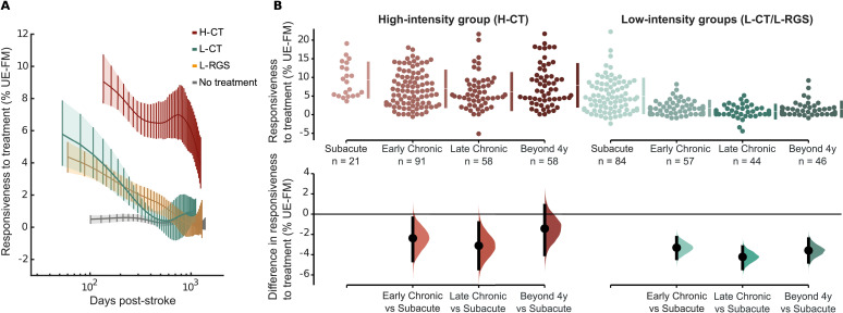
All groups that received treatment showed significant improvements (all treatment groups UE-FM; mean 5±5 SD; no treatment group UE-FM: mean 0±1 SD; p value <0.001, two-sided Mann-Whitney test). We observed that this improvement was proportional to training intensity, that is, the high-intensity group showed higher responsiveness to treatment at all measurement points as compared to both low-intensity groups (difference in responsiveness between H-CT and L-CT/L-RGS; mean 4±1 SE for subacute; mean 5±1 SE for early chronic; mean 5±1 SE for late chronic; mean 6±1 SE for beyond 4y; p<0.001, two-sided Mann-Whitney test) representing a clinically meaningful change for the non-subacute patients.8 The analysis of the effect of chronicity showed a consistent decrease in responsiveness to the three types of treatment (figure 1A). Patients who started treatment in the subacute phase (<6 months) showed the largest improvement in comparison to patients at chronic stages for both high-intensity (the mean differences in responsiveness between subacute and all other chronicity groups ranged from −1 to −3; all p<0.01, two-sided Mann-Whitney test) and low-intensity treatments (the mean differences in responsiveness between subacute and all other chronicity groups ranged from −3 to −4; all p<0.01, two-sided Mann-Whitney test) as shown in figure 1B. The decrease of responsiveness extended beyond the subacute phase and was invariant to training intensity, that is, those who started low-intensity treatment earlier at the chronic stage (6–18 months) also displayed higher gains as compared to those who started later (difference in responsiveness between early and late chronic; mean 1±0 SE; p<0.05, two-sided Mann-Whitney test). Interestingly, a late start did not attenuate the patient’s responsiveness to the high-intensity programme (difference in responsiveness between early and late chronic; mean 1±1 SE; p=0.38, two-sided Mann-Whitney test).
Figure 1.
Recovery dynamics across chronicity for low- and high-intensity interventions. (A) Comparison of patients’ responsiveness to high-intensity and low-intensity treatment: high-intensity conventional treatment (H-CT), low-intensity conventional treatment (L-CT), low-intensity computer-based neurorehabilitation (L-RGS) and a no treatment group (follow-up measurement after a period of no treatment). The patient’s responsiveness to treatment is expressed by the normalised recovery rate (weekly improvement normalised by potential recovery) on the UE-FM scale,6 by the patient’s chronicity at the time of the recruitment. Solid lines indicate the estimated averages and 95% bootstrapped CIs based on the Whittaker smoothing algorithm.6 (B) (Top) Dot plot showing the distribution of normalised recovery rates to high and pooled low-intensity treatment at different stages of chronicity. (Bottom) Estimation plots indicate the effect sizes of the mean differences between the responsiveness at the subacute (horizontal line) or the responsiveness at later stages. RGS, Rehabilitation Gaming System; UE-FM, upper extremity section of the Fugl-Meyer.
Discussion
In this retrospective analysis, we showed that, in both the QSUL and the RGS cohorts, responsiveness to treatment was present at practically all stages post-stroke, demonstrating a gradually declining impact with chronicity and modulation of the responsiveness by the intensity of the intervention. Our findings also support an early rehabilitation start regardless of intensity (ie, at the subacute stage). Beyond this general effect, the high-intensity approach showed a consistent higher impact over low-intensity rehabilitation protocols (L-RGS and L-CT) at all stages post-stroke. Indeed, the advantageous impact of high-intensity rehabilitation on recovery may compensate for a late start. For instance, patients who underwent high-intensity training displayed enhanced responsiveness even when treatment was delivered with a 2-year delay compared with low-intensity interventions. Notice, however, that the difference in intensity between the two rehabilitation approaches was prominent. While the QSUL programme consisted of 6 hours daily, the low-intensity group underwent 0.5–1 hour daily training for only 3 days a week. The lack of proportionality between the groups suggests a non-linear interaction between treatment type, intensity, chronicity and responsiveness.9 This analysis lacks a control group receiving no rehabilitation treatment at early stages post-stroke; thus, we cannot rule out the effect of standard treatment on the patients’ responsiveness to additional treatments. Future studies should investigate and model these relationships to unmask the precise type and intensity of treatment to promote recovery at each stage post-stroke. Altogether, these findings suggest that current stroke guidelines must be revised to incorporate high-intensity rehabilitation protocols across the entire continuum of chronicity.10 Currently, patients obtain only 22 min of treatment,11 with less treatment time at later phases post-stroke. Most importantly, we need to find effective solutions to deliver individualised high-intensity rehabilitation protocols. The increase in stroke survivors and the associated burden poses a challenge for the limited resources of our current healthcare system and asks for the translation of effective principles of neurorehabilitation to scalable technologies to deliver sustainable long-term care addressing the socioeconomic burden of stroke.
Footnotes
Twitter: @dr_nickward, @MartinaMaierBCN
Contributors: BRB, NW and PFMJV planned the analysis. All authors contributed to data acquisition. BRB analysed the data. All authors wrote the draft paper. PFMJV submitted the study and is responsible for the overall content as guarantor.
Funding: This study was supported by the cRGS project under the grant agreement H2020-EU, ID: 840052, and by the RGS@home project from H2020-EU, EIT Health, ID: 19 277.
Competing interests: PFMJV is the founder and interim CEO of Eodyne Systems S.L., which aims at bringing scientifically validated neurorehabilitation technology to society.
Provenance and peer review: Not commissioned; externally peer reviewed.
Ethics statements
Patient consent for publication
Not required.
References
- 1. Ward NS. Restoring brain function after stroke - bridging the gap between animals and humans. Nat Rev Neurol 2017;13:244–55. 10.1038/nrneurol.2017.34 [DOI] [PubMed] [Google Scholar]
- 2. Bernhardt J, Hayward KS, Dancause N. A stroke recovery trial development framework: consensus-based core recommendations from the second stroke recovery and rehabilitation roundtable. Neurorehabil Neural Repair 2019;14:792–802. [DOI] [PubMed] [Google Scholar]
- 3. Wang H, Camicia M, Terdiman J, et al. Daily treatment time and functional gains of stroke patients during inpatient rehabilitation. Pm R 2013;5:122–8. 10.1016/j.pmrj.2012.08.013 [DOI] [PubMed] [Google Scholar]
- 4. Dromerick AW, Lang CE, Birkenmeier RL, et al. Very early Constraint-Induced movement during stroke rehabilitation (vectors): a single-center RCT. Neurology 2009;73:195–201. 10.1212/WNL.0b013e3181ab2b27 [DOI] [PMC free article] [PubMed] [Google Scholar]
- 5. Ward NS, Brander F, Kelly K. Intensive upper limb neurorehabilitation in chronic stroke: outcomes from the Queen square programme. J Neurol Neurosurg Psychiatry 2019;90:498–506. 10.1136/jnnp-2018-319954 [DOI] [PubMed] [Google Scholar]
- 6. Ballester BR, Maier M, Duff A, et al. A critical time window for recovery extends beyond one-year post-stroke. J Neurophysiol 2019;122:350–7. 10.1152/jn.00762.2018 [DOI] [PMC free article] [PubMed] [Google Scholar]
- 7. Bernhardt J, Hayward KS, Kwakkel G, et al. Agreed definitions and a shared vision for new standards in stroke recovery research: the stroke recovery and rehabilitation roundtable taskforce. Neurorehabil Neural Repair 2017;31:793–9. 10.1177/1545968317732668 [DOI] [PubMed] [Google Scholar]
- 8. Page SJ, Fulk GD, Boyne P. Clinically important differences for the upper-extremity Fugl-Meyer scale in people with minimal to moderate impairment due to chronic stroke. Phys Ther 2012;92:791–8. 10.2522/ptj.20110009 [DOI] [PubMed] [Google Scholar]
- 9. Selles RW, Andrinopoulou E-R, Nijland RH, et al. Computerised patient-specific prediction of the recovery profile of upper limb capacity within stroke services: the next step. J Neurol Neurosurg Psychiatry 2021;92:574–81. 10.1136/jnnp-2020-324637 [DOI] [PMC free article] [PubMed] [Google Scholar]
- 10. Ganesh A, Gutnikov SA, Rothwell PM, et al. Late functional improvement after lacunar stroke: a population-based study. J Neurol Neurosurg Psychiatry 2018;89:1301–7. 10.1136/jnnp-2018-318434 [DOI] [PMC free article] [PubMed] [Google Scholar]
- 11. Veerbeek JM, van Wegen E, van Peppen R, et al. What is the evidence for physical therapy poststroke? A systematic review and meta-analysis. PLoS One 2014;9:e87987. 10.1371/journal.pone.0087987 [DOI] [PMC free article] [PubMed] [Google Scholar]


