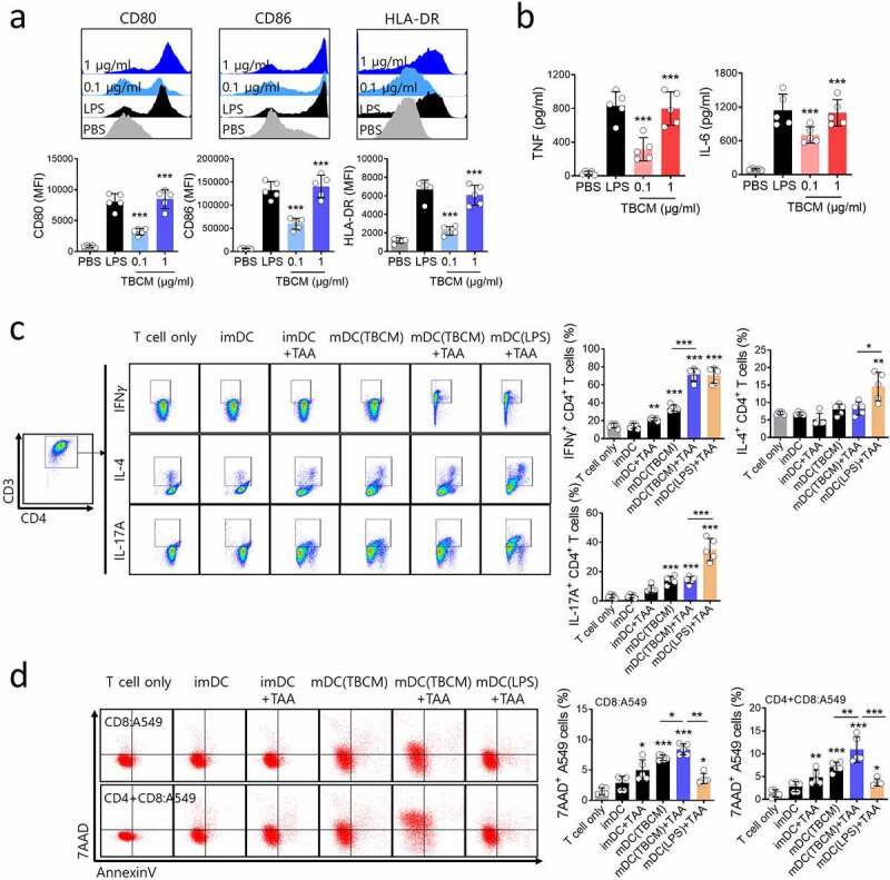
Figure 5.

TBCM-induced human DCs enhances antigen-specific T cell cytotoxic response against human cancer cell. (a) Human DCs were activated for 24 h with TBCM (1 μg/ml) or LPS (0.1 μg/ml) and analyzed for the surface expression marker by flow cytometry. (b) Cytokine levels in the culture supernatant were measured by ELISA. (c) A549 cell lysates (tumor-associated antigens, TAAs)-, TBCM- or LPS-activated human DCs were cocultured with CD4+ T cells (DC:T cell = 1:10) isolated from PBMCs for 72 h. Then, intracellular cytokine production in CD4+ T cells were assessed by flow cytometry. (d) TAAs-, TBCM- or LPS-activated human DCs were cocultured with CD8+ T cells (DC:T cell = 1:10) in the presence or absence of CD4+ T cells for five days. Thereafter, the cells were cocultured with CFSE-labeled A549 cells (E:T = 5:1) for 48 h, and antigen-specific cytotoxicity of T cells were measured by cell apoptosis assay. Significant differences (*p 0.05, **p 0.01, ***p 0.001) among the different groups are shown in the related figures, and the data are presented as the means s.e.m. of five independent experiments.
