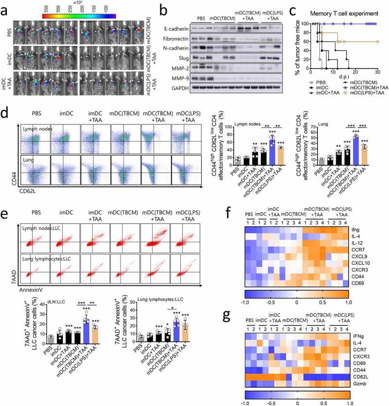
Figure 8.

TBCM-induced DCs generate memory T cells and exert sustained tumor prevention effects. (a) C57BL/6 mice were vaccinated with BMDCs twice at intervals of one week. Seven weeks after the last DC injection, LLC cancer cells were injected intravenously. Seven days after the cancer cell injection, mice were observed for tumor size measurement using an IVIS imaging system 100 and sacrificed for tissue analysis. Representative pictures show luciferase-positive LLC cancer cells one week after cancer cell injection. (b) Protein expression of EMT-related proteins and matrix metalloproteinases MMP-2 and MMP-9 in lung tissue was assessed by immunoblotting assay. (c) After mice were vaccinated with DCs twice at intervals of one week, mice were subcutaneously injected with LLC cancer cells and observed for tumor incidence. (d) One week after cancer cell injection (see a), the population of CD44high CD62Llow CD4+ T cells in the lymph nodes and lung was measured by flow cytometry. (e) One week after cancer cell injection (see a), lung lymphocytes and lymph nodes were cocultured with CFSE-labeled LLC cancer cells (E:T = 5:1), and cytotoxicity against LLC cells was assessed by flow cytometry. (f,g) The transcription level of mRNA in lymph nodes and lung lymphocytes was quantified by RT–qPCR. Significant differences (*p 0.05, **p 0.01, and ***p 0.001) among the different groups are shown in the related figures, and the data are presented as the means s.e.m. of five independent experiments.
