Abstract
We report the first case of onychomycosis caused by Kloeckera apiculata in a woman with multiple sclerosis. Video-dermoscopic examination showed a spiked pattern and distal irregular aspect. Colonies on Sabouraud agar were white, creamy, and smooth. A microscopic examination showed blastoconidia. MALDI-TOF confirmed Kloeckera apiculata as the causal agent.
Keywords: Onychomycosis, Multiple sclerosis, Kloeckera apiculata, Dermatophytome
Established Facts
Dermatophytes are the most common causal agents of onychomycosis.
Candida spp. and non-dermatophyte molds can also affect the nails.
Novel Insights
We talked about Kloeckera apiculata, a yeast with uncommon human affection.
We found a few case reports of this agent of onychomycosis in immunocompetent patients but none in patients with multiple sclerosis.
Introduction
Onychomycosis is the fungal infection of nail plate [1, 2]. Dermatophytes are the most common causal agents [3]; however, Candida spp. and non-dermatophyte molds can also affect the nails [4].
Kloeckera apiculata is the anomorphic phase of Hanseniaspora uvarum; a yeast in the Ascomycetes group in the Hemiascomycetes class, from Endomycetales Saccharomycetaceae order. Hanseniaspora genus included 6 species; only K. apis and K. apiculata have been isolated in humans. K. apiculata has been found in soil, water, grapes, bananas, must, cocoa, cucumber in brine, bivalve molluscs, crabs, prawns, and fruit flies [5, 6, 7, 8, 9, 10]. It has also been described in the fermentation processes of some wines [11]. We found only 2 reports about isolation in healthy humans and none in multiple sclerosis patients.
Case Report/Case Presentation
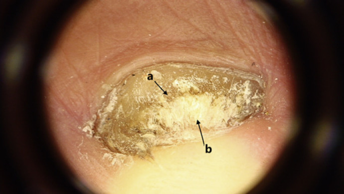
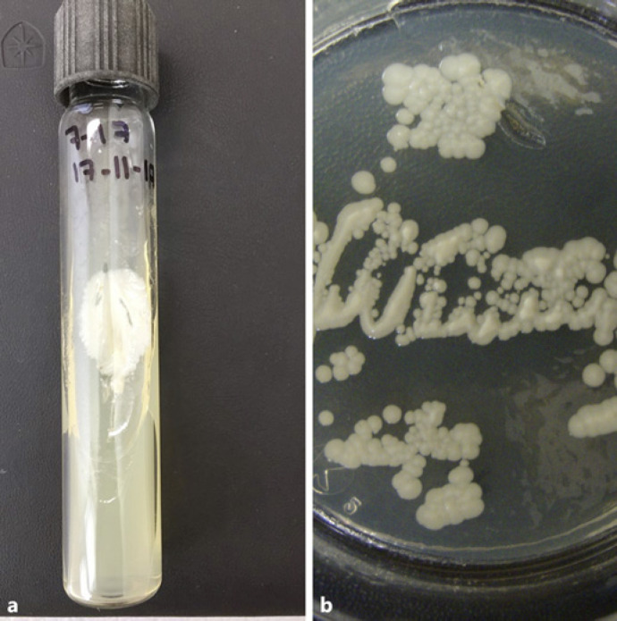
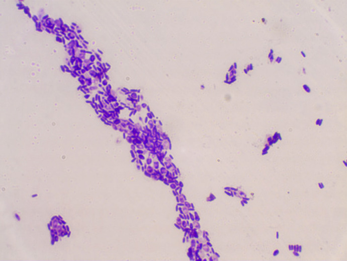
A 38-year-old Mexican woman with a medical history of 2 years of multiple sclerosis relapsing-remitting, treated with gamma interferon (IFN-γ), was attended due to onychodystrophy of the first toenail, at the Mycology Section at “Dr. Manuel Gea González” General Hospital. Dermatological examination showed a yellow-white depigmentation of the nail plate and distal subungual hyperkeratosis of the left first toenail. Video-dermatoscopic examination showed spiked pattern and distal irregular aspect (ruin appearance) (shown in Fig. 1). On microscopic direct examination of nail clippings, with Chlorazol-black-E® stain, thin, long, and septate hyphae were observed. Later, 2 cultures of the nail scales were performed on Sabouraud and Mycosel agar (Beckton-Dickinson®), with 3 weeks apart. In both, the same yeast growth was obtained (white, creamy, and smooth colonies) (shown in Fig. 2), and microscopic examination showed blastoconidia, which were similar to the morphological aspect of Candida spp. (shown in Fig. 3). CHROMagar-Candida identified C. glabrata. MALDI-TOF (matrix-assisted laser desorption/ionization time-off light mass spectrometry, VITEK MS, BioMérieux, France) confirmed Kloeckera apiculata as causal agent. The patient received an occlusive cure with urea 40% ointment and later a ciclopirox nail lacquer, currently with clinical improvement.
Fig. 1.
Video-dermatoscopic examination: spiked pattern (a), and distal irregular aspect (ruin appearance) (×40) (b).
Fig. 2.
Kloeckera apiculata colonies on Sabouraud agar a week after the first culture (a) and 3 weeks after the first isolation (b).
Fig. 3.
Microscopic examination of Kloeckera apiculata colonies (Gram stain, ×40).
Discussion/Conclusions
Multiple sclerosis is an inflammatory demyelinating disease affecting the central nervous system [12]. In Latin America, the epidemiological evidence indicates an increased prevalence of multiple sclerosis [13]. We did not find any case reports about onychomycosis in multiple sclerosis patients. Onychomycosis represents 30% of the superficial mycosis and 50% of all nail disorders. Trichophyton rubrum is the most frequent agent [14]. Non-dermatophyte molds represent 2–12% of onychomycosis [15]. The infection in humans by Kloeckera apiculata is uncommon. Emmanouil-Nikoloussi et al. [16] reported the first case of isolation of Kloeckera apiculata from lesions of the buccal mucosa in a 70-year-old woman. It was identified by Api-20C Aux-system, they described a yeast with a thick cell wall in which the outer surface appeared fuzzy and a bilateral budding and ascospores. García-Martos et al. [17] reported 3 cases of infection by Kloeckera apiculata; in a 9-year-old immunocompetent male with gastroenteritis, in the fecal sample that grew yeasts on blood agar. The colonies were cultivated on Sabouraud dextrose agar and CHROMagar-Candida incubated at 30°C. The other 2 patients were a 39-year-old man and a 36-year-old woman with onycholysis and dark brown pigmentation affecting nails of the fourth and second fingers of their right hands. A sample was identified on Sabouraud dextrose agar, CHROMagar-Candida and Sabouraud dextrose agar with cycloheximide; incubated at 30°C. After 72 h in all 3 patients, the growth of colonies was observed on Sabouraud agar; these were creamy-white, waxy looking, smooth, with unbroken border colonies; in the CHROMagar-Candida, colonies were small, dark lilac colored, with a halo around. Under microscopic observation, yeasts were seen as small and lemon shaped, and identified as Kloeckera apiculata by fermentation and assimilation of carbohydrates using a microgallery ATB ID 32C (Bio-Mérieux, France) [17].
In our patient, on clinical examination of the nail, hyperkeratosis was observed, and the nail clapping confirmed a fungal infection. Characteristics of culture and microscopic morphology are similar to Candida genus, like the previous reports. However, these 2 reports had different method of the yeast identification; in our case, we performed spectrometry (MALDI-TOF®). Onychomycosis by Kloeckera apiculata has not been described before in a multiple sclerosis patient.
In conclusion, we report an unusual first isolation of Kloeckera apiculata in a patient with onychomycosis and multiple sclerosis. We point out the identification of this yeast with mass spectrometry.
Statement of Ethics
The written informed consent was obtained from the patient for publication of this case report and any accompanying images. The ethical approval was not required in accordance with local guidelines.
Conflict of Interest Statement
We declare that we do not have any conflict of interest. Dr. Arenas is an editorial board member of “Skin Appendage Disorders.”
Funding Sources
We do not have funding of any research relevant to the study, including sponsor names and explanations of the roles of these sources in the preparation of data or the manuscript.
Author Contributions
C.D. Sánchez-Cardenas involved in description of case and literature search. D.C. Vega-Sánchez involved in diagnostic test and literature search. T.R. González-Suárez involved in description of case and literature search. J. Flores-Rivera involved in diagnostic test and literature search. R.G. Arenas involved in edition of the final version. T. Corona involved in edition of the final version.
References
- 1.Sigurgeirsson B, Baran R. The prevalence of onychomycosis in the global population: a literature study. J Eur Acad Dermatol Venereol. 2014;28((11)):1480–91. doi: 10.1111/jdv.12323. [DOI] [PubMed] [Google Scholar]
- 2.Eisman S, Sinclair R. Fungal nail infection: diagnosis and management. BMJ. 2014;24((348)):g1800. doi: 10.1136/bmj.g1800. [DOI] [PubMed] [Google Scholar]
- 3.Arenas R. Dermatofitosis en México. Rev Iberoam Micol. 2002;19:63–7. [PubMed] [Google Scholar]
- 4.Moreno G, Arenas R. Other fungi causing onychomycosis. Clin Dermatol. 2010;28((2)):160–3. doi: 10.1016/j.clindermatol.2009.12.009. [DOI] [PubMed] [Google Scholar]
- 5.De Araujo FV, Soares CA, Hagler AN, Mendonça-Hagler LC. Ascomycetous yeast communities of marine invertebrates in a southeast Brazilian mangrove ecosystem. Antonie Van Leeuwenhoek. 1995;68((2)):91–9. doi: 10.1007/BF00873096. [DOI] [PubMed] [Google Scholar]
- 6.Morais PB, Martins MB, Klaczko LB, Mendonça-Hagler LC, Hagler AN. Yeast succession in the Amazon fruit Parahancornia amapa as resource partitioning among Drosophila spp. Appl Environ Microbiol. 1995;61((12)):4251–7. doi: 10.1128/aem.61.12.4251-4257.1995. [DOI] [PMC free article] [PubMed] [Google Scholar]
- 7.Pagnocca F, Mendonca-Hagler L. Yeast associated with he white shrimp Penaeus schmitti, sediment, and water os Sepetiba Bay, Rio de Janeiro Brasil. Yeast. 1989;5:479–83. [PubMed] [Google Scholar]
- 8.Bilbao A, Irastorza A, Dueñas M, Fernandez K. The effect of temperature on the growth of strains of Kloeckera apiculata and Saccharomyces cerevisiae in apple juice fermentation. Lett Appl Microbiol. 1997;24((1)):37–9. doi: 10.1046/j.1472-765x.1997.00340.x. [DOI] [PubMed] [Google Scholar]
- 9.Lachance MA. Yeast communities in a natural tequila fermentation. Antonie Van Leeuwenhoek. 1995;68((2)):151–60. doi: 10.1007/BF00873100. [DOI] [PubMed] [Google Scholar]
- 10.Suzzi G, Romano P, Westall F, Vannini L. The flocculation of wine yeasts: biochemical and morphological characteristics in Kloeckera apiculata. Antonie Van Leeuwenhoek. 1996;69((3)):273–7. doi: 10.1007/BF00399616. [DOI] [PubMed] [Google Scholar]
- 11.García-Marquéz E. Microorganismos del Jerez. Microbiologia. 1995;11:51–8. [PubMed] [Google Scholar]
- 12.Katz Sand I. Classification, diagnosis, and differential diagnosis of multiple sclerosis. Curr Opin Neurol. 2015;28((3)):193–205. doi: 10.1097/WCO.0000000000000206. [DOI] [PubMed] [Google Scholar]
- 13.Corona T, Román GC. Multiple sclerosis in Latin America. Neuroepidemiology. 2006;26((1)):1–3. doi: 10.1159/000089230. [DOI] [PubMed] [Google Scholar]
- 14.Shemer A, Trau H, Davidovici B, Amichai B, Grunwald M. Onychomycosis: rationalization of tropical treatment. Isr Med Assoc J. 2008;10:415–6. [PubMed] [Google Scholar]
- 15.Ramani R, Srinivas CR, Ramani A, Kumari TG, Shivananda PG. Molds in onychomycosis. Int J Dermatol. 1993;32((12)):877–8. doi: 10.1111/j.1365-4362.1993.tb01403.x. [DOI] [PubMed] [Google Scholar]
- 16.Emmanouil-Nikoloussi E, Kanellaki-Kyparissi M, Papavassiliou P, Koliakos K, Dermentzopoulou M, Foroglou C. Hanseniaspora uvarum the ultrastructural morphology of a rare ascomycete, isolated from oral thrush. Bull Gr Int Rech Sci Stomatol Odontol. 1994;37:13–7. [PubMed] [Google Scholar]
- 17.García-Martos P, Hernández-Molina JM, Galán F, Ruiz-Henestrosa JR, García-Agudo R, Palomo MJ, et al. Isolation of Hanseniaspora uvarum (Kloeckera apiculata) in humans. Mycopathologia. 1998;144((2)):73–5. doi: 10.1023/a:1006900909455. [DOI] [PubMed] [Google Scholar]


