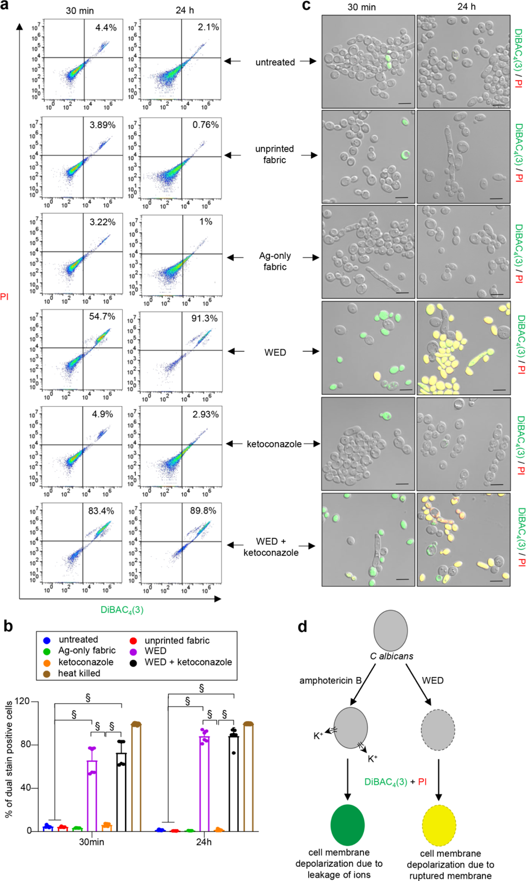
Figure 6: WED depolarized Candida albicans cell membrane.

(a) Representative scatter plots for flow cytometry analysis of DiBAC4(3)+ and PI+ population. C albicans cells (untreated or with respective treatments) were cultured in YPD broth. At respective time intervals, an aliquot was taken and stained with DiBAC4(3) and PI. After 30 mins of dual staining, cells were washed and processed for flow cytometry analysis. Scatter plots with DiBAC4(3)+ and PI+ population were plotted. (b) Graphical representation of flow cytometry analysis. Dual stain positive populations, analyzed using FlowJo software, were plotted in this graph. n = 6. §P < 0.0001 (Two-way ANOVA followed by post-hoc Sidak multiple comparison test). Data are represented as the mean ± SD. (c) Representative images of C albicans cells confirming membrane depolarization after treatment with WED, alone or in combination with ketoconazole. Aforementioned samples remaining after flow cytometry were observed at 63X magnification. Scale bar represents 5 µm. Display settings for all images were kept same. (d) Schematic representation of possible mechanism for WED mediated cell membrane depolarization in C albicans cells. Black border on cell represents cell membrane. Dotted border represents damaged cell membrane. Amphotericin B causes cell membrane depolarization by forming pores causing rapid leakage of ions, especially potassium ions (K+), without damaging the cell membrane. Hence cells treated with amphotericin B get stained with DiBAC4(3) only and give a green fluorescence signal. On the other hand, WED treated cells are observed to take up both fluorescent stains [DiBAC4(3) and PI] as witnessed by a yellow fluorescence signal. This indicates WED causes cell membrane depolarization by cell membrane damage.
