Abstract
Endothelial dysfunction (ED) is a hallmark of atherosclerosis and is influenced by well-defined risk factors, including hypoxia, dyslipidemia, inflammation, and oscillatory flow. However, the individual and combined contributions to the molecular underpinnings of ED remain elusive. We used global gene expression in human coronary artery endothelial cells to identify gene pathways and cellular processes in response to chemical hypoxia, oxidized lipids, IL-1β induced inflammation, oscillatory flow, and these combined stimuli. We found that clustering of the surrogate risk factors differed from the sum of the individual insults that gave rise to emergent phenotypes such as cell proliferation. We validated these observations in samples of human coronary artery atherosclerotic plaques analyzed using single-cell RNA sequencing. Our findings suggest a hierarchical interaction between surrogates of CV risk factors and the advent of emergent phenotypes in response to combined stimulation in endothelial cells that may influence ED.
Subject terms: Atherosclerosis, Vascular diseases
Introduction
Chronic exposure to well-known cardiovascular (CV) risk factors, including lipids, high blood pressure, tobacco usage, physical inactivity, and diabetes, profoundly affects endothelial function1. Surrogates of cardiovascular risk factors are instrumental as biomarkers, drug targets, and tools to investigate their influence on endothelial dysfunction (ED), a critical element for vascular diseases. A large body of evidence suggests that CV risk factors have individual or cumulative effects contributing to the development or worsening of ED, although the complex interplay leading to vascular derangements is poorly understood2. Abnormal blood flow, associated with arterial branching and vessel wall irregularities3,4, chronic inflammatory states5,6, increased oxidized lipids7,8 exposure or hypoxia9,10 have been shown to affect endothelial and vascular function.
The heterogeneity of endothelial cells (EC) between the vascular bed and tissues must also be considered, especially in light of recent data obtained from systematic global gene expression analyses using single-cell RNA-seq11,12. In the murine EC atlas, 78 sub- clusters of ECs were identified based on more than 32,000 ECs from tissues, including brain, testis, liver, spleen, small intestine, colon, skeletal muscle, and heart. These data indicate that the tissue source, as opposed to the type of vascular bed in the blood vessel network, is a critical component that explains most of the heterogeneity of the EC molecular signature12. In this context, a significant limitation of current knowledge is the lack of studies using representative ECs from diverse tissues or associated with a particular phenotype. This is highlighted by the frequent use of human umbilical vein endothelial cells (HUVEC) as a model. These cells are easily accessible and facilitate comparing results across studies; however, they are limited to capturing the broad tissue specificities or represent the phenotype of interest. Indeed, HUVEC represents about 44% of the 360 datasets available in The Endothelial Cell Database (EndoDB)13 repository (https://endotheliomics.shinyapps.io/endodb/)13.
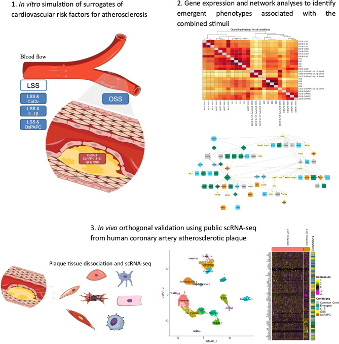
To address some of these questions, we used human coronary artery endothelial cells (HCAEC) to model the effect of surrogate CV risk factors using global endothelial gene expression. We evaluated the roles of chemical hypoxia (CoCl2), oxidized lipids (OxPAPC), inflammation (IL-1β), and oscillatory shear stress (OSS) in global gene expression using laminar shear stress (LSS) as a reference to establish the hierarchy of cellular response to these stimuli and the impact of the combined effect of all stimuli, which is frequently observed in patients with cardiovascular disease (Fig. 1). Our findings revealed new insights into stimulus-dependent EC gene expression and cellular processes associated with the individual or combined disturbances that may contribute to ED.
Figure 1.
Overall workflow for the identification and validation of emergent phenotypes associated to the combined exposure to surrogates of CV risk factors such as hypoxia (CoCl2), Oxidized lipids (OxPAPC), inflammation (IL-1β) and OSS on human coronary artery endothelial cells (HCAEC). 1 Experimental design for in vitro exposure of HCAEC to individual or combined stimuli. 2 Global gene expression and gene regulatory network analyses. 3 Orthogonal validation using scRNA-seq data from human coronary atherosclerotics plaques (dataset GSE131778). The cartoons were created using the Mind the Graph platform (www.mindthegraph.com) and data presented in the manuscript to illustrate the overall workflow.
Results
The effect of surrogate CV risk factors on HCAEC global transcriptomics
We assessed the global transcriptomics in HCAEC exposed individually to CoCl2, OxPAPC, IL-1β, and OSS or the combined effect of all stimuli using the cells maintained on LSS as the reference (Supplementary Fig. 1a). We first verified the ability of each stimulus to activate or inhibit downstream associated biomarkers: activation of HIF-1α in response to chemical hypoxia (CoCl2, 150 μM); increased ATF3 and reduced CD36 mRNA expression by OxPAPC (50 μg/ml); increased VCAM-1 protein expression by IL-1β (10 ng/ml)-induced inflammation and misalignment of the ECs exposed to OSS (± 5 dyn/cm2 at 1 Hz). As shown in Suppl. Figure 1b-e, these surrogates of cardiovascular risk factors altered downstream endothelial biomarkers as expected under the present experimental conditions.
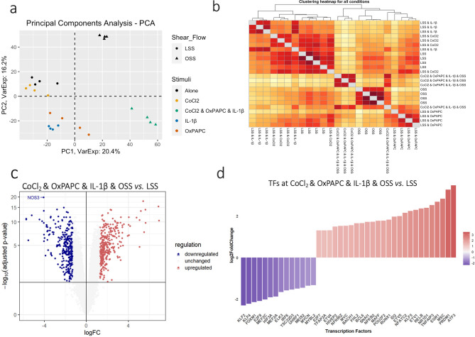
The effects of each stimulus or the combined exposure on HCAEC global gene expression assessed by principal component analysis (PCA) are shown in Fig. 2a. PC1 and PC2 explain almost 37% of the variance of the expression data in response to all experimental perturbations. The PC1 highlights the critical influence of flow patterns on ECs, dividing the data between samples exposed to either LSS or OSS, while PC2 separates the influence of inflammation, oxidized lipids, and the combined stimuli from all other conditions. Examination of PC1 and PC2 indicates that the combined stimulus perturbation is separated from all individual perturbations, consistent with the appearance of emergent phenotypes not anticipated by the sum of individual stimuli. The HCAEC exposed to LSS clustered with CoCl2, whereas the OxPAPC and IL-1β clustered together in a third group, leaving OSS samples isolated as the fourth group (Fig. 2a). The heatmap in Fig. 2b reveals the details of the hierarchical contribution of each condition using the Euclidean distance and the LSS as reference. The two-dimensional graph depicts the samples with the combined stimulus as the most distant (yellow), consistent with the PCA (Fig. 2a). Next, OSS and IL-1β were the individual stimuli more distant from the LSS, followed by OxPAPC and CoCl2 stimuli. These findings suggest that the more distant the samples are from the LSS, the more significant each stimulus's impact modulates the global HCAEC transcriptome. This image is consistent with a hierarchical influence of the various stimuli to alter gene expression, causing or altering endothelial dysfunction (ED). CoCl2, OxPAPC, IL-1β, OSS, and the combined exposure display a graded impact on changing global gene expression (Fig. 2b).
Figure 2.
Gene expression profile revealed the hierarchical contribution of the CVD risk factors CoCl2, OxPAPC, IL-1β and OSS and key TFs relevant to endothelial function. (a) Principal Component Analysis (PCA) of gene expression profile from each sample showed that the PC1 explained 20.4% of the variation while PC2 16.2%. (b) Hierarchical clustering heatmap using Euclidean distance as a measured parameter revealed details of hierarchical contribution by each condition according to their distance to the LSS. (c) Volcano plot showed the differentially expressed genes (DEGs) when comparing the clustered risk factors vs. LSS. The DEGs were considered as adjusted p-value < 0.05 and |Log2foldchange|> 1.3. (d) TF identified among the DEGs and their respective log2 foldchange. Genes in blue are downregulated and in red are upregulated.
We identified 620 differentially expressed genes (DEGs) between the two extreme conditions, the combined stimuli (CoCl2 & OxPAPC & IL-1β & OSS) versus the LSS control group. There were 297 DEGs that were downregulated and 323 that were upregulated (Fig. 2c), in which the NOS3 gene, a canonical vasodilator associated with the nitric oxide (NO) production14, was the most significant downregulated gene. This finding is consistent with the notion that decreased NO production is associated with ED (Fig. 2c). The list of all DEGs is displayed in Supplementary Table 1.
Among the DEGs, we observed 40 differentially expressed transcription factors (TFs) (14 downregulated and 26 upregulated) that may be critical to governing the combined stimulus-induced downstream responses in HCAEC (Fig. 2d). The shear stress-sensitive TFs KLF2 and KLF4 were downregulated, while the TFs NFKBIZ, NFKB2, NFATC2, and RELB and their chemokine targets CXCL1/3/4/5/6/8 were upregulated (Supplementary Table 1). MYC, associated with proliferation, cell growth, and differentiation, was upregulated in the combined stimulus group15,16. EGR1 that is known to mediate downstream hypoxia response, was also upregulated17. Although the upregulation of HIF1A gene expression was not observed, its protein was increased, as shown in Supplementary Fig. 1b. ATF3 gene, known to be activated by OxPAPC18, was upregulated in the combined stimulus group, similar to observed when we exposed static ECs only to OxPAPC (Supplementary Fig. 1c).
The HCAEC global gene expression response to the combined stimulus is different from the sum of each stimulus
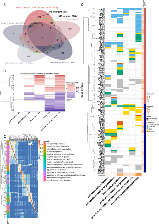
Using LSS as a reference, we compared the HCAEC exposed to each stimulus and the combined stimulus for 48 h (Supplementary Fig. 2a-b). Considering the differential expression analysis of individual stimuli, we identified 8, 179, 275, and 412 DEGs associated with CoCl2, OxPAPC, IL-1β-induced inflammation, and OSS, respectively (Fig. 3a, Supplementary Fig. 2c-g). Among the 620 DEGs affected by the combined stimulus condition compared to the control LSS, 349 were also affected by one or more individual perturbations (Fig. 3a,b [within the dashed green line]). In contrast, 271 DEGs were exclusively associated with the combined stimulus condition. Among the common 349 DEGs, we observed that OxPAPC uniquely modulated 37 DEGs, 90 were associated with IL-1β-induced-inflammation stimulus, and 146 were modulated only by OSS (Fig. 3a [green squares] and Supplementary Fig. 2c).
Figure 3.
Dysregulated DEGs distribution on individual and clustered risk factors followed by GO map of biological process enriched for the 620 DEGs of the clustered risk factors vs. LSS. (a) Venn diagram of DEGs from each condition. The DEGs were considered as adjusted p-value ≤ 0.05 and |Log2foldchange|≥ 1.3; black circle and black arrow regard the unique DEGs from the clustered risk condition; dashed green circle and green arrow regard to the commonly shared genes of the clustered risk factors with each stimulus; dashed black line for unique stimuli-dependent genes, 146 DEGs for OSS, 90 for IL-1β and 37 for OxPAPC. (b) Heatmap of 349 DEGs shared among the DEGs from each stimulus. (c) Heatmap of the similarity matrix of enriched GO terms (biological process) for the clustered risk factors DEGs. Redundancy analysis was applied to identify and keep the most representative term from the redundant terms (cutoff = 0.7 and adjusted p-value ≤ 0.05). (d) Canonical enriched terms in CoCl2 & IL-1β & OxPAPC & OSS vs. LSS (121 DEGs) and the
source of the stimulus individual (represented by colours light blue, yellow and orange), common (grey), more than one stimulus, and if it is emergent, meaning that the gene is only altered if all four stimuli are combined (green). Gene score and enrichment risk score were calculated by − log10(adjusted p-value).
The overall influence of each stimulus is depicted in Fig. 3b, indicating that the global gene expression changes individually where the CoCl2 stimuli were the smallest. In contrast, OxPAPC and OSS had a greater influence on suppressing gene expression, and IL-1β- induced inflammation primarily increased gene expression. Interestingly, E2F8 and ABCG2 were both downregulated, and they were the only DEGs modulated in all conditions examined (Fig. 3b). The TF E2F8 regulates angiogenesis through activation of VEGFA in cooperation with HIF-1α19, while ABCG2 is associated with the cardiac repair of myocardial infarction and ECs survival20.
In summary, the individual stimuli did not recapitulate the condition in which all stimuli were combined. Moreover, 44% of DEGs (271) were exclusively associated with the clustered condition, an emerging pattern likely to be lost when each stimulus is assessed individually.
DEGs and canonical pathways that may contribute to CV risk factors associated with ED
We used Gene Ontology (GO) enrichment analysis to gain insight into biological processes on the 620 DEGs induced by the combined stimulus condition. We grouped the GO terms based on their semantic similarities to minimize the redundancy among the enriched sets. Then, the most representative term was selected according to the highest score within each group of redundant terms. The similarity matrix with the redundant GO terms and their representative term (parent) are represented in Fig. 3c as a heatmap. The findings suggested enriched canonical pathways associated with ED and atherosclerotic plaque formation. The representative groups were as follows: leukocyte migration (green cluster), response to lipopolysaccharide (LPS) and smooth muscle cell proliferation (purple and pink clusters); NO metabolic process, positive regulation of vasculature development, and reactive oxygen species (ROS) biosynthetic process (blue cluster); and cell-substrate adhesion and extracellular matrix (ECM) organization (orange to brown clusters). The GO terms associated with each representative group are displayed in Supplementary Fig. 3a. Figure 3d illustrates six representative processes, showing the DEGs involved and how they are affected by each stimulus or the combined condition. The complete map, including the 15 grouped GO terms, is presented in Supplementary Fig. 3b. It is important to note that the specific DEGs are not always the same even though each stimulus or combined effect influences common processes.
Nitric oxide (NO) and ROS
Decreased NO bioavailability and increased ROS production pathways are enriched in our data and are a hallmark of endothelial dysfunction21,22 (Fig. 3c,d, and Supplementary Fig. 3a and b). In the NO metabolic process term, the ARG2 gene, upregulated in the clustered condition, is known for suppressing NO production by competing with the L-arginine substrate23. Downstream, L-arginine production might also be impaired by the downregulation of the enzyme responsible for its synthesis (ASS1, arginosuccinate synthase)24. NOS3 and its primary activator KLF2 were both downregulated in the clustered condition in comparison to LSS. GCH1 associated with positive regulation of NOS325 was also downregulated. CYP1B1 was also downregulated in the clustered condition, and its inhibition was associated with increased oxidative stress and decreased eNOS26. ARG2, NOS3, KLF2, and CYP1B1 were only modulated by OSS stimulus (Fig. 3d from top to bottom, in yellow), while ASS1 (in gray) was suppressed by the stimuli OxPAPC, IL-1β, and OSS. The upregulation of pro-inflammatory genes such as ICAM1, TLR2, and, especially, PTGS2, with its peroxidase activity, was previously associated with induction of oxidative stress27 and, consequently, increased ROS production (Fig. 3d top, in light blue). Interestingly, except for the ASS1 gene, genes regulated by NO production were mainly affected by the OSS stimulus (Fig. 3d bottom, in yellow), while genes responsible for oxidative stress induction were predominantly modulated by IL-1β inflammatory stimulus (Fig. 3d top, in light blue).
Inflammatory response and adhesion molecules
ECs respond to LPS via the activation of cytokines, chemokines, and adhesion molecules28,29. We observed critical pro-inflammatory gene activation, including IL6, CXCL1/3/5/6, TLR2, NFKB2, and RIPK2 (Fig. 3d top, in light blue). Adhesion molecules such as E-selectin (SELE—in gray), ICAM1, CD44, SDC4, and CSF1 were upregulated and are associated with leukocyte adhesion and transmigration consistent with the growth plaque phenotype30 (Fig. 3d, in light blue). Even though IL-1β induces the majority of the DEGs involved in inflammatory pathways, we also observed a pro-inflammatory role of OSS (CX3CL1, SELE, MCP1 (CCL2), and OxPAPC (CXCL8) (Fig. 3d middle, in gray).
Interestingly, the emergent genes involved in cell-substrate adhesion, leukocyte migration, and response to LPS terms were not directly related to activation of the immune system except by CCL5 and IL36B, both upregulated (Fig. 3d top, in green). The upregulated genes ABCA1 and TRIB1 were associated with increased cholesterol uptake31,32. PD-L2 (PDCD1LG2), upregulated, and CD58, downregulated, were associated with suppression of T cell-mediated immune responses33,34 and stimulation of T cell, respectively35,36 indicating negative feedback on T cell inflammatory response. Finally, we also observed a cell survival phenotype with upregulation of DAPK2, BCL2, MUC4, which primarily promotes cell growth and apoptosis resistance37–39, followed by the upregulation of PI3K (PIK3CD), which is associated with cell growth, proliferation, and metabolism40,41 (Fig. 3d top to middle, in green). It appears that the emergent DEGs, in addition to their role in inflammatory modulation, are associated with lipid uptake, cell growth, and survival.
Vessel remodeling and angiogenesis
The natural progression of atherosclerotic lesions is accompanied by varying degrees of matrix synthesis and breakdown, contributing to vascular remodelling42,43. Our data showed that ECM components and matrix-degrading proteases were modulated: elastin (ELN), fibulin (FBLN2), and fibrillins (FBN2) were all downregulated, while laminin (LAMC2) and collagen (COL27A1) were upregulated; metalloproteinases (MMP1/10) were upregulated, whereas disintegrin and metalloproteinase (ADAM15/19) were both downregulated. These genes were modulated primarily by more than one condition (Fig. 3d and Supplementary Fig. 3b, in gray) except by MMP10 and FBLN2 that were uniquely modulated by OxPAPC and ADAM15/19 by OSS.
Increased angiogenesis processes44,45 follow excessive expansive remodeling, intimal thickening, and ischemia. In this context, we observed activation of angiogenic genes (PGF, PTGS2, ADM, CEACAM1, and ANGPT2) in the clustered condition compared to LSS (Fig. 3d top to the middle). PGF was modulated only by the combined stimuli, whereas PTGS2 was IL-1β stimulus-dependent. ADM and CEACAM1 were regulated by OSS, whereas IL-1β and OxPAPC modulated ANGPT2. Interestingly, the activation of the vasculature development pathways occurred mainly by genes modulated by OSS (CXCR4 and EGR1 were upregulated, and NOS3, KLF2, and KLF4 were downregulated) (Fig. 3d top and bottom, in yellow). Indeed, it has been demonstrated that OSS stimulates angiogenesis while LSS stabilizes vessel remodeling by inhibiting angiogenesis sprouting46,47. Our data suggest that OSS plays a critical role in regulating vasculature development rather than any other stimulus alone or in combination.
Coagulation and thrombogenesis
The shift from low-risk to high-risk plaques usually accompanies ECM degradation, plaque erosion, or rupture with thrombosis of the plaque surface48–50. Three critical genes associated with coagulation cascade were activated and depicted in the positive regulation of vasculature development and cell- substrate adhesion terms (Fig. 3d). Tissue factor (F3 in gray), upregulated by OxPAPC and IL-1β, initiates blood coagulation by forming a complex with circulating factor VII or VIIa51; heparanase (HPSE in blue), upregulated by IL-1β, is a pro-coagulant that increases the generation of activation factor X in the presence of tissue factor and activation factor VII52, and F2RL1, an emergent gene in green, is involved in the coagulation of factor X53. Anti-thrombogenic genes were downregulated, including thrombomodulin (THBD) and plasminogen activator (PLAT), both modulated by OSS (Fig. 3d and Supplementary Fig. 2c). Our data suggest that pro-coagulant factors were activated primarily by inflammatory IL-1β stimulus while OSS suppresses the anti- coagulant genes.
Overall, these data demonstrate that global gene expression response to key surrogates of CV risk factors recapitulates known ED biological processes. Moreover, we provide a yet undescribed context-dependent view regarding the role of individual stimuli and an emergent pattern that arises from the combined stimulus condition, which is usually present in cardiovascular patients.
Transcriptional regulatory ED network
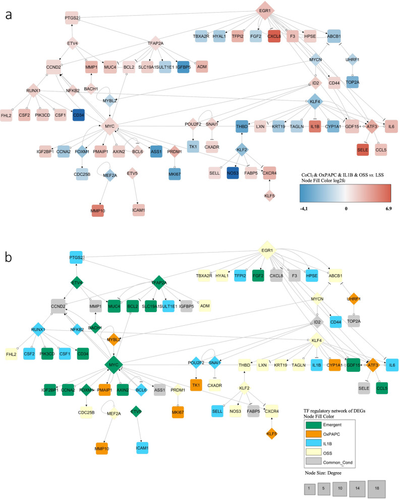
To understand the functional relationship between the combined versus the individual surrogates of CV risk factors in HCAEC response, we generated a gene regulatory network to map the TFs to their target DEGs using the TRRUST database54. We first used the 620 DEGs from the combined condition, in which the TF-DEG network was composed of 71 DEGs (22 TFs among them) with 94 interactions (Fig. 4a). The three highest connected nodes were EGR1, MYC, and KLF4, with 18, 17, and 11 pairs of interactions, respectively. In Fig. 4b, we highlight the role of each condition on these responses: OSS (yellow), the emergent effect from the combined stimulus condition (green), and IL-1β-induced inflammation (blue) were critical factors modulating 19, 17, and 16 genes, respectively, of the generated gene regulatory network. The TF-DEG regulatory networks of the individual stimuli are displayed in Supplementary Fig. 4.
Figure 4.
TF-DEGs regulatory network of the combined stimuli condition. (a) Using the TRRUST database (version 2), the transcription factors (TFs) were mapped to their published transcriptional targets. The identified transcriptional network was further filtered based on whether the TF and their targets were a DEG in CoCl2 & OxPAPC & IL-1β & OSS vs. LSS (adjusted p-value ≤ 0.05 and |Log2foldchange|≥ 1.3). The diamond shape is TF and the square is the DEG target. The edges mean arrow (activation), circle (unknow) and bar (repression). Node size according to the degree. Genes in blue are downregulated, genes in red upregulated. (b) TF-DEGs regulatory network and the condition which they also are differentially expressed. Green is a modulated emergent gene; Orange is an OxPAPC modulated gene; Light blue is an IL-1β modulated gene; Yellow is an OSS modulated gene; Gray is when the gene is modulated in more than one condition.
EGR1, KLF4, and KLF2 are key TFs nodes modulated by OSS
Among the individual stimuli, OSS had a significant effect on ED, and it appears that the TFs that play a Critical role are EGR1 (upregulated) and KLF2, and KLF4 (downregulated) (Fig. 4a,b, in yellow). EGR1 targets include the upregulated genes PTGS2, CCND2, TFPI2, CXCL8, F3, HPSE, GDF15, ATF3, and IL6, and the downregulated genes were TBXA2R, HYAL1, FGF2, and ABCB1 (Fig. 4a). Nevertheless, only CCND2, TBXA2R, HYAL1, and ABCB1 were differentially expressed by the OSS condition (Fig. 4b and Supplement Fig. 4d). This analysis inferred the genes regulated by a given TF and confirmed whether its targets were differentially expressed by the stimuli modulating the TF (Fig. 4b).
EGR1 was the most critical hub in the network. This TF has been recently identified as the most enriched in regulatory regions in human vein and artery endothelial cells subjected to OSS55 and has been predicted to act as a significant regulator of ECs under OSS56. KLF2 and KLF4 are significant regulators of anti-inflammatory response and maintenance of vascular integrity57. Their repression may increase leakage, formation of blood clots, and altered vascular tone57. Inflammatory KLF4 targets were all upregulated: LXN, IL1B, and IL6 (Fig. 4a). LXN was the only one modulated by OSS. KLF2 pro-atherogenic target CXCR4 was upregulated while the anti-atherogenic target NOS3 was downregulated, and they were all OSS stimulus-dependent (Fig. 4a,b). KLF2 and KLF4 regulated the anti-thrombogenic gene THBD (Fig. 4a).
Our data are consistent with a large body of evidence indicating that EGR1, KLF4, and KLF2 are master regulators of ECs under shear stress55,57,58. These TFs increase NOS3 expression, reduce oxidative stress, and inhibit chemokines receptors like CXCR4. A more detailed and similar network with all OSS stimulus-dependent DEGs is shown in Supplementary Fig. 4d.
Proliferative emergent behavior and its impact on inflammatory stimuli
The combined stimulation condition uncovered the MYC node. This response suggests that clustering of risk factors elicits emergent properties that the individual perturbations cannot recapitulate. MYC targets comprise CCND2, CCNA2, FOXM1, and BCL2 associated with cell cycle and apoptosis resistance16,38, where CCND2 and BCL2 are upregulated and CCNA2 and FOXM1 are downregulated (Fig. 4a). The emergent hubs include MYC and TFAP2A, shown in green in Fig. 3b. The IL-1β-stimulated TFs RUNX1 and BCL6 promote angiogenesis59,60 (Fig. 4a,b). The RUNX1 targets CSF1/2 and PIK3CD are involved in cell survival, growth, proliferation, and inflammatory responses. CSF1/2 were activated by IL-1β stimulus, while PIK3CD (upregulated) is an emergent gene and well-known activator of the mTOR pathway signalling61,62 (Fig. 4a,b).
Curiously, when we performed the TF-DEGs gene regulatory network for each stimulus, the critical network was driven by IL-1β-inflammation stimulus, where the NFKB1 was the central hub (Supplementary Fig. 4c). Nevertheless, NFKB1 was not differentially expressed in the clustered condition, nor were many of its targets (BMP2, CXCL2, LCN2, VCAM1, and IRF1). Furthermore, most of the NFKB1 gene targets had their log2 fold-change decreased in the clustered condition compared to the IL-1β- inflammation stimulus alone (Supplementary Fig. 4e). It appears that the clustered condition suppresses part of the inflammatory response, and it gives space to a proliferative behavior and EC survival phenotype.
Validation of the HCAEC data using single-cell RNA-seq from human coronary atherosclerotic plaque
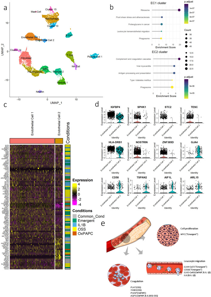
Our in vitro data indicated that the combined stimulus condition is critical to uncover emergent phenotypes that may disrupt the endothelial cell compensatory mechanisms leading to disease states. We then determined whether this was the case in an in vivo chronic and inflammatory atherosclerotic plaque condition. We re-analyzed a public single-cell RNA-seq dataset obtained from human coronary artery atherosclerotic lesions from patients with clustered risk factors (GSE131778)63. We analyzed 10,671 cells and identified 17 cell clusters (Fig. 5a and Supplementary Fig. 5a). We used PECAM1 (CD31) expression to identify the ECs clusters (Supplementary Fig. 5b and Supplementary Table 2). The cell type clusters were annotated by the top defining genes in each cluster (Supplementary Fig. 5c).
Figure 5.
scRNA-seq of human coronary artery atherosclerotic plaque identified two endothelial cells (EC) clusters which are defined by the clustered risk factors DEGs. (a) UMAP visualization of clustering identified 17 cell populations (n = 10,671 cells). (b) KEGG enrichment analysis of the two EC clusters using the cluster marker differentially expressed genes between a cluster and all remaining cells. DEG were considered for adjusted p-value ≤ 0.05 and |Log2foldchange|≥ 0.25. (c) Heatmap of the 103 clustered risk factors DEGs expression levels from each endothelial cell of the two clusters followed by the condition in which they are differentially expressed. (d) Violin plot of emergent DEGs which are differentially expressed in EC2 vs. EC1. (e) Schematic illustrating the key dysregulated pathway in the clustered risk factors which are recapitulated in human atherosclerotic plaque. (e) Created in the Mind the Graph platform (www.mindthegraph.com).
We first performed a pathway enrichment analysis using the DEGs between each cluster against all the remaining ones. The DEGs (843) observed in the EC1 cluster were mainly involved in ribosome-associated pathways, fluid shear stress in atherosclerosis, cancer proteoglycan, and leukocyte transendothelial migration. In contrast, the DEGs (644) in the EC2 cluster were related to coagulation cascade, viral myocarditis, and type I diabetes mellitus (Fig. 5b and Supplementary Table 2).
Our primary goal was to determine whether the DEGs associated with the combined stimuli were involved in the transcriptomic profile associated with the two EC clusters (843 and 644 DEGs for clusters EC1 and EC2, respectively) and uncover the potential role of the emergent genes in these processes. First, 17% of the DEGs previously identified (103/620 DEGs of the combined stimulus condition) were among the DEGs defining these two EC clusters. Figure 5c shows how these 103 DEGs were influenced by each stimulus in both EC clusters based on our in vitro assays. The OSS stimulus modulated 32 genes, 31 genes displayed the emergent pattern, followed by 22 differentially expressed genes in more than one condition, and 13 and 5 DEGs were modulated only by IL-1β and OxPAPC, respectively (Fig. 5c). Interestingly, it appears that these validated 103 DEGs are critical genes defining the EC1 and EC2 clusters in the heatmap (Fig. 5c middle to bottom). For example, IGFBP4, PLVAP, and AQP1 are less expressed in EC2 than EC1, while ARL15, GJA5, and GJA4 are more expressed in EC2 than EC1 (Fig. 5c).
Next, we identified 673 DEGs between EC cluster 2 vs. 1 (Supplementary Fig. 5d and Supplementary Table 3). Examining them against the 620 DEGs from the in vitro combined stimuli condition, we identified 71, with 23 displaying the emergent pattern. To highlight these 23 emergent DEGs in human coronary artery plaque, we plotted them in each EC cluster (Fig. 5d and the complete list in Supplementary Fig. 5e). IGFBP4, SPHK1, STC2, TESC, HLA-DRB1, and NOSTRIN were downregulated in EC2 compared to EC1. These genes are involved with a broad array of cellular functions. IGFBP4 and STC2, for example, have been associated with cell growth and glucose uptake64 and angiogenesis induction65, respectively. SPHK1 was associated with endothelial permeability66, while TESC was associated with cardiomyocyte hypertrophy67. HLA-DRB1 was associated with ED in patients with rheumatoid arthritis68, while NOSTRIN was associated with decreased eNOS activity69 (Fig. 5d). MYC was upregulated at EC1 only compared to all other clusters (Fig. 5c and Supplementary Fig. 5f.).
In contrast, we observed that GJA4, CD58, TSPAN2, AIF1L, and ARL15 were upregulated in EC2 compared to EC1. GJA4 and CD58 have been associated with increased cell–cell junction and leukocyte migration70,71. ARL15 overexpression showed a protective effect on the endothelium by increasing NO and decreasing ROS72 production. TSPAN2 appears to play a role in the migration of ECs and SMCs73 and has been strongly associated with atherosclerosis in large arteries74. However, the role of TSPAN2 remains to be elucidated73. AIF1L was associated with increased necrotic core and is a marker of plaque vulnerability75.
Understanding the molecular architecture underlying the EC clusters' biological function improves upon evaluating the behavior of the genes in response to the individual stimuli in vitro. PLAT and THBD (OSS) and AQP1 (OxPAPC & IL-1β & OSS) were downregulated in EC2 compared to the EC1 cluster (Supplementary Fig. 5f.), suggesting that these cells are more susceptible to thrombosis, consistent with data shown in Fig. 5b. The decreased expression of genes associated with cell–cell junction in EC1, such as GJA4 (emergent) and GJA5 (OxPAPC & IL-1β), are consistent with increased adhesion and migration of leukocytes pathway, enriched in the EC1 cluster (Fig. 5b).
These findings suggest that HCAECs under the combined surrogates of risk factors recapitulated, at least in part, the changes in canonical pathways observed in vivo in human coronary atherosclerotic plaque samples analyzed by single-cell RNA-seq. The 103 DEGs from the in vitro experiment overlapped with the DEGs from human atherosclerotic plaque ECs, allowing us to infer the pathways that are preferentially influenced by each stimulus or by the combined stimulation in vitro, further supporting our model. The composition of ECs on the plaque suggests that one cluster displays an inflamed and proliferative susceptible phenotype with increased leukocyte migration (EC1), whereas the cells are more prone to thrombosis EC2 cluster (Fig. 5e).
Discussion
We showed that DEGs observed in ECs from human coronary artery plaques can be modeled in vitro by exposing HCAEC to surrogates of the main CV risk factors applied individually or in combination. Using this approach, we provide new insights about the ED molecular architecture and the gene networks and inferred cellular processes modified by individual surrogates of CV risk factors, which display a hierarchical influence pattern. Furthermore, we demonstrated for the first time that the combined exposure to surrogates of CV risk factors, as seen in cardiovascular patients, give rise to emergent properties with unique modulation of gene networks and processes, such as the activation of cellular proliferative pathways, that is not recapitulated by the sum of influences from the individual perturbations.
It is well-known that hypoxia44,76, OxPAPC77,78, IL-1β1,6 and OSS79,80 are major players in endothelial function/dysfunction. Hypoxia and HIF signaling stimulate EC survival and growth, cell invasion, and glucose metabolism, contributing, in general, to angiogenesis81,82. Chemical hypoxia also increases HIF-1α83 by preventing its degradation, as we showed in Supplementary Fig. 1b, even though we did not observe differences in HIF-1α expression under this stimulus. It is also known that OSS alters EC metabolism via activating HIF-1α, which induces cell proliferation and inflammation by activating glycolytic enzymes84. Interestingly, CoCl2 or OSS alone did not achieve this proliferative and angiogenic phenotype, only observed when the stimuli were combined. This response may be explained by the EC compensatory capacity's cumulative perturbations and exhaustion, giving rise to altered phenotypes.
The modification of an established response to individual perturbations or the appearance of an emergent phenotype associated with the combined stimulation were also detected in other canonical responses. We recapitulated the activation of critical inflammatory markers including NF-κβ (NFKB1), ICAM1, E-selectin (SELE), and IL-8 (CXCL8) that was observed in LSS cultured HUVEC exposed to IL-1β and TNF-α 85. However, when the IL-1β was combined with other stimuli, NFKB1 no longer changed its expression (Supplementary Fig. 4e), and neither did several of its pro-inflammatory targets (BMP2, CXCL2, LCN2, VCAM1, and IRF1). Interestingly, this partial suppression of inflammatory response was accompanied by the emergence of a proliferative and cell survival program activated by the proto-oncogenes MYC and BCL2, with the former being an emergent hub on the TF-DEGs network (Fig. 4b). MYC is an exciting target because it has been associated with atherosclerosis by promoting smooth muscle cell proliferation86,87, and we showed that it is also upregulated in ECs from human coronary atherosclerotic plaques. We were only able to model this phenomenon because we analyzed the individual and the combined contribution of the stimuli, a likely in vivo scenario where multiple CV risk factors coexist.
Alsaigh et al.88 characterized ECs from human carotid artery atherosclerotic plaques using scRNA-seq similarly to the analysis we performed in the endothelial cells from coronary plaques data from a previously reported study63. They also identified two EC clusters, one localized on the atherosclerotic core (AC) displaying comparable gene expression changes that we observed in the EC2 cluster (ACKR1, IL6, IGFBP4, HLA- DRA, HLA-DRB1, and NOSTRIN were downregulated while MPZL2, GJA5, ELN, END1, RHOB were upregulated) (Supplementary Fig. 5d-f). In contrast, the EC cluster from the adjacent region to the carotid plaque called the proximal adjacent (PA) region, behaved as the EC1 cluster described here. They found that the DEGs that define the AC cluster were enriched in epithelial-mesenchymal transition, angiogenesis, coagulation cascade, and hypoxia pathways, whereas the inflammatory response, MYC targets, and E2F targets pathways defined the PA cluster88. Altogether, the results from datasets of scRNA-seq from atherosclerotic plaques in different vascular beds suggest different transition states of ED throughout atherosclerotic lesions. These observations are consistent with our findings, indicating a hierarchy among the effects of different stimuli and the appearance of emergent phenotypes when the stimuli are combined. Thus, varying with the presence and the intensity of the different pathologic stimuli, as seen in different patients, may give rise to several subsets of phenotypes.
Using the global gene expression patterns from in vitro perturbations of ECs and data from human coronary artery plaques, we identified the emergent properties that can arise from multiple non-linear interactions and feedback loops governing EC function/dysfunction. The reductionist in vitro approach was informative for identifying the hierarchy of the various stimuli in altered global gene expression and gene networks, enabling inferences regarding ED processes, including leukocyte adhesion, thrombogenesis, and cell proliferation. Notably, the genes associated with the latter were only identified by the combination of surrogates of CV risk factors, highlighting the difficulties in recapitulating components of complex phenotypes in vitro. The effects of CV risk factor clustering surrogates on ECs are different from the sum of the individual responses, and approximately 44% of DEGs and associated gene pathways are unique to the clustering condition.
Our results suggest that changes in gene networks and inferred cellular processes observed in coronary artery atherosclerotic plaques could be modeled and further understood using in vitro approaches where individual. The combined contribution of different factors can be carefully managed dissected to uncover hierarchic patterns and emergent properties. These emergent properties may have significant consequences for fundamental investigations of endothelial (dys)function associated with multiple interactions. One may speculate that, regardless of these characteristics, testing additional samples from different beds and conditions may enable prediction and identification of the main hubs or gene networks associated with specific or combined stimuli enabling the design of targeted therapeutic approaches to prevent or retard plaque development. In summary, we provided evidence of a hierarchical effect between surrogates of CV risk factors and the advent of emergent phenotypes in response to combined stimulation in ECs that may influence ED.
Methods
Cell culture and stimulations
HCAEC were acquired from LONZA. The cells were grown in EBM-2MV medium (LONZA) and incubated at 37 °C, 5% CO2. All experiments were carried out between passages 4 and 6. HCAEC were previously treated with cobalt chloride (CoCl2, Sigma-Aldrich), OxPAPC (1-palmitoyl-2- arachidonoyl-sn-glycero-3-phosphorylcholine) (SINAPSE), interleukin 1β (IL-1β) (Thermo Fisher Scientific), laminar shear stress (LSS), or oscillatory shear (OSS) to confirm each stimulus individually. As soon as the cells reached 100% confluence (static condition except by shear stress), they were subsequently individually treated with CoCl2 (150 μM), OxPAPC (curve concentration of 25, 50, and 100 μg/ml) or IL-1β (10 ng/ml) and cultured for 48 h. To confirm the effect of each stimulus, we used the protein expression of VCAM for the IL-1β stimulus, CD36, and ATF3 gene expression for treatment with OxPAPC, and protein expression for HIF1-α to confirm chemical hypoxia induced by CoCl2.
For experiments with shear stress, we used the IBIDI equipment (IBIDI). Using the Pump Control software, this equipment simulates, controls, and quantifies the shear force applied to ECs throughout the experiment. The ECs were grown on μ-Slides I Luer 0.4 (IBIDI) coated with 0.1% gelatin. HCAECs were grown to 100% confluence (approximately 100,000 cells/cm2) on the slides and incubated overnight under standard static cell culture conditions. The ECs were exposed to a unidirectional atheroprotective shear flow with 20 dynes/cm2 (LSS) and an atherogenic, bidirectional at 1 Hz (0 ~ ± 5 dynes/cm2) shear flow (OSS) for 48 h. We used the cell alignment as a positive effect of the shear forces.
Once all the stimuli were confirmed, we further experimented with the individual or combined factors as follows: i) cultivation of ECs in μ-Slides I Luer 0.4 overnight; the next day, the cells were coupled to the perfusion set containing 12 ml of EBM-2MV medium; ii) addition of stimuli: IL-1β (10 ng/ml), OxPAPC (50 μg/ml), or CoCl2 (150 μM) individually or combined to the perfusion set and integrated into the pump; iii) the ECs were subjected to either the LSS or OSS for 48 h (Supplementary Fig. 1a). The total RNA was collected after each experiment.
Reverse-transcriptase quantitative polymerase chain reaction (RT-qPCR)
The ECs were lysed with 1 mL of TRIzol Reagent (Invitrogen). Total RNA after extraction and purification was reverse transcribed using SuperScript IV Reverse Transcriptase (Thermo Fisher Scientific). Real-time PCR was performed using SYBR-Green (Roche) with the QuantiStudio 12 K Flex system (Applied Biosystems). We used GAPDH Ct values to normalize ΔCt. The variation in gene expression between samples was calculated using the ΔΔCt method. The primers are as follows:
ATF3 Fow./Rev.: GCAAAGTGCCGAAACAAGAAG/GCTTCTCCGACTCTTTCTGC
CD36 Fow./Rev.: AGATGCAGCCTCATTTCCAC/AGATGCAGCCTCATTTCCAC
Protein extraction and western blotting
The cells were lysed in RIPA buffer (150 mM NaCl, 0.5% sodium deoxycholate, 1% Triton X-100, 0.1% SDS, 50 mM Tris–HCl, pH 8) containing protease and phosphatase inhibitors (Sigma Aldrich), and proteins were quantified using a BCA protein assay kit (Pierce Biotechnology). Equivalent amounts of protein were solubilized in sample buffer (0.5% SDS, 10% glycerol, 0.05% bromophenol blue, 50 mM dithiothreitol, 50 mM Tris, pH 6.8) and subjected to polyacrylamide gel electrophoresis in the presence of sodium dodecyl sulfate using polyacrylamide gels of different concentrations. After electrophoresis, proteins were transferred to polyvinylidene fluoride membranes (PVDF 0.45, Amersham Hybond P). Membranes containing the transferred proteins were incubated first in blocking solution (5% bovine serum albumin and 0.1% Tween 20 in TBS, pH 7.4) for 1 h at room temperature, followed by incubation for 16–18 h in specific primary antibody diluted in blocking solution at 4 °C. The following primary antibodies were used: anti-HIF-1α (1:1000, #3619 Cell Signaling Technology) and anti-VCAM-1 (1:1000, #TA50391, Origene). The membranes were also incubated with primary antibodies specific for GAPDH (1:2000, #ab22555 Abcam) or beta-actin (1:2000, #ab8227 Abcam) used as internal controls (housekeeping). After incubation for 1 h at room temperature with secondary antibody conjugated to horseradish peroxidase, the bound primary antibody was detected using a chemiluminescent image analyzer (ImageQuant LAS 4000 mini, GE HealthCare), and the images were quantified by densitometry using ImageJ software (Wayne Rasband).
Microarray gene expression profiling
We used the RNeasy Micro Kit (Qiagen) for total RNA isolation to increase the efficiency of RNA extraction from samples with little material, such as those grown in IBIDI. We follow the manufacturer's protocol (RNeasy Micro Handbook). To generate global gene expression (> 20,000 genes), we used the Human Clariom S Assays (Thermo Fisher Scientific) that detect only the exons present in all expressed isoforms of transcripts known as exons constituting locus of a single gene. The samples were processed in a plate matrix in the GeneTitan TM Microarray System (ThermoFisherScientific). The entire protocol for processing and running the samples followed the standard protocol (GeneChip TM WT PLUS Reagent Kit Manual Target Preparation for GeneChip TM Whole Transcript (WT) Expression Arrays).
Microarray analysis and quality control
Using the 24 CEL files generated from the GeneTitan TM Microarray System, the quality control of each file was checked before further analysis. The package used was the array QualityMetrics89 to identify abnormal signal intensities (saturation or noise), an average of the variance of the intensity of this signal, and possible outliers. To calculate abnormal intensities, we used the Kolmogorov–Smirnov test between the distribution of the intensity matrix of each array (sample) and the distribution of the grouped data. Another analysis was the standard deviation of the intensities in the matrixes after normalization and transformation to a logarithm scale to identify possible signal saturation. Finally, Hoeffding's D statistic was performed in the joint distribution of M and A for each matrix to identify outliers. M and A were defined as follows: M = log2 (I1) − log2 (I2) A = 1/2 (log2 (I1) + log2 (I2)), where I1 is the intensity of the studied matrix and I2 is the intensity of a "pseudo" matrix that consists of the median between the matrixes. D > 0.15 was considered a threshold to remove outliers. All samples passed the quality control check.
Differential expression analysis
The analysis of differential gene expression was performed with the limma package90. The analysis was performed using the logarithmic matrix of the signal strength adjusted in a linear model, using the lmFit () function. We apply Bayes empirical statistics to this linear model, calculating a moderate t-test for each gene, calculating the t-value and its corresponding p-value for each gene. Concomitantly, an adjustment is made by multiple tests. In this case, the adjustment was made by BH, which is the method of Benjamini and Hochberg to control the false discovery rate. We consider DEGs to be those with an adjusted p-value < 0.05 and the absolute value of |Log2Foldchange|> 1.3. The analysis of the transcriptome data was performed using the R platform to analyze and visualize the molecular profile of gene expression, differential expression analysis, pathway enrichment analysis, and GO for biological processes.
GO-term enrichment and Kyoto Encyclopedia of Genes and Genomes (KEGG) pathway enrichment analysis
We performed an over-representation analysis to determine whether known biological processes or gene pathways are enriched using a list of DEGs. GO terms and KEGG pathway enrichment analysis was performed using the clusterProfiler91 Bioconductor package. Redundancy analysis was applied to identify and keep the most representative GO terms from the redundant terms (cutoff = 0.9 and adjusted p-value ≤ 0.05). We calculate a score based on the − log10 (adjusted p- value) of the significantly enriched pathways and biological processes. Distances between points represent the similarity between terms. GO terms and KEGG enriched pathways were considered with an adjusted p-value less than or equal to 0.05.
Transcriptional gene regulatory network
Using the TRRUST database (version 2), the TFs were mapped to their published transcriptional targets. The identified transcriptional network was further filtered based on whether the TF and targets were a DEG (adjusted p-value ≤ 0.05 and |Log2Foldchange|≥ 1.3). Node size varied according to the degree. Cytoscape (version 3.7.2) was used to topologically visualize the TF- DEGs regulatory network and Network Analyzer tool to identify the hubs based on high degree and high closeness centrality values.
Single-cell RNA-seq public data analysis
The public data (GSE131778) for the scRNA-seq of human coronary artery plaques63 are available at the Gene Expression Omnibus database. We used the cell gene expression matrixes to further analysis using the R package Seurat version 3.2.292. We trimmed the cells expressing fewer than 200 genes and genes expressed in fewer than three cells. We filtered out cells that had unique gene counts (nFeature_RNA) over 2,500 or less than 200. Cells containing > 5% mitochondrial genes were presumed to be of poor quality and were also discarded. Cell counts were log-normalized by a size factor of 10,000 RNA counts and then scaled. Next, PCA was applied to the scaled data for dimensionality reduction. We used the FindNeighbors() and FindClusters() functions on the Euclidean distance PCA space to refine the edge weights between any two cells to iteratively group cells with a similar expression pattern. We used UMAP to visualize the identified clusters of cells. We applied the FindMarkers () function to identify the markers that defined each cluster, which automatically, via differential expression analysis, compared a cluster against all others. We kept as gene markers p-value ≤ 0.05 and average |Log2Foldchange|≥ 0.25. The top ten cell markers were used to manually annotated each cell type.
Statistical analysis
For all statistical analyses, R software (version 4.1.0) was used. For qPCR and western blot, we used Student’s t-test to compare two groups and one-way analysis of variance for multiple comparisons. Statistical significance was considered at p < 0.05.
Supplementary Information
Acknowledgements
IPS is a recipient of a fellowship from the Coordination for the Improvement of Higher Education Personnel (CAPES 88882.328126/2019-01). JEK is supported by grants from the Sao Paulo Research Foundation Fundação de Amparo a Pesquisa do Estado de São Paulo [FAPESP-INCT—20214/50889-7 and 2013/17368-0] and Conselho Nacional de Desenvolvimento Científico e Tecnológico—CNPq [INCT—465586/2014-7 and 309179/2013-0].
Abbreviations
- ATF3
Activating transcription factor 3
- CCL2
C–C motif chemokine 2
- CV
Cardiovascular
- CXCL1
C-X-C motif chemokine 1
- CXCL8
C-X-C motif chemokine 8
- CXCR4
C-X-C chemokine receptor type 4
- CXCR7
C-X-C chemokine receptor type 7
- CXCL12
C-X-C motif chemokine 12
- DEGs
Differentially expressed genes
- ECs
Endothelial cells
- ECM
Extracellular matrix
- ED
Endothelial dysfunction
- FGF2
Fibroblast growth factor 2
- GJA4
Gap junction alpha-4 protein
- GJA5
Gap junction alpha-5 protein
- GO
Gene ontoloty
- HCAEC
Human coronary artery endothelial cell
- HIF-1α
Hypoxia-inducible factor 1-alpha
- ICAM1
Intercellular adhesion molecule 1
- IL-1β
Interleukin 1 beta
- KEGG
Kyoto encyclopedia of genes and genomes pathway
- KLF2
Kruppel-like Factor 2
- KLF4
Kruppel-like Factor 4
- LSS
Laminar shear stress
- MMP1
Matrix metalloproteinase-1
- MMP9
Matrix metalloproteinase-9
- MCP-1
Monocyte chemoat- tractant protein-1
- NOS3
Nitric oxide synthase 3
- OSS
Oscillatory shear stress
- OxPAPC
Oxidezed 1-palmitoyl-2-arachidonyl-sn-glycero-3-phospholcholine
- PECAM1
Platelet endothelial cell adhesion molecule-1
- PI3K
Phosphatidylinositol 3-kinase
- ROS
Reactive oxygen species
- SELE
E-selectin
- scRNA-seq
Single-cell RNA-seq
- SMCs
Smooth muscle cells
- TFs
Transcription factors
- THBD
Thrombomodulin
- TNF-α
Tumour necrosis factor alpha
Author contributions
Conception and design: I.P.S., M.H.F.A., S.K.T., and J.E.K.; Data collection: I.P.S., M.H.F.A., and M.V.R.; Writing the article: I.P.S. and J.E.K.; Critical revision of the article: I.P.S., and J.E.K.; Statistical analysis: I.P.S.; obtained funding: J.E.K.; overall responsibility: J.E.K.
Data availability
The raw data (CEL files) of the microarray expression data were deposited at the GEO database with the reference (GSE181822).
Competing interests
The authors declare no competing interests.
Footnotes
Publisher's note
Springer Nature remains neutral with regard to jurisdictional claims in published maps and institutional affiliations.
Supplementary Information
The online version contains supplementary material available at 10.1038/s41598-022-05404-7.
References
- 1.Xu S, et al. Endothelial dysfunction in atherosclerotic cardiovascular diseases and beyond: From mechanism to pharmacotherapies. Pharmacol. Rev. 2021;73:924–967. doi: 10.1124/pharmrev.120.000096. [DOI] [PubMed] [Google Scholar]
- 2.McCarron JG, et al. Heterogeneity and emergent behaviour in the vascular endothelium. Curr. Opin. Pharmacol. 2019;45:23–32. doi: 10.1016/j.coph.2019.03.008. [DOI] [PMC free article] [PubMed] [Google Scholar]
- 3.Timmins LH, et al. Oscillatory wall shear stress is a dominant flow characteristic affecting lesion progression patterns and plaque vulnerability in patients with coronary artery disease. J. R. Soc. Interface. 2017;14:20160972. doi: 10.1098/rsif.2016.0972. [DOI] [PMC free article] [PubMed] [Google Scholar]
- 4.Peiffer, V., Sherwin, S. J. & Weinberg, P. D. Does low and oscillatory wall shear stress correlate spatially with early atherosclerosis? A systematic review. Cardiovasc. Res. (2013). [DOI] [PMC free article] [PubMed]
- 5.Cheng C, et al. Atherosclerotic lesion size and vulnerability are determined by patterns of fluid shear stress. Circulation. 2006;113:2744–2753. doi: 10.1161/CIRCULATIONAHA.105.590018. [DOI] [PubMed] [Google Scholar]
- 6.Galkina E, Ley K. Immune and inflammatory mechanisms of atherosclerosis (*) Annu Rev Immunol. 2009;27:165–197. doi: 10.1146/annurev.immunol.021908.132620. [DOI] [PMC free article] [PubMed] [Google Scholar]
- 7.Furnkranz, A. et al. Oxidized phospholipids trigger atherogenic inflammation in murine arteries. Arterioscler. Thromb. Vasc. Biol. (2005) [DOI] [PubMed]
- 8.Miller YI, Shyy JYJ. Context-dependent role of oxidized lipids and lipoproteins in inflammation. Trends Endocrinol. Metab. 2017;28:143–152. doi: 10.1016/j.tem.2016.11.002. [DOI] [PMC free article] [PubMed] [Google Scholar]
- 9.Folco, E. J. et al. Hypoxia but not inflammation augments glucose uptake in human macrophages: Implications for imaging atherosclerosis with 18fluorine-labeled 2-deoxy-D-glucose positron emission tomography. J. Am. Coll. Cardiol. (2011). [DOI] [PubMed]
- 10.Wong BW, Marsch E, Treps L, Baes M, Carmeliet P. Endothelial cell metabolism in health and disease: Impact of hypoxia. EMBO J. 2017;36:2187–2203. doi: 10.15252/embj.201696150. [DOI] [PMC free article] [PubMed] [Google Scholar]
- 11.Paik DT, et al. Single-cell RNA sequencing unveils unique transcriptomic signatures of organ-specific endothelial cells. Circulation. 2020;142:1848–1862. doi: 10.1161/CIRCULATIONAHA.119.041433. [DOI] [PMC free article] [PubMed] [Google Scholar]
- 12.Kalucka J, et al. Single-cell transcriptome atlas of murine endothelial cells. Cell. 2020;180:764–779.e20. doi: 10.1016/j.cell.2020.01.015. [DOI] [PubMed] [Google Scholar]
- 13.Khan, S. et al. EndoDB: A database of endothelial cell transcriptomics data. Nucleic Acids Res. (2019). [DOI] [PMC free article] [PubMed]
- 14.Gkaliagkousi, E. & Ferro, A. Nitric oxide signalling in the regulation of cardiovascular and platelet function. Front. Biosci. (2011). [DOI] [PubMed]
- 15.Evan, G. et al. Integrated control of cell proliferation and cell death by the c-myc oncogene. Philos. Trans. R. Soc. Lond. Ser. B Biol. Sci. (1994). [DOI] [PubMed]
- 16.Bouchard, C., Marquardt, J., Brás, A., Medema, R. H. & Eilers, M. Myc-induced proliferation and transformation require Akt-mediated phosphorylation of FoxO proteins. EMBO J. (2004). [DOI] [PMC free article] [PubMed]
- 17.Sperandio, S. et al. The transcription factor Egr1 regulates the HIF-1α gene during hypoxia. Mol. Carcinog. (2009). [DOI] [PubMed]
- 18.Aung, H. H. et al. Induction of ATF3 gene network by triglyceride-rich lipoprotein lipolysis products increases vascular apoptosis and inflammation. Arterioscler. Thromb. Vasc. Biol. (2013). [DOI] [PMC free article] [PubMed]
- 19.Weijts, B. G. M. W. et al. E2F7 and E2F8 promote angiogenesis through transcriptional activation of VEGFA in cooperation with HIF1. EMBO J. (2012). [DOI] [PMC free article] [PubMed]
- 20.Higashikuni, Y. et al. The ATP-binding cassette transporter ABCG2 protects against pressure overload-induced cardiac hypertrophy and heart failure by promoting angiogenesis and antioxidant response. Arterioscler. Thromb. Vasc. Biol. (2012). [DOI] [PubMed]
- 21.Münzel T, Daiber A, Ullrich V, Mülsch A. Vascular consequences of endothelial nitric oxide synthase uncoupling for the activity and expression of the soluble guanylyl cyclase and the CGMP-dependent protein kinase. Arterioscler. Thromb. Vasc. Biol. 2005;25:1551–1557. doi: 10.1161/01.ATV.0000168896.64927.bb. [DOI] [PubMed] [Google Scholar]
- 22.Incalza, M. A. et al. Oxidative stress and reactive oxygen species in endothelial dysfunction associated with cardiovascular and metabolic diseases. Vasc. Pharmacol. (2018). [DOI] [PubMed]
- 23.Keshet, R. & Erez, A. Arginine and the metabolic regulation of nitric oxide synthesis in cancer. DMM Dis. Models Mech. (2018). [DOI] [PMC free article] [PubMed]
- 24.Gyeong, I. M., Kim, I. S., Lee, B. H. & Yong, C. B. Endothelial argininosuccinate synthetase 1 regulates nitric oxide production and monocyte adhesion under static and laminar shear stress conditions. J. Biol. Chem. (2011). [DOI] [PMC free article] [PubMed]
- 25.Gao, H., Wu, G., Spencer, T. E., Johnson, G. A. & Bazer, F. W. Select nutrients in the ovine uterine lumen. V. Nitric oxide synthase, GTP cyclohydrolase, and ornithine decarboxylase in ovine uteri and peri-implantation conceptuses. Biol. Reprod. (2009). [DOI] [PubMed]
- 26.Tang, Y. et al. CYP1B1 and endothelial nitric oxide synthase combine to sustain proangiogenic functions of endothelial cells under hyperoxic stress. Am. J. Physiol. Cell Physiol. (2010). [DOI] [PMC free article] [PubMed]
- 27.Muñoz, M. et al. COX-2 is involved in vascular oxidative stress and endothelial dysfunction of renal interlobar arteries from obese Zucker rats. Free Radic. Biol. Med. (2015). [DOI] [PubMed]
- 28.Menden, H., Tate, E., Hogg, N. & Sampath, V. LPS-mediated endothelial activation in pulmonary endothelial cells: Role of Nox2-dependent IKK-β phosphorylation. Am. J. Physiol. Lung Cell. Mol. Physiol. (2013). [DOI] [PMC free article] [PubMed]
- 29.Dayang, E. Z. et al. Identification of LPS-activated endothelial subpopulations with distinct inflammatory phenotypes and regulatory signaling mechanisms. Front. Immunol. (2019). [DOI] [PMC free article] [PubMed]
- 30.Muller, W. A. Getting leukocytes to the site of inflammation. Vet. Pathol. (2013). [DOI] [PMC free article] [PubMed]
- 31.Stamatikos A, et al. ABCA1 overexpression in endothelial cells in vitro enhances ApoAI-mediated cholesterol efflux and decreases inflammation. Hum. Gene Ther. 2019;30:236–248. doi: 10.1089/hum.2018.120. [DOI] [PMC free article] [PubMed] [Google Scholar]
- 32.Johnston, J. M. et al. Myeloid tribbles 1 induces early atherosclerosis via enhanced foam cell expansion. Preprint at 10.1101/615872v1 (2019). [DOI] [PMC free article] [PubMed]
- 33.Rodig, N. et al. Endothelial expression of PD-L1 and PD-L2 down-regulates CD8+ T cell activation and cytolysis. Eur. J. Immunol. (2003). [DOI] [PubMed]
- 34.Ahmad, S. M. et al. The inhibitory checkpoint, PD-L2, is a target for effector T cells: Novel possibilities for immune therapy. Oncoimmunology (2018). [DOI] [PMC free article] [PubMed]
- 35.Savage, C. O. S., Hughes, C. C. W., McIntyre, B. W., Picard, J. K. & Pober, J. S. Human CD4+ T cells proliferate to HLA-DR+ allogeneic vascular endothelium: Identification of accessory interactions. Transplantation (1993). [DOI] [PubMed]
- 36.Leitner, J., Herndler-Brandstetter, D., Zlabinger, G. J., Grubeck-Loebenstein, B. & Steinberger, P. CD58/CD2 is the primary costimulatory pathway in human CD28 − CD8 + T cells. J. Immunol. (2015). [DOI] [PubMed]
- 37.Szyniarowski, P. et al. A comprehensive siRNA screen for kinases that suppress macroautophagy in optimal growth conditions. Autophagy (2011). [DOI] [PubMed]
- 38.Ruvolo, P. P., Deng, X. & May, W. S. Phosphorylation of Bcl2 and regulation of apoptosis. Leukemia (2001). [DOI] [PubMed]
- 39.Carraway, K. L., Theodoropoulos, G., Kozloski, G. A. & Carothers Carraway, C. A. Muc4/MUC4 functions and regulation in cancer. Future Oncol. (2009). [DOI] [PMC free article] [PubMed]
- 40.Kumar, A. & Carrera, A. C. New functions for PI3K in the control of cell division. Cell Cycle (2007). [DOI] [PubMed]
- 41.Di-Luoffo, M., Ben-Meriem, Z., Lefebvre, P., Delarue, M. & Guillermet-Guibert, J. PI3K functions as a hub in mechanotransduction. Trends Biochem. Sci. (2021). [DOI] [PubMed]
- 42.Lacolley P, Regnault V, Segers P, Laurent S. Vascular smooth muscle cells and arterial stiffening: Relevance in development, aging, and disease. Physiol. Rev. 2017;97:1555–1617. doi: 10.1152/physrev.00003.2017. [DOI] [PubMed] [Google Scholar]
- 43.Lu P, Takai K, Weaver VM, Werb Z. Extracellular matrix degradation and remodeling in development and disease. Cold Spring Harb. Perspect. Biol. 2011;3:1–24. doi: 10.1101/cshperspect.a005058. [DOI] [PMC free article] [PubMed] [Google Scholar]
- 44.Sluimer, J. C. et al. Hypoxia, hypoxia-inducible transcription factor, and macrophages in human atherosclerotic plaques are correlated with intraplaque angiogenesis. J. Am. Coll. Cardiol. (2008). [DOI] [PubMed]
- 45.De Vries MR, Simons KH, Jukema JW, Braun J, Quax PHA. Vein graft failure: From pathophysiology to clinical outcomes. Nat. Rev. Cardiol. 2016;13:451–470. doi: 10.1038/nrcardio.2016.76. [DOI] [PubMed] [Google Scholar]
- 46.Tressel, S. L., Huang, R. & Jo, H. Oscillatory shear stress promotes endothelial cell migration and angiogenesis. FASEB J. (2006).
- 47.Tressel, S. L., Huang, R. P., Tomsen, N. & Jo, H. Laminar shear inhibits tubule formation and migration of endothelial cells by an angiopoietin-2-dependent mechanism. Arterioscler. Thromb. Vasc. Biol. (2007). [DOI] [PMC free article] [PubMed]
- 48.Burke, A. P. & Virmani, R. Pathophysiology of acute myocardial infarction. Med. Clin. N. Am. (2007). [DOI] [PubMed]
- 49.Virdis, A., Ghiadoni, L., Giannarelli, C. & Taddei, S. Endothelial dysfunction and vascular disease in later life. Maturitas (2010). [DOI] [PubMed]
- 50.Vaughan, D. E. PAI-1 and atherothrombosis. J. Thromb. Haemost. (2005). [DOI] [PubMed]
- 51.Bogdanov, V. Y. et al. Alternatively spliced human tissue factor: A circulating, soluble, thrombogenic protein. Nat. Med. (2003). [DOI] [PubMed]
- 52.Ziliotto, N. et al. Expression profiles of the internal jugular and saphenous veins: Focus on hemostasis genes. Thromb. Res. (2020). [DOI] [PubMed]
- 53.Keshava, S. et al. Endothelial cell protein C receptor opposes mesothelioma growth driven by tissue factor. Cancer Res. (2013). [DOI] [PMC free article] [PubMed]
- 54.Han, H. et al. TRRUST v2: An expanded reference database of human and mouse transcriptional regulatory interactions. Nucleic Acids Res. (2018). [DOI] [PMC free article] [PubMed]
- 55.Bondareva O, et al. Identification of atheroprone shear stress responsive regulatory elements in endothelial cells. Cardiovasc. Res. 2019;115:1487–1499. doi: 10.1093/cvr/cvz027. [DOI] [PubMed] [Google Scholar]
- 56.Ajami NE, et al. Systems biology analysis of longitudinal functional response of endothelial cells to shear stress. Proc. Natl. Acad. Sci. 2017;114:201707517. doi: 10.1073/pnas.1707517114. [DOI] [PMC free article] [PubMed] [Google Scholar]
- 57.Sangwung P, et al. KLF2 and KLF4 control endothelial identity and vascular integrity. JCI Insight. 2017;2:e91700. doi: 10.1172/jci.insight.91700. [DOI] [PMC free article] [PubMed] [Google Scholar]
- 58.Andueza, A. et al. Endothelial reprogramming by disturbed flow revealed by single-cell RNA and chromatin accessibility study. Cell Rep. (2020). [DOI] [PMC free article] [PubMed]
- 59.Buchberger, E. et al. Inhibition of the transcriptional repressor complex Bcl-6/BCoR induces endothelial sprouting but does not promote tumor growth. Oncotarget (2017). [DOI] [PMC free article] [PubMed]
- 60.Whitmore, H. A. B. et al. TNF-α signaling regulates RUNX1 function in endothelial cells. FASEB J. (2021). [DOI] [PMC free article] [PubMed]
- 61.Keppler-Noreuil, K. M., Parker, V. E. R., Darling, T. N. & Martinez-Agosto, J. A. Somatic overgrowth disorders of the PI3K/AKT/mTOR pathway & therapeutic strategies. Am. J. Med. Genet. Part C Semin. Med. Genet. (2016). [DOI] [PMC free article] [PubMed]
- 62.Akhoondi S, et al. Identification of Fat4 as a candidate tumor suppressor gene in breast cancers. Nature. 2014;9:1–3. doi: 10.1002/ijc.23775. [DOI] [PMC free article] [PubMed] [Google Scholar]
- 63.Wirka RC, et al. Atheroprotective roles of smooth muscle cell phenotypic modulation and the TCF21 disease gene as revealed by single-cell analysis. Nat. Med. 2019;25:1280–1289. doi: 10.1038/s41591-019-0512-5. [DOI] [PMC free article] [PubMed] [Google Scholar]
- 64.Giannini, S., Cresci, B., Manuelli, C., Pala, L. & Rotella, C. M. Diabetic microangiopathy: IGFBP control endothelial cell growth by a common mechanism in spite of their species specificity and tissue peculiarity. J. Endocrinol. Investig. (2006). [DOI] [PubMed]
- 65.Law, A. Y. S. & Wong, C. K. C. Stanniocalcin-1 and -2 promote angiogenic sprouting in HUVECs via VEGF/VEGFR2 and angiopoietin signaling pathways. Mol. Cell. Endocrinol. (2013). [DOI] [PubMed]
- 66.Tauseef, M. et al. Activation of sphingosine kinase-1 reverses the increase in lung vascular permeability through sphingosine-1-phosphate receptor signaling in endothelial cells. Circ. Res. (2008). [DOI] [PMC free article] [PubMed]
- 67.Kobayashi, S., Nakamura, T. Y. & Wakabayashi, S. Calcineurin B homologous protein 3 negatively regulates cardiomyocyte hypertrophy via inhibition of glycogen synthase kinase 3 phosphorylation. J. Mol. Cell. Cardiol. (2015). [DOI] [PubMed]
- 68.Gonzalez-Gay, M. A., Gonzalez-Juanatey, C. & Ollier, W. E. Endothelial dysfunction in rheumatoid arthritis: Influence of HLA-DRB1 alleles. Autoimmun. Rev. (2004). [DOI] [PubMed]
- 69.Kovacevic, I. et al. The F-BAR protein NOSTRIN dictates the localization of the muscarinic M3 receptor and regulates cardiovascular function. Circ. Res. (2015). [DOI] [PubMed]
- 70.Wong CW, et al. Connexin37 protects against atherosclerosis by regulating monocyte adhesion. Nat. Med. 2006 doi: 10.1038/nm1441. [DOI] [PubMed] [Google Scholar]
- 71.Mestas, J. & Hughes, C. C. W. Endothelial cell costimulation of T cell activation through CD58-CD2 interactions involves lipid raft aggregation. J. Immunol. (2001). [DOI] [PubMed]
- 72.Shen, J. et al. ARL15 overexpression attenuates high glucose-induced impairment of insulin signaling and oxidative stress in human umbilical vein endothelial cells. Life Sci. (2019). [DOI] [PubMed]
- 73.Zhang, Y. et al. Migrasome and tetraspanins in vascular homeostasis: Concept, present, and future. Front. Cell Dev. Biol. (2020). [DOI] [PMC free article] [PubMed]
- 74.Rosand, J. et al. Loci associated with ischaemic stroke and its subtypes (SiGN): A genome-wide association study. Lancet Neurol. (2016). [DOI] [PMC free article] [PubMed]
- 75.Liu, P., Li, W., Hu, Y. & Jiang, Y. Absence of AIF1L contributes to cell migration and a poor prognosis of breast cancer. Onco. Targets. Ther. (2018). [DOI] [PMC free article] [PubMed]
- 76.Tawakol, A. et al. HIF-1α and PFKFB3 mediate a tight relationship between proinflammatory activation and anerobic metabolism in atherosclerotic macrophages. Arterioscler. Thromb. Vasc. Biol. (2015). [DOI] [PMC free article] [PubMed]
- 77.Hitzel, J. et al. Oxidized phospholipids regulate amino acid metabolism through MTHFD2 to facilitate nucleotide release in endothelial cells. Nat. Commun. (2018). [DOI] [PMC free article] [PubMed]
- 78.Karki P, Birukov KG. Lipid mediators in the regulation of endothelial barriers. Tissue Barriers. 2018;6:1–16. doi: 10.1080/21688370.2017.1385573. [DOI] [PMC free article] [PubMed] [Google Scholar]
- 79.Peiffer V, Sherwin SJ, Weinberg PD. Does low and oscillatory wall shear stress correlate spatially with early atherosclerosis? A systematic review. Cardiovasc. Res. 2013;99:242–250. doi: 10.1093/cvr/cvt044. [DOI] [PMC free article] [PubMed] [Google Scholar]
- 80.Souilhol C, et al. Endothelial responses to shear stress in atherosclerosis: A novel role for developmental genes. Nat. Rev. Cardiol. 2020;17:52–63. doi: 10.1038/s41569-019-0239-5. [DOI] [PubMed] [Google Scholar]
- 81.Wong BW, Marsch E, Treps L, Baes M, Carmeliet P. Endothelial cell metabolism in health and disease: Impact of hypoxia. EMBO. J. 2017;36:2187–2203. doi: 10.15252/embj.201696150. [DOI] [PMC free article] [PubMed] [Google Scholar]
- 82.Semenza, G. L. Hypoxia-inducible factor 1: Master regulator of O2 homeostasis. Curr. Opin. Genet. Dev. (1998). [DOI] [PubMed]
- 83.Li, Q., Ma, R. & Zhang, M. CoCl2 increases the expression of hypoxic markers HIF-1α, VEGF and CXCR4 in breast cancer MCF-7 cells. Oncol. Lett. (2018). [DOI] [PMC free article] [PubMed]
- 84.Wu, D. et al. HIF-1α is required for disturbed flow-induced metabolic reprogramming in human and porcine vascular endothelium. eLife vol. 6 (2017). [DOI] [PMC free article] [PubMed]
- 85.Sheikh S, et al. Differing mechanisms of leukocyte recruitment and sensitivity to conditioning by shear stress for endothelial cells treated with tumour necrosis factor-α or interleukin-1β. Br. J. Pharmacol. 2005;145:1052–1061. doi: 10.1038/sj.bjp.0706281. [DOI] [PMC free article] [PubMed] [Google Scholar]
- 86.Marin, M. L., Gordon, R. E., Veith, F. J., Tulchin, N. & Panetta, T. F. Distribution of c-myc oncoprotein in healthy and atherosclerotic human carotid arteries. J. Vasc. Surg. (1993). [PubMed]
- 87.Komuro, I., Kurihara, H., Sugiyama, T., Takaku, F. & Yazaki, Y. Endothelin stimulates c-fos and c-myc expression and proliferation of vascular smooth muscle cells. FEBS Lett. (1988). [DOI] [PubMed]
- 88.Alsaigh, T., Evans, D., Frankel, D. & Torkamani, A. Decoding the transcriptome of atherosclerotic plaque at single-cell resolution. Present at 10.1101/2020.03.03.968123v1.full (2020). [DOI] [PMC free article] [PubMed]
- 89.Kauffmann, A., Gentleman, R. & Huber, W. arrayQualityMetrics—A bioconductor package for quality assessment of microarray data. Bioinformatics (2009). [DOI] [PMC free article] [PubMed]
- 90.Ritchie, M. E. et al. Limma powers differential expression analyses for RNA-sequencing and microarray studies. Nucleic Acids Res. (2015). [DOI] [PMC free article] [PubMed]
- 91.Yu, G., Wang, L. G., Han, Y. & He, Q. Y. ClusterProfiler: An R package for comparing biological themes among gene clusters. Omi. A J. Integr. Biol. (2012). [DOI] [PMC free article] [PubMed]
- 92.Butler, A. and R. S. Seurat—Documentation. Bioconductor (2017).
Associated Data
This section collects any data citations, data availability statements, or supplementary materials included in this article.
Supplementary Materials
Data Availability Statement
The raw data (CEL files) of the microarray expression data were deposited at the GEO database with the reference (GSE181822).


