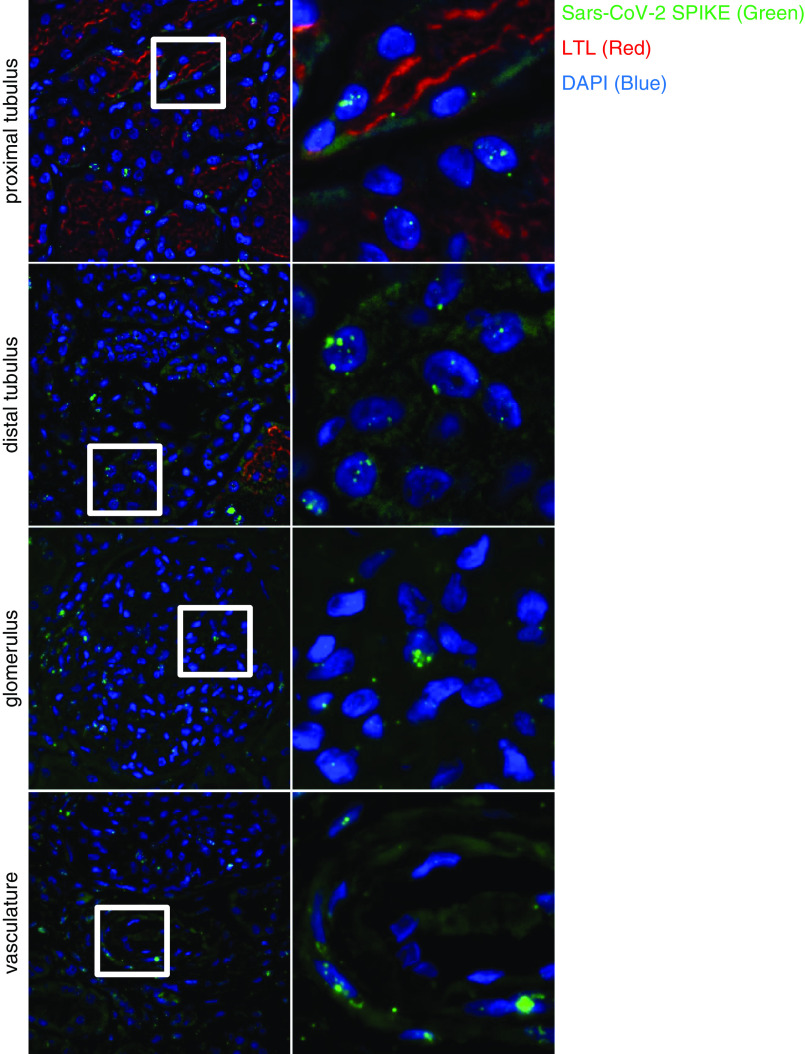
Figure 4.
Detection and spatial distribution of viral RNA using fluorescence in situ hybridization. The first (overview) and second (targeted zoom) columns display positive signal for viral RNA in different renal compartments, including proximal and distal tubules, glomeruli, and vessels. nCoV2019-S RNA is in green; Lotus tetragonolobus lectin (LTL) is in red; DAPI is in blue (patient 4).

