Abstract
Objective:
To evaluate the effects of different concentrations of chlorhexidine on the decline in force of orthodontic elastics.
Materials and Methods:
In a laboratory study, five groups of samples were tested, with one control group represented by distilled water (group 1) and four experimental groups: 0.12% manipulated chlorhexidine (group 2), 0.2% manipulated chlorhexidine (group 3), 0.12% chlorhexidine gluconate–based oral solution (0.12% Periogard; group 4), and 0.2% Cleanform mouthwash (formula and action; group 5). The test groups were submersed in artificial saliva at 37°C. Templates were used and submerged in the chlorhexidine solutions for 30 seconds twice a day. Force was measured with a digital dynamometer at six different time intervals: 0, 1, 7, 14, 21, and 28 days.
Results:
No statistical differences were found among the groups in the initial period, at 24 hours, and at 7 days (P > .05). There were statistical differences between groups 2 and 5 at 14 days of the experiment and between group 1 and the others at 28 days. In the initial period, the force was statistically higher than it was at any of the other periods of the experiment (P < .05).
Conclusion:
In the present study, chlorhexidine showed no significant influence on the force degradation of the chain elastics tested.
Keywords: Degradation, Elastomeric chains, Chlorhexidine
INTRODUCTION
Use of an orthodontic appliance demands that the wearer take special care because the presence of this device in the oral cavity leads to greater accumulation of bacterial plaque around brackets and bands.1,2 Considering that deficient oral hygiene generally is a reason why it is difficult to achieve successful orthodontic treatment, it is necessary for the dentist to implement an individualized model of a program of preventive education for each patient.3 In individuals who cannot or are unable to perform good oral hygiene, in addition to mechanical control, it is important to implement chemical plaque control. Among the antiseptics for oral use, chlorhexidine is one of the most powerful and most studied antimicrobial agents.4–6
Orthodontic movement comes from the application of force on a dental unit by means of accessories, such as brackets, springs, bands, wires, and elastics.7,8 Elastic devices are important sources for transmission of force to teeth but are not considered ideal because the force they generate diminishes as a function of the time of activation, oral medium, and other factors related to diet.9
The action of chlorhexidine on the mechanical properties of these elastics, such as the force degradation over the course of time, has hardly been discussed in the literature. Therefore, the aim of this study was to determine the effects of different concentrations of chlorhexidine on the decline in force of these elastics.
MATERIALS AND METHODS
A laboratory study was conducted to test the degradation of orthodontic chain elastics under the influence of chlorhexidine. Five groups of samples were tested, and each of these groups was composed of a total of 18 orthodontic chain elastics. The samples evaluated were distilled water (control group; group 1), 0.12% manipulated chlorhexidine (group 2), 0.2% manipulated chlorhexidine (group 3), 0.12% Periogard (Colgate, São Paulo, SP, Brazil; group 4), and 0.2% Cleanform mouthwash (Fórmula e Ação, São Paulo, Brazil; group 5). In another five receptacles, one for each group mentioned above, artificial saliva was reserved for immersion of the samples.
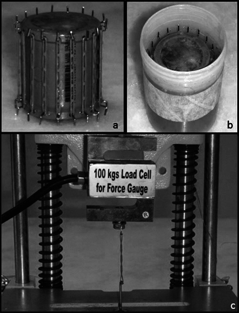
To fabricate the personalized templates, polyvinyl chloride (PVC) tubes were used, in which small orifices were made to insert the supporting rods for the orthodontic chain elastics. Self-polymerizing acrylic resin was injected into the PVC tube to fix the rods. The orifices were separated by a horizontal mean distance of 0.5 cm. On the test specimen, the elastics were put into place and stretched along a vertical distance of 23.5 mm. The elastics used were of the short-spacing type from Morelli (Sorocaba, Brazil). As they are presented in a single continuous chain, the elastomeric chains were cut to a standard, so that a total of five links were left free, with two links being responsible for fixation onto the template (Figure 1).
Figure 1. .
(a) Test specimen with stretched chain elastics. (b) Test specimen dipped in artificial saliva. (c) Digital dynamometer (Instrutherm DD-300), measurement of chain elastic.
The templates enabled the elastomeric chain to be immersed in the artificial saliva solution during the 28 days the laboratory study lasted. The devices containing the activated elastic segments were kept immersed in artificial saliva from Splabor (São Paulo, Brazil) at a controlled temperature of 37 ± 1°C, ideal for reproducing conditions in the oral cavity. This temperature was controlled by means of a thermostat and digital thermometer (Splabor). The chain elastics were removed from the receptacle containing artificial saliva and were dipped in the corresponding chlorhexidine solutions, leaving them immersed for 30 seconds twice a day with an interval of 12 hours between immersions. In the moments immediately before measurements, the devices were removed from the receptacle, force measurements were taken, and they were then replaced in the receptacles.
Six force measurements were taken during the experimental period at the following time intervals: initial (0), 1, 7, 14, 21, and 28 days. These measurements were taken with a digital dynamometer (Instrutherm DD-300, São Paulo, Brazil; Figure 1).
The chain elastics were removed from the templates and placed on the dynamometer, previously calibrated with regard to the distance of 23.5 mm of the templates. This guaranteed greater reliability of the data obtained. After each measurement, the force measurer was restarted, and the values were noted on a control sheet.
After all of the groups had been measured, the elastics were fixed on their respective templates and inserted in the receptacles of artificial saliva, which were put into the oven (Splabor). The level of saliva in the receptacle was verified every day, so that the elastics would be covered by this solution at all times.
Statistical analyses were performed with the program SPSS 13.0 (SPSS Inc, Chicago, Ill). Descriptive statistical analysis including mean and standard deviation were calculated for the groups evaluated. The values for the quantity of force released were submitted to the analysis of variance to determine whether there were statistical differences among the groups, and afterward, the Tukey test was performed.
RESULTS
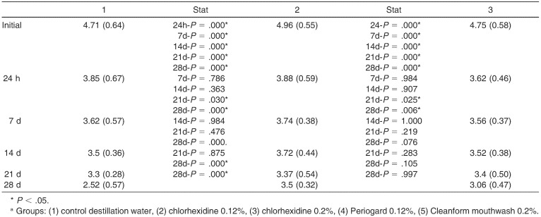
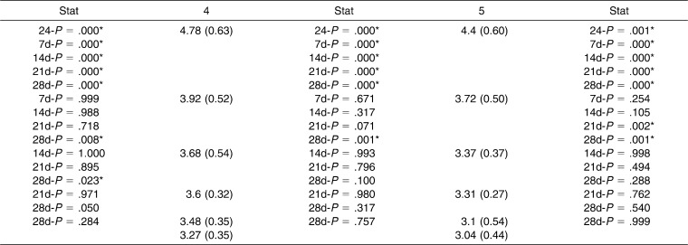
When the groups were compared with one another in the same period, no statistical differences among the groups were found in the initial, 24-hour, and 7-day time intervals (P > .05). There were statistical differences between groups 2 and 5 at 14 days of the experiment and between group 1 and the others at 28 days (Table 1).
Table 1. .
Mean Values (kgf), Standard Deviation, and Statistical Analysis (Comparison Between Groups by Time) of the Groups Evaluateda
Table 1. .
Extended
When adopting the mean values, it may be observed that at the end of the 28 days, the control group had a lower force value than the chlorhexidine test groups. Among these, the two 0.12% chlorhexidine groups obtained the highest values of decline in force.
When the groups were evaluated individually comparing the factor time, the force was statistically higher in the initial period than that of all the other experimental periods (P < .05; Table 2).
Table 2. .
Comparisons Between the Groups Individually at Different Times of Evaluation (kgf)a
Table 2. .
Extended
DISCUSSION
Orthodontic accessories adhered to the tooth surfaces make it difficult to perform oral hygiene and act as additional bacterial plaque retainers, leading to enamel demineralization of enamel, causing white stains, dental caries, and gingivitis.1
In dentistry, chlorhexidine acts in preventive manner in the reduction of bacterial plaque, such as in physically handicapped persons with motor limitations, mentally retarded and geriatric patients, and patients using orthodontic appliances.3,6 It is a synthetic antimicrobial agent that presents a high level of activity without, however, having the secondary effects that most antimicrobial agents have.10,11
In orthodontic patients, in addition to the factor time, exposure to water, alterations in temperature, and enzymes and other chemical substances, such as chlorhexidine, may have a significant influence on the amount of force released by orthodontic elastics.12 For this reason, it is imperative to know the action of chlorhexidine on this material to enable safe and satisfactory orthodontic treatment.
Most orthodontic accessories used for applying forces and performing movement do not provide stable force. With the variation of time, the intensity of force initially used diminishes, and thereby tooth movement is compromised and can be reduced or even interrupted. Elastic materials present this peculiarity, called force degradation.13
Years ago, Baty et al.14 and De Genova et al.15 evaluated the behavior of elastics over a period of 3 weeks. Here, the period of 4 weeks was selected because it coincides with the time interval frequently occurring between orthodontic consultations, the same period observed by Motta et al.16 Moreover, conducting this test in vitro generates initial results in the interaction between a material and the biologic tissue in a short and timely manner, reducing the need for tests in animals.17
When evaluated in a humid medium, the force degradation released by synthetic elastic materials is significantly greater than it is when this is done in a dry environment18,19; therefore, the chain elastic segments were kept immersed in artificial saliva. Because it concerns a simulation of the oral cavity, the temperature at which the elastics was maintained was 37 ± 1°C, as this is the body temperature and also because it is known that temperature participates in the force degradation released by the elastics.20 Another factor that influences mechanical behavior is the size of chain elastics—described as short, medium, or long—as well as their configuration.21 In this study, the short chain elastic, without spaces between the links, was adopted, since this maintain a higher percentage of force over the course of time.9
In this study, the results demonstrated that when using elastics with the short configuration, groups 3 and 5 (0.2% chlorhexidine) maintained a higher percentage of force during the course of 28 days in comparison with groups 2 and 4 (0.12% chlorhexidine), which underwent greater force degradation in the same period of time. This implication arises from the action of 0.12% chlorhexidine on the force degradation of the elastics at its highest values of force at the end of the study, when compared with the composition of 0.2% of the samples. It was also observed that the control group had its lowest value of force in comparison with the test groups at the end of the 28 days. Although there was a difference in the values among the groups, the results obtained point out that chlorhexidine does not have a significant influence on the force degradation of the chain elastics (Table 1).
Gioka et al.22 evaluated the relaxation of force of latex elastics during a period of 24 hours of extension and to do this estimated the extension necessary to achieve the related force. It was concluded that extension of the elastic to achieve the related force varies between 2.7 and 5 times the original length.22 In this study, the latex elastics showed a force of relaxation on the order of 25%, which consists of a component with an elevated initial decline and a latent part with a reduced rate. Most of the relaxation occurred within the first 3 to 5 hours after extension, irrespective of the manufacturer, size, or level of force of the elastic. In the present study, the large amount of decline in force within the first 24 hours was followed by mainly consistent levels of force up to 4 weeks. In the times after the initial moment, the intensity of force varied discretely within each group at the different times. Right from the first hour, the elastics of all the groups presented statistically lower values when compared with the respective values of force initially released, corroborating the findings of previous studies.
Therefore, knowledge of the alterations in the mechanical properties of chain elastics when elongated and exposed to the action of chlorhexidine is necessary to combine these accessories with the use of this solution. Nevertheless, this interaction had no significant influence on the force degradation of the elastics.
CONCLUSIONS
Chlorhexidine does not act significantly on the force degradation of orthodontic chain elastics.
The two 0.12% chlorhexidine groups obtained the highest values of decline in elastic force.
REFERENCES
- 1.Ai H, Lu HF, Liang HY, et al. Influences of bracket bonding on mutans streptococcus in plaque detected by real time fluorescence-quantitative polymerase chain reaction. Chin Med J (Engl) 2005;118:2005–2010. [PubMed] [Google Scholar]
- 2.dos Santos RL, Pithon MM, Vaitsman DS, Araujo MT, de Souza MM, Nojima MG. Long-term fluoride release from resin-reinforced orthodontic cements following recharge with fluoride solution. Braz Dent J. 2010;21:98–103. doi: 10.1590/s0103-64402010000200002. [DOI] [PubMed] [Google Scholar]
- 3.Oltramari-Navarro PV, Titarelli JM, Marsicano JA, et al. Effectiveness of 0.50% and 0.75% chlorhexidine dentifrices in orthodontic patients: a double-blind and randomized controlled trial. Am J Orthod Dentofacial Orthop. 2009;136:651–656. doi: 10.1016/j.ajodo.2008.01.017. [DOI] [PubMed] [Google Scholar]
- 4.Du MQ, Tai BJ, Jiang H, Lo EC, Fan MW, Bian Z. A two-year randomized clinical trial of chlorhexidine varnish on dental caries in Chinese preschool children. J Dent Res. 2006;85:557–559. doi: 10.1177/154405910608500615. [DOI] [PubMed] [Google Scholar]
- 5.Lobo PL, de Carvalho CB, Fonseca SG, et al. Sodium fluoride and chlorhexidine effect in the inhibition of mutans streptococci in children with dental caries: a randomized, double-blind clinical trial. Oral Microbiol Immunol. 2008;23:486–491. doi: 10.1111/j.1399-302X.2008.00458.x. [DOI] [PubMed] [Google Scholar]
- 6.Ribeiro LG, Hashizume LN, Maltz M. Effect of different 1% chlorhexidine varnish regimens on mutans streptococci levels in saliva and dental biofilm. Am J Dent. 2008;21:295–299. [PubMed] [Google Scholar]
- 7.Karras JC, Miller JR, Hodges JS, Beyer JP, Larson BE. Effect of alendronate on orthodontic tooth movement in rats. Am J Orthod Dentofacial Orthop. 2009;136:843–847. doi: 10.1016/j.ajodo.2007.11.035. [DOI] [PubMed] [Google Scholar]
- 8.Shibazaki T, Yozgatian JH, Zeredo JL, et al. Effect of celecoxib on emotional stress and pain-related behaviors evoked by experimental tooth movement in the rat. Angle Orthod. 2009;79:1169–1174. doi: 10.2319/121108-629R.1. [DOI] [PubMed] [Google Scholar]
- 9.Araujo FBC, Ursi WJS. Estudo da degradação da força gerada por elásticos ortodônticos sintéticos. R Dental Press Ortodon Ortop Facial. 2006;11:52–61. [Google Scholar]
- 10.Adler MT, Brigger KR, Bishop KD, Mastrobattista JM. Comparison of bactericidal properties of alcohol-based chlorhexidine versus povidone-iodine prior to amniocentesis. Am J Perinatol. 2012;29:455–458. doi: 10.1055/s-0032-1304827. [DOI] [PubMed] [Google Scholar]
- 11.Sanders TH, Hawken SM. Chlorhexidine burns after shoulder arthroscopy. Am J Orthop. 2012;41:172–174. [PubMed] [Google Scholar]
- 12.Beattie S, Monaghan P. An in vitro study simulating effects of daily diet and patient elastic band change compliance on orthodontic latex elastics. Angle Orthod. 2004;74:234–239. doi: 10.1043/0003-3219(2004)074<0234:AIVSSE>2.0.CO;2. [DOI] [PubMed] [Google Scholar]
- 13.Loriato LB, Machado AW, Pacheco W. Clinical and biomechanical aspects of elastics in orthodontics. R Clin Ortodon Dental Press. 2006;5 [Google Scholar]
- 14.Baty DL, Volz JE, von Fraunhofer JA. Force delivery properties of colored elastomeric modules. Am J Orthod Dentofacial Orthop. 1994;106:40–46. doi: 10.1016/S0889-5406(94)70019-2. [DOI] [PubMed] [Google Scholar]
- 15.De Genova DC, McInnes-Ledoux P, Weinberg R, Shaye R. Force degradation of orthodontic elastomeric chains-: a product comparison study. Am J Orthod. 1985;87:377–384. doi: 10.1016/0002-9416(85)90197-6. [DOI] [PubMed] [Google Scholar]
- 16.Motta AFJ, Cury-Saramago AA, Nojima LI. In vitro evaluation of force delivered by elastic chains [in Portuguese] Dental Press J Orthod [online] 2011;16(6) [Google Scholar]
- 17.Daguano JKMF, Santos C, Rogero SO. Citotoxicity analysis of bioceramics for use in systems of implantations. Revista Matéria. 2007;12:134–139. [Google Scholar]
- 18.Kanchana P, Godfrey K. Calibration of force extension and force degradation characteristics of orthodontic latex elastics. Am J Orthod Dentofacial Orthop. 2000;118:280–287. doi: 10.1067/mod.2000.104493. [DOI] [PubMed] [Google Scholar]
- 19.Ash JL, Nikolai RJ. Relaxation of orthodontic elastomeric chains and modules in vitro and in vivo. J Dent Res. 1978;57:685–690. doi: 10.1177/00220345780570050301. [DOI] [PubMed] [Google Scholar]
- 20.Hwang CJ, Cha JY. Mechanical and biological comparison of latex and silicone rubber bands. Am J Orthod Dentofacial Orthop. 2003;124:379–386. doi: 10.1016/s0889-5406(03)00564-x. [DOI] [PubMed] [Google Scholar]
- 21.Eliades T, Eliades G, Silikas N, Watts DC. Tensile properties of orthodontic elastomeric chains. Eur J Orthod. 2004;26:157–162. doi: 10.1093/ejo/26.2.157. [DOI] [PubMed] [Google Scholar]
- 22.Gioka C, Zinelis S, Eliades T, Eliades G. Orthodontic latex elastics: a force relaxation study. Angle Orthod. 2006;76:475–479. doi: 10.1043/0003-3219(2006)076[0475:OLE]2.0.CO;2. [DOI] [PubMed] [Google Scholar]


