Figure 2.

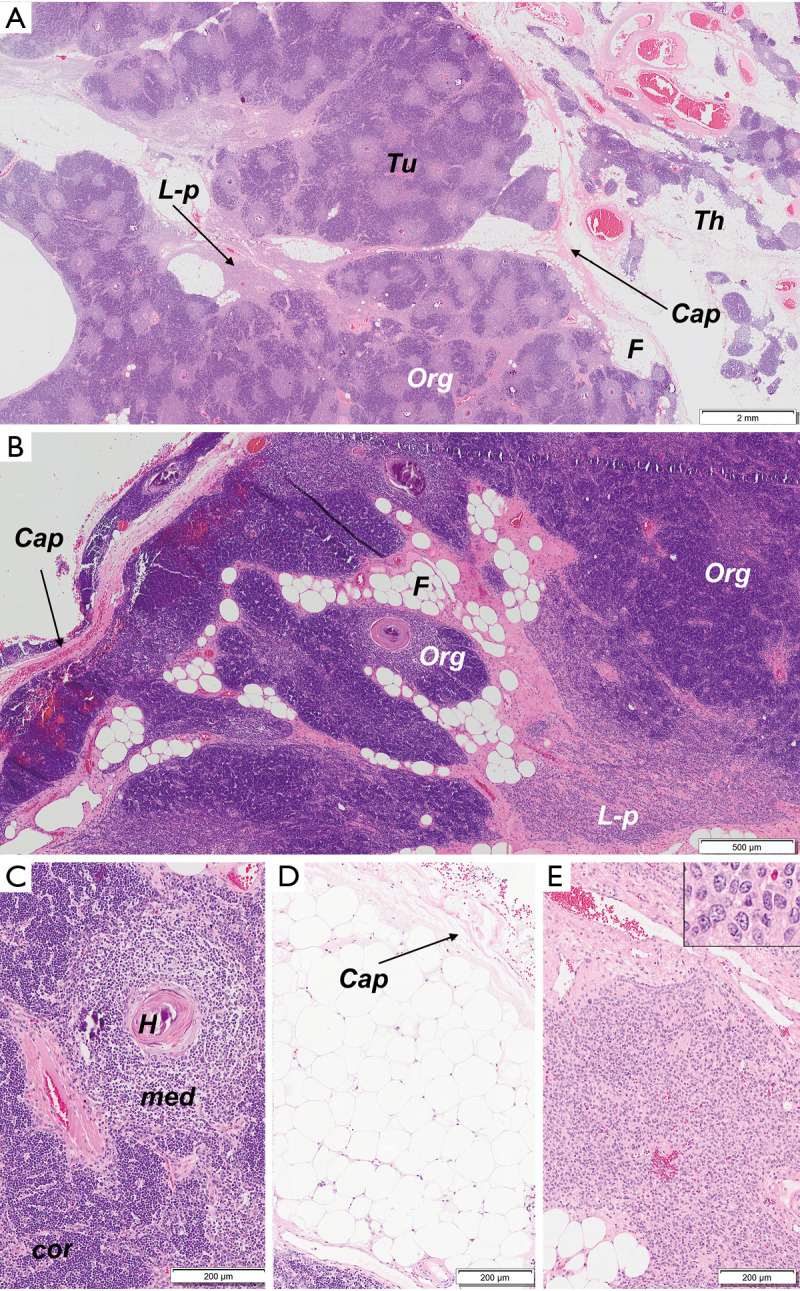
Microscopic features of the mediastinal tumor. (A) Low-power magnification of a tumor (Tu) and normal thymus (Th) separated by fibrous capsule (Cap). Three elements could be identified: an organoid component (Org), lymphocytes-poor, epithelial component (L-p) and fatty tissue (F). There was no evidence of invasion outside the capsule. (B) Medium-power magnification of the tumor - organoid component (Org), lymphocyte-poor, epithelial (L-p) component and fatty tissue (F). All elements were covered by a fibrous capsule (Cap). A small rim of compressed normal thymic tissue was visible outside the capsule. (C) The organoid component (Org) reproduced the morphology of the normal thymus and contained light staining, medullary (med) areas with Hassall corpuscles (H) and dark staining, cortical (cor) areas. (D) Normal-looking, mature fat inside the tumor. (E) The lymphocytes-poor component comprised of densely packed epithelial cells with slight atypia (inset). [Hematoxylin and eosin stain, magnification ×5 (A), ×20 (B), ×40 (C,D,E) and ×200 (inset)].
