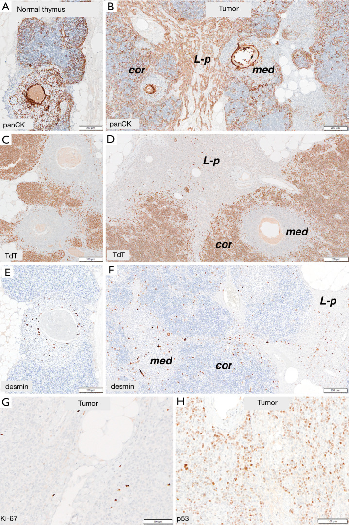
Figure 3.
Immunohistochemical features of the normal thymic tissue (A,C,E) and the tumor (B,D,F,G,H). (A,B) Pancytokeratin stain—distribution of positive cells in the tumor (B) was similar to the normal thymus (A). Keratin-positive cells are concentrated at the periphery of cortical areas (cor) and dispersed in medullary regions (med) of the organoid component, while they formed sheets in the lymphocyte-poor component (L-p). (C,D) TdT-expression in immature T-lymphocytes was seen only in cortical areas of the normal thymus (C) and the tumor (D). There was no expression of TdT in lymphocyte-poor component. (E,F) Anti-desmin reaction revealed myoid cells dispersed in medullary regions of the thymus (E), numerous myoid cells were also found in medullary regions (med) of the organoid component and lymphocyte-poor component (L-p). (G,H) Very low Ki-67 index (G) and significant p53-index (H) in lymphocyte-poor epithelial component (A, B: AE1AE3, magnification ×40; C, D: TdT, ×40; E, F: Desmin, ×40; E: Ki-67, ×100; H: p53, ×100).

