Abstract
Background
Telomere is essential for chromosomal stability and its length has been proven to be related to prognosis in many malignant tumors. This study aims to investigate the relevance of telomere length with clinical and pathologic features and its prognostic value in colorectal cancer (CRC).
Methods
Telomere status of CRC and adenoma cells were measured by telomere-specific quantitative fluorescent in situ hybridization (Q-FISH). The relative telomere length (RTL) was calculated as the mean telomere fluorescent intensity units (TFUs) in carcinoma cell divided by the TFU in cancer-associated fibroblast cell (CAF).
Results
One hundred CRC patients, who were received surgery treatment during 2013 to 2014 and fifty-seven patients who underwent the examination of colonoscope and were confirmed as adenoma were enrolled. TFUs of carcinoma cell and CAF were statistically significantly lower than in adjacent mucosa cell (P=0.0079). Although there was no difference between the three kinds of adenoma cells (P=0.5457), TFU in adenoma cells was significantly lower than in CAF (P<0.0001) and independent with age. TFU and the RTL were statistically significantly lower in adenoma cells than in carcinoma (all P<0.0001). TFU of carcinoma cell in distant metastases patients were significantly lower than that without distant metastases patients (P=0.002). When cut by the median value of TFU of carcinoma cell and RTL, patients with a lower TFU or RTL had statistically significantly poorer overall survival (OS) (P=0.0027, HR: 4.6, 95% CI: 1.9–11.0; P=0.0163, HR: 2.95, 95% CI: 1.22–7.12) and disease-free survival (DFS) (P=0.0057, HR: 3.14, 95% CI: 1.40–7.06; P=0.0271, HR: 2.49, 95% CI: 1.11–5.59, respectively) than those patients with higher TFU or RTL. On multivariate analysis, the TFU of carcinoma cell was proved to be an independent prognostic value both for OS and DFS (P=0.0005, HR: 4.975, 95% CI: 1.616–15.385; P=0.007, HR: 3.57, 95% CI: 1.410–9.010).
Conclusions
The length of telomere in carcinoma and adenoma cells were consistently shorter and the telomere changes were early carcinogenesis event, even the epithelial cells were morphologically not malignant. The length of telomere was associated with tumor metastases and prognosis, suggesting telomere probably was an important cue of the biological behavior of CRC.
Keywords: Colorectal cancer (CRC), telomere, prognosis, quantitative fluorescent in situ hybridization (Q-FISH)
Introduction
Colorectal cancer (CRC) is the third most common cancer and the second cause of cancer death worldwide and 883,200 deaths are estimated to have occurred in 2018 (1). Great progress has made, such as increased awareness and early detection, improved treatments on surgery and chemotherapy, which have all contributed to prolonged survival though, CRC is still the most common cause of cancer-related deaths. CRC involves multi-step transformation of normal colon tissue to precancerous polyps or adenomas and finally to a malignant neoplasm. Not all polyps or adenomas are evolved into CRC, but for most colorectal malignancies, clinical and epidemiological evidences clearly indicate that they are the precursor lesions (2). In the progression of adenoma-carcinoma transition, different genetic and epigenetic events are involved (3).
Telomere is repetitive DNA sequences (TTAGGG) located at the end of chromosomes and its main functions is stabilizing chromosomes by protecting them from end-to-end fusion and DNA degradation (4,5). Telomere undergoes a progressive shortening during each cell-replication cycle because of incomplete DNA replication of a lagging strand (so called as the end-replication problems) and telomere shortening induces somatic cells to undergo senescence and apoptosis (4). So, in this sense, telomere may act as a tumor-suppressing mechanism, preventing cells from uncontrolled division. However, continuous erosion of telomere may impair its function in protecting chromosome ends, leading to chromosomal instability, a key event in the initiation of carcinogenesis and cancer progression (6-8).
As we known, chromosomal instability and microsatellite instability were considered as the two main pathways of CRC carcinogenesis (9). Telomere shortening is identified as an early event in CRC carcinogenesis. Of note, most (approximately 85%) of CRCs are associated with chromosomal instability and telomere dysfunction is considered as a fundamental player in this process (10). Telomere shortening was frequently found in adenoma-carcinoma transition and telomere dysfunction initiated cancer formation by induction of chromosomal instability (11-13). While most studies identified telomere shortening as a critical initial event in carcinogenesis, the role of telomere length in cancer cells as a marker of disease progression was controversial (14). In fact, no agreement concerning the role of telomere length as a marker of disease progression has been reached in CRC. A few researches indicated that telomere length in cancer tissue was significantly longer in the late stage of colorectal tumors (15,16), other studies had not indicated any correlation between telomere length and stage (17-19). In one study, shorter telomere length (TL) was observed in tumors with earlier stage, but not in those with advanced stage (20). As for a prognostic role, telomere length hadn’t been confirmed either. Only in some of these studies indicated longer telomeres were associated with poor clinical outcome (16,19,21).
In tumor microenvironment, cancer-associated fibroblast cell (CAF) constituted the majority cell type and was considered as a crucial role in CRC, such as tumor growth, progression, metastasis, angiogenesis and immune responses (22). CAF was originally activated by tumor cells from a subpopulation of fibroblasts and was consider as prognostic biomarkers in cancers (23). In prostate cancer cell, shorter telomere length in cancer-associated stromal cell (fibroblasts and smooth muscle) were prognostic marker for progress to metastasis and die of their prostate cancer (24).
Considering the relevance of telomere in carcinogenesis and its role as prognostic biomarkers, our main aim in this work was to highlight the changes of telomere in difference kinds of adenomas and to clarify the relevance between the clinical and pathologic features and its prognostic value in CRC. With this objective, we evaluated the telomere status of CRC and adenoma by telomere-specific quantitative fluorescent in situ hybridization (Q-FISH) with which could provide single cell resolution of telomere length while maintaining tissue architecture. We present the following article in accordance with the REMARK reporting checklist (available at https://dx.doi.org/10.21037/tcr-20-3341).
Methods
Patients
One hundred patients with CRC, who were received surgery treatment at the Department of Colorectal Surgery, the first affiliated hospital, ZheJiang University School of Medicine during 2015 to 2016, were enrolled in the present study. Biopsy tissues which were obtained from fifty-seven patients who underwent the examination of colonoscope during 2013 and were confirmed as adenoma were also used. Form the reports of pathological diagnosis, sixteen were serrated adenoma, nineteen were tubular adenoma and twenty-two were villous adenoma. Patients received (neo)adjuvant radiotherapy or chemotherapy before operation were excluded. The study was conducted in accordance with the Declaration of Helsinki (as revised in 2013). The study was approved by the ethics committee of the first affiliated hospital, ZheJiang University School of Medicine (No.2020-656) and informed consent was taken from all the patients.
Q-FISH
Before Q-FISH, the pathological diagnosis was confirmed by a pathologist under a microscope though the standard H.E sections. Paraffin-embedded, 4-µm-thick sections adjacent to the tissue used for H.E. sections were obtained. After dewaxing in xylene hydration in alcohol series (100%; 90%; 70%), sections were boiled in microwave oven for 10 minutes in citrate-buffer (0.01 M, PH: 6.0). Rinsed in phosphate buffer saline (PBS), sections were then incubated in preheated pepsin solution for 5 minutes (37 °C). After dehydrated the slides following the alcohol series (70%; 90%; 100%) and air dried the sections about 5minutes until all alcohol was gone, 15 µL hybridisation mix [10 mM Tris, 2.14 mM MgCl2; 0.5% Blocking reagent (Roche), 0.1 nM Cy3 conjugated telomere peptide nucleic acid probe (sequence: Cy3-OO-(CCCTAA)3, catalog#F1002, Panagene Inc., Korea] were applied and covered with a coverslip. For hybridization, slides were denatured at 80 °C for 3 min and incubated in a humid chamber for two hours. At last, the sides were rinsed in wash buffer (0.1% bovine serum albumin and 10mMTris, 70% Formamide (Sigma-aldrich, catalog#34724-1L-R), TBS-T (TBS plus 0.1% tween-100) and PBS, and applied about 30µl mounting solution Antifade+4’,6-diamidino-2- phenylindole (DAPI) (catalog#H-1200, Vector) and covered with a coverslip.
During each experiment, a slide of normal colon mucosa was used as internal control. Pictures were taken under a fluorescence microscope (100× objective lens and 10× ocular lens, Olympus BX51, Japan) with the software of NIS-Elements. Both Cy3 (telomere signal) and DAPI (nuclear stain signal) images were captured under the same microscope field. The exposure time was held constant at 150 ms for all Cy3 images to keep the signals within the linear range of the camera and enable comparison of telomere signal. At least ten pictures form different sites of the sample were taken. Telomere length were measured in carcinoma cells and CAF cells. At least 50 carcinoma or adenoma cells, CAF cells of the total telomere fluorescent intensity units (TFUs) were quantitatively measured by the software of TFL-TeloV2.
Statistical analysis
Investigators were blinded to the pathological information of any patient at the time of analysis. The relative telomere length (RTL) was calculated as the TFU in carcinoma cell or adjacent mucosa divided by the TFU in CAF cell. TFUs and RTL were expressed as median (interquartile range). An interquartile range was defined as the distribution of TFU values between 25th and 75th percentiles. Statistical analyses were conducted on natural data by using Mann-Whitney tests. Differences between TFU in more than two parameters were analyzed using non-parametrical ANOVA (Kruskal-Wallis test). The relationship between the patient age at diagnosis and TFU values was calculated by Pearson correlation coefficient. Overall survival (OS) was defined as time from study enrolment until death or last follow-up. Disease-free survival (DFS) was defined as time from study enrolment until cancer recurrence or metastases or death without evidence of recurrence or a second primary tumor, or date of last visit. The median values were used as cut-off. Based on the cut-off, CRC patients were stratified into two groups. Survival curves were plotted using Kaplan–Meier method and log-rank test was used for comparison. Multivariate analysis was performed by multivariate Cox model. Statistical analyses were conducted using Graphpad Prism 8 (GraphPad Software, Inc., La Jolla, CA, USA) and SPSS (version 19.0) software (IBM Corporation, Armonk, NY, USA). P values <0.05 were considered statistically significant.
Results
Clinical characteristics of CRC patients
The description of the population including fundamental characteristics of CRC and background variables were summarized in Table 1. The median age for the group of CRC patients was 61 years (range, 33–88 years); of those, 45% were men and 55% were women. As for the 57 adenoma patients, 16 were serrated adenomas, 19 were tubular adenoma, 22 were villous adenoma. The median age was 56 years (range, 31–65 years), 31 (54%) were men and 26 (46%) were women.
Table 1. Clinical and pathologic features of colorectal cancer patients.
| Feature (N=100) | Variable | N | TFU of tumor | P | TFU of CAF | P | RTL | P |
|---|---|---|---|---|---|---|---|---|
| Age (years) | <65 | 64 | 1,535 (572–4,897) | 0.245 | 2,885 (1,873–3,913) | 0.046 | 0.73 (0.25–2.5) | 0.815 |
| ≥65 | 36 | 2,718 (576–7,514) | 2,317 (1,309–3,223) | 0.99 (0.28–1.87) | ||||
| Gender | Male | 45 | 2,082 (457–5,779) | 0.967 | 2,600 (1,764–3,738) | 0.386 | 0.71 (0.20–2.1) | 0.406 |
| Female | 55 | 1,876 (655–4,315) | 2,271 (1,575–3,337) | 0.92 (0.37–2.0) | ||||
| Tumor location | Right colon | 26 | 1,292 (334–5,726) | 0.505 | 1,855 (739–2,831) | 0.127 | 1.07 (0.31–3.20) | 0.657 |
| Left colon | 74 | 2,082 (616–5,486) | 2,437 (1,776–3,506) | 0.92 (0.25–1.96) | ||||
| Differentiation | Well | 54 | 2,189 (691–5,779) | 0.260 | 2,437 (1767–3,542) | 0.303 | 0.94 (0.29–2.30) | 0.879 |
| Moderate–poor | 46 | 1,085 (481–5,709) | 2,162 (1,268–2,993) | 0.92 (0.18–1.92) | ||||
| T stage | T1-2 | 38 | 2,066 (698–6,235) | 0.643 | 2,599 (1,964–3,581) | 0.212 | 0.71 (0.25–2.53) | 0.587 |
| T3-4 | 62 | 1,879 (523–5,450) | 2,254 (1,340–3,484) | 0.94 (0.29–1.96) | ||||
| Lymph node metastasis | Negative | 52 | 1,820 (669–4,715) | 0.776 | 2,440 (1,764–3,463) | 0.849 | 0.74 (0.26–1.97) | 0.581 |
| Positive | 48 | 2,198 (516–7,500) | 2,395 (1,529–3,508) | 0.84 (0.29–2.15) | ||||
| Perineural invasion | Negative | 97 | 1,994 (626–5,508) | 0.021 | 2,451 (1,810–3,508) | 0.001 | 0.89 (0.26–2.10) | 0.577 |
| Positive | 3 | 360 (130–499) | 404 (384–506) | 0.71 (0.34–0.75) | ||||
| lymphovascular invasion | Negative | 90 | 2,136 (631–5,666) | 0.037 | 2,427 (1,776–3,451) | 0.815 | 0.93 (0.29–2.53) | 0.0662 |
| Positive | 10 | 676 (130–1,989) | 2,173 (1,042–4,876) | 0.35 (0.13–1.07) | ||||
| Distant metastases | Negative | 89 | 2,314 (684–5,802) | 0.002 | 2,550 (1,728–3,574) | 0.208 | 0.98 (0.31–2.17) | 0.020 |
| Positive | 11 | 557 (130–956) | 2,156 (1,261–2,427) | 0.23 (0.08–0.89) | ||||
| Dukes stage | A | 29 | 1,943 (719–5,248) | 0.023 | 2,550 (1,882–4,010) | 0.447 | 0.74 (0.22–2.07) | 0.072 |
| B | 21 | 1,765 (703–5,125) | 2,368 (1,106–3,334) | 0.99 (0.32–2.68) | ||||
| C | 39 | 2,759 (617–7,771) | 2,799 (1,703–3,871) | 1.08 (0.48–2.34) | ||||
| D | 11 | 557 (130–956) | 2,156 (1,261–2,427) | 0.23 (0.08–0.89) |
TFU, telomere fluorescent intensity units; CAF, cancer-associated fibroblast cell; RTL, relative telomere length.
All patients were subject to a follow-up process, and the examinations including abdomen contrast-enhanced computed tomography and colonoscopy were appointed at 1 month after the treatment. The last update of patient follow-up for this study was December 2020 and none was lost to follow up. All the patients were followed up for at least 2 years and the median follow-up time was 46 months. During the follow-up, 20 (20%) patients were dead and 5 (5%) patients were suffered from distant metastasis or local recurrence. The 1 and 3-year OS rates of all the patients were estimated as 97% and 72%.
TFU in tumor, adenoma and adjacent mucosa cell
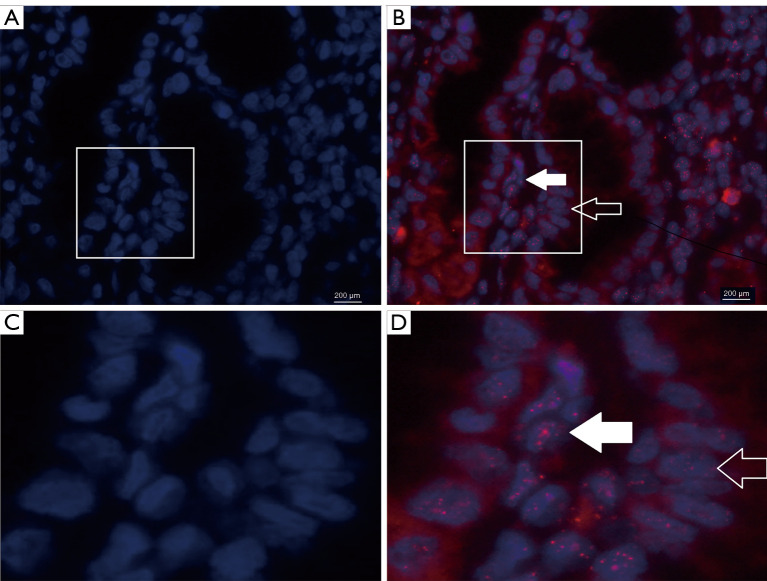
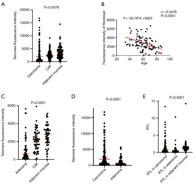
The representative images of telomere fluorescence in situ hybridization in CRC were shown in Figure 1. TFUs of carcinoma cells and CAF were statistically significantly lower than in adjacent mucosa [median (interquartile range): 1,968 (572–5,519) vs. 3,410 (1,845–4,719) vs. 3,425 (1,890–4,727), P=0.0079, Figure 2A]. Although there was no significant difference between age in carcinoma cell and RTL, TFUs in CAF were lower in age ≥ 65 years [2,885 (1,873–3,913) vs. 2,317 (1,309–3,223), P=0.046, Table 1]. What’s more, an inverse relationship between TFU in CAF and age was recorded (R= −0.4476 and P<0.0001, Y = −50.76*X + 5,825, Figure 2B), with a decrease telomere at a rate of 50.76 TFU each year. However, neither TFU nor the RTL in carcinoma cell and in adjacent mucosa, the inverse correlations hadn’t been found (r=0.064, P=0.5236; r=0.1650, P=0.1009 and r=−0.019, P=0.8599; r=−0.043, P=0.6737). TFUs in carcinoma and in CAF cells and the RTL did not significantly differ with gender, tumor location and tumor differentiation (Table 1).
Figure 1.
Representative images of telomere fluorescence in situ hybridization in different tissue types. (C) and (D) were the partial enlarged drawing of (A) and (B), respectively. (A) and (C), nuclei were counterstained with DAPI (blue florescence). (B) and (D). Telomere length was reflected by the total red fluorescence intensity in nuclei. In CRC tissue, the CAF cells (filled arrows) show bigger, more numerous and intense red signal red signal than carcinoma cells (open arrows), reflecting longer telomere length in CAF. CAF, cancer-associated fibroblast cell.
Figure 2.
TFU and RTL changes in carcinoma, adenoma and cancer-associated fibroblast (CAF) cells. (A) TFUs in carcinoma cells were statistically significantly lower than in CAF. (B) There was an inverse relationship between TFU in CAF and age, with a decrease telomere at a rate of 50.76 TFUs each year. (C) TFU in adenoma cells was significantly lower than in CAF. (D,E) Both TFU and the RTL were statistically significantly lower in adenoma cells. TFU, telomere fluorescent intensity units; RTL, relative telomere length; CAF, cancer-associated fibroblast cell.
We did not find any difference between the three kinds of adenoma cells (serrated, tubular and villous adenoma) [538 (143–2,187) vs. 526 (380–2,322) vs. 688 (153–1,276), P=0.5457]. Consistent with carcinoma cells, TFU in adenoma cells was significantly lower than in CAF and in adjacent mucosa [607 (246–1,413) vs. 2,178 (1,307–2,964) vs. 2,810 (1,916–3,602), P<0.0001, Figure 2C] and also independent with age. TFU in adenoma cells were statistically significantly lower than in carcinoma [607 (246–1,413) vs. 1,968 (572–5,519), P<0.0001; Figure 2D]. RTLs in carcinoma, adenoma and adjacent mucosa cell were also compared and RTL in adenoma and adjacent mucosa cell were significantly lower [0.826 (0.26–1.97) vs. 0.362 (0.11–0.81) vs. 1.353 (1.003–1.588), P<0.0001, respectively. Figure 2E]. To exclude the other influencing factors, TFU of CAF in carcinoma tissue and adenoma tissue were compared, no differences were found [2,395 (1,678–3,497) vs. 2,178 (1,307–2,964), P=0.1206].
TFU, RTL and tumor progression
According to T staging, patients were divided into two groups: T1–2 group and T3–4 group. TFU did not statistically significantly differ between the different tumor T stages [2,066 (698–6,235) vs. 1,879 (523–5,450), P=0.643]. TFU did not differ between lymph node metastasis either [1,820 (669–4,715) vs. 2,198 (516–7,500), P=0.776]. 3 patients were confirmed as perineural invasion and 10 as lymphovascular invasion. TFU of carcinoma cell in perineural invasion and lymphovascular invasion were significantly lower [360 (130–499) vs. 1,994 (626–5,508), P=0.021; 676 (130–1,989) vs. 2,136 (631–5,666), P=0.037, respectively]. TFU of CAF in perineural invasion patients were significantly lower too (404 (384–506) vs. 2,451 (1,810–3,508), P=0.001]. 11 patients suffering from distant metastases in this study and TFU of carcinoma cell in distant metastases patients were significantly lower than that without distant metastases patients [557 (130–956) vs. 2,314 (684–5,802), P=0.002]. By Dukes stage, 29 belong to stage A, 21 stage B, 39 stage C and 11 stage D and TFU was significantly lower than other stage patients [1,943 (719–5,248) vs. 1,765 (703–5,125) vs. 2,759 (617–7,771) vs. 557 (130–956), for all, P=0.023 and A and D, P=0.0352; B and D, P=0.0417; C and D, P=0.0124]. However, except that patients with distant metastases had significantly lower RTL [0.23 (0.08–0.89) vs. 0.98 (0.31–2.17), P=0.020], neither TFU in CAF nor the RTL had relevant to tumor progression (Table 1).
Lower TFU and RTL were associated with poor prognosis
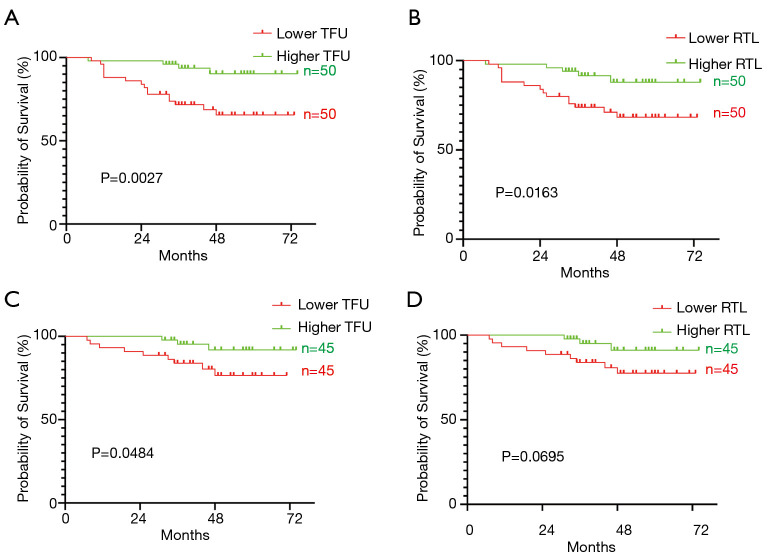
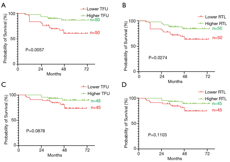
Kaplan-Meier method and the log-rank test was used for comparison of outcomes and the median values of the TFU of carcinoma cell (1909), CAF (2399), adjacent mucosa (3281) and RTL (0.826) were used as cutoff. Patients with a lower TFU or RTL had statistically significantly poorer OS than those patients with higher TFU or RTL (P=0.0027, HR: 4.6, 95% CI: 1.9–11.0; P=0.0163, HR: 2.95, 95% CI: 1.22–7.12, respectively. Figure 3A,B). Given the Dukes D stage patients had much lower TFU and 72.7% (8/11) were dead during follow up. To test the prognostic value, we excluded Dukes D stage patients and found that only lower TFU still predicted a poorer OS (P=0.0484, HR: 3.4, 95% CI: 1.1–10.7; Figure 3C), although the difference in RTL for OS trend to be significant (P=0.0695, HR: 2.86, 95% CI: 0.92–8.87. Figure 3D). As for DFS, lower TFU or RTL also had a poor prognosis (P=0.0057, HR: 3.14, 95% CI: 1.40–7.06; P=0.0271, HR: 2.49, 95% CI: 1.11–5.59, respectively. Figure 4A,B). After excluded Dukes D stage patients, prognosis of patients with lower TFU trend to be poor (P=0.0878, HR: 2.64, 95% CI: 0.92–7.52. Figure 4C) but the difference in RTL was not significant (P=0.1103, HR: 2.484, 95% CI: 0.87–7.08. Figure 4D). However, TFU of CAF and adjacent mucosa did not show any prognostic value, neither for OS (P=0.3011, HR: 1.592, 95% CI: .0.66–3.8; P=0.8672, HR: 1.078, 95% CI: 0.45–2.6) nor DFS (P=0.1836, HR: 1.731, 95% CI: 0.77–3.9; P=0.3024, HR: 1.532, 95% CI: 0.68–3.45). Other clinicopathological parameters, such as tumor differentiation, T stage, lymph node metastasis and distant metastases were also associated with prognosis and gender, age and tumor location were not (Tables 2,3).
Figure 3.
Lower TFU and RTL were associated with OS. (A) Patients with a lower TFU had statistically significantly poorer OS than those patients with higher TFU. (B) Lower RTL patients had statistically significantly poorer OS. (C) After excluded Dukes D stage patients, lower TFU still predicted a poorer OS. (D) Lower RTL was associated with short OS, although the difference was not significant. TFU, telomere fluorescent intensity units; RTL, relative telomere length; OS, overall survival.
Figure 4.
Lower TFU and RTL were associated with DFS. (A) Patients with a lower TFU were associated with short DFS. (B) Lower RTL patients had statistically significantly poorer DFS. (C) After excluded Dukes D stage patients, lower TFU was associated with short DFS, although the difference was not significant. (D) Lower RTL was not associated with DFS when Dukes D stage patients were taken off. TFU, telomere fluorescent intensity units; RTL, relative telomere length; DFS, disease-free survival.
Table 2. Univariate and multivariate cox regression analysis of OS for univariate and multivariate.
| Variable | Univariable analysis | Multivariable analysis | |||
|---|---|---|---|---|---|
| P | Relative risk (95% CI) | P | Relative risk (95% CI) | ||
| Gender | 0.4018 | 1.452 (0.60–3.5) | – | – | |
| Age | 0.9730 | 1.016 (0.41–2.5) | – | – | |
| Tumor location | 0.8141 | – | – | – | |
| Differentiation | 0.0469 | 2.793 (1.014–7.693) | – | – | |
| T stage | 0.0268 | 6.941 (2.579–18.68) | – | – | |
| Lymph node metastasis | 0.0002 | 5.617 (2.300–13.72) | 0.045 | 3.139 (1.026–9.60) | |
| Distant metastases | <0.0001 | 172.2 (30.43–974.8) | 0.001 | 5.475 (2.027–14.792) | |
| TFU of carcinoma cell | 0.0027 | 4.6 (1.9–11.0) | 0.005 | 4.975 (1.616–15.385) | |
| RTL | 0.0163 | 2.95(1.22–7.12) | – | – | |
OS, overall survival; RTL, relative telomere length.
Table 3. Univariate and Multivariate cox regression analysis of DFS for univariate and multivariate.
| Variable | Univariable analysis | Multivariable analysis | |||
|---|---|---|---|---|---|
| P | Relative risk (95% CI) | P | Relative risk (95% CI) | ||
| Gender | 0.6663 | 1.190 (0.53–2.66) | – | – | |
| Age | 0.9906 | 1.005 (0.43–2.34) | – | – | |
| Tumor location | 0.2684 | 1.598 (0.62–4.09) | – | – | |
| Differentiation | 0.0099 | 3.545 (1.36–9.28) | – | – | |
| T stage | 0.0111 | 2.908 (1.28–6.63) | – | – | |
| Lymph node metastasis | 0.0011 | 3.914 (1.72–8.90) | – | – | |
| Distant metastases | <0.0001 | 374.3 (72.2–1941) | 0.001 | 10.11 (3.896–26.22) | |
| TFU of carcinoma cell | 0.0057 | 3.14 (1.40–7.06) | 0.007 | 3.57 (1.410–9.010) | |
| RTL | 0.0271 | 2.49 (1.11–5.59) | – | – | |
DFS, disease-free survival; RTL, relative telomere length.
In the next step, TFU of carcinoma cell and RTL, together with tumor differentiation, T stage, lymph node metastasis and distant metastases were included in multivariate Cox proportional hazards analysis and TFU of carcinoma cell (P=0.0005, HR: 4.975, 95% CI: 1.616–15.39), lymph node metastasis (P=0.045, HR: 3.139, 95% CI: 1.026–9.60) and distant metastases (P=0.001, HR: 5.475, 95% CI: 2.027–14.792) turn out to be independent prognostic factors for OS (Table 2) and TFU of carcinoma cell (P=0.007, HR: 3.57, 95% CI: 1.410–9.010) and distant metastases (P=0.001, HR: 10.11, 95% CI: 3.896–26.22) were independent prognostic factors for DFS (Table 3).
Discussion
In present study, we demonstrated that TFUs in carcinoma and adenoma cells were consistently lower than in CAF and the TFUs in adenoma cell were even lower than in carcinoma. This result supported the hypothesis that telomere changes were early carcinogenesis event, even the epithelial cells were morphologically not malignant. TFUs in carcinoma and RTL were associated with tumor metastases and patients who had low TFU or RTL had significantly poor prognosis.
Telomere is progressive shortening because of the end- replication problems and continuous erosion of telomere may impair its function in protecting chromosome ends, leading to genetic instability (6-8). Chromosomal instability and microsatellite instability are identified as the two main pathways of CRC carcinogenesis (9,25). CRC were thought to arise from the adenoma-carcinoma sequence by sequential accumulation of genetic alterations (26). However, the changes of telomere in the adenoma-carcinoma sequence has not been well established. Consistent with previous reports, it seemed certain that telomere length in CRC was shorter than in normal mucosa (11-13,15-20). Evidences showed telomere length was shorter in low-grade and high-grade dysplasia than in carcinoma (27). Our results shown that telomere in adenomas were shorted than CAF and carcinoma cell, which were consistent with Nirosha Suraweera’s observation: adenoma telomere length was significantly shorter than matched normal mucosa, more prevalent tumor telomere shortening than carcinoma patients, indicated that shortened telomeres and telomere maintenance engagement occur early in adenomatous polyp development (28). Clinically, only a small fraction of adenomas actually become malignant, it is hard to predict which adenomas will progress. Bertorelle et al. argued that aggressive polyps had shorter telomeres than carcinoma free adenomas and telomere could distinguish malignant from benign adenomas (10). Another research also observed that different adenoma had its own mechanism: tubular adenomas were associated with mutations of APC, KRAS, and p53, whereas serrated adenomas progressed through microsatellite instability and BRAF mutations (9). However, as for telomere length, tubular and serrated adenoma were found to be similar (11). Our results were the same, the difference of telomere length in different adenoma subtypes was not significant (date not shown), suggesting telomere shorting was common phenomenon in adenoma. Given the intensive cell proliferation with telomere loss and the relatively longer telomere length, mechanisms of telomeric sequences synthesis, such as up-regulation of telomerase, should be involved. Indeed, the trend of growing telomerase activity in the adenoma-carcinoma sequence, as well as the decrease in telomere length has been reported (29). Taken together, our results strongly supported the concept that telomere erosion is a critical initial event in colorectal carcinogenesis. Further studies are required to better understand the role of telomere in the pathological progression in colorectal neoplasia.
CAF constituted the majority cell type in tumor microenvironment and play a crucial role in tumor growth, progression, metastasis, angiogenesis (22). We measured the telomere of CAF in carcinoma and adenoma simultaneously. As mentioned above, telomere was progressively shortened with each successive cell division and therefor associated with aging (4). Our results supported this concept and TFUs in CAF were lower in age ≥65 years, but not in carcinoma cell. What’s more, the TFUs in CAF were found to be inversely correlated with age, which was in line with previous studies found in normal mucosa tissue (16,18,30). This negative correlation has also been demonstrated in other malignancies, such as breast cancer (31). To the best of our knowledge, this study is the first to report that telomere changes in CAF and record the inverse relationship between TFU in CAF and age.
The relationship between telomere and CRC progression was not entirely clear. Some investigators identified that telomeres were longer in late stage cases (16,21), others could not confirm the correlation between telomere length and disease stage (17-19,29). Our date showed that TFU of carcinoma cell and RTL were stable in Dukes A, B and C, when patients with distant metastasis (Dukes D), TFU and RTL were striking shorted. This observation was also reported in a previous research that TL was statistically significantly shorter in liver-metastasis tumors than primary tumor and the adjacent non-cancerous liver tissues (20). It has been found that depending on the drug combination, chemotherapy exerted a degree of transient telomere shortening effect and could be recovered to normal TL in around 2 years (32). So the short telomeres in liver metastases tissue were conjectured due to the treatment in CRC, since all the patients with distant metastases underwent various regimens of chemotherapy (20). Our date had a discrepancy between them, because all the patients involved in this research didn’t receive any chemotherapy at all. The clinical outcome of different tumor location is varying and prognosis of colon cancer is better than that of rectal cancer (33). Currently, it’s under debate whether rectal cancer is actually a distinct entity. In relation to telomere length, it is noteworthy that, although previous results demonstrated that telomere length differed according to tumor location, being longer in rectal cancers (18,21,29), the relationship between telomere length and tumor location was still controversial. We did not find any relationship between telomere length and tumor location just like other studies (15,16,19,34).
We demonstrated a shorted OS and DFS in CRC patients with a decreased TFU and RTL. To minimize the influence of Dukes D stage patients (lower TFU, RTL and high dead rate), lower TFU was still associated with poor OS after excluded them. Some studies had been Long telomere length in cancer tissues as predictors of poorer survival (10,15,34). The reason for aggressive tumor exhibited constantly critically short telomere was speculated as: malignant cells undergoing rapidly cell division required high level of telomerase activity, however, the cellular machinery could not be always guaranteed. The prognostic value of telomere in CRC was discordant in relation to the telomere alterations and prognosis. Some studies found no evidence of cancer RTL associated with disease-free survival or OS, some even opposite to our observation (16,19-20,21,28,30,35). Taken together, the use of telomere as clinical prognostic parameter is still a challenge. Additional mechanism, such as telomere maintenance, telomerase activity alteration and genome instability, should be investigated to completely interpret the different clinical prognosis based on the telomere status.
There are some limitations in this study. First of all, this is a single-center study and the number of patients is relatively larger than most of other studies, but it is nonetheless limited by sample size, particularly as adenoma were further divided into smaller subgroups. Secondly, our survey also has inherent limitations that relate to the applied methods. We can measure TFUs in single adenoma cell and single carcinoma cell, but Q-FISH is not a high-throughput method for large-scale epidemiologic study.
Conclusions
In conclusion, the present study demonstrated that telomere in carcinoma and adenoma cells were consistently shorter, indicating the telomere changes were early carcinogenesis event, even the epithelial cells were morphologically not malignant. Telomere in carcinoma was associated with tumor metastases and was associated with clinical prognosis.
Acknowledgments
We thank Dr Xiaosun Liu for his constructive review and English editing of this manuscript.
Funding: None.
Ethical Statement: The authors are accountable for all aspects of the work in ensuring that questions related to the accuracy or integrity of any part of the work are appropriately investigated and resolved. The study was conducted in accordance with the Declaration of Helsinki (as revised in 2013). The study was approved by the ethics committee of the first affiliated hospital, ZheJiang University School of Medicine (No.2020-656) and informed consent was taken from all the patients.
Footnotes
Reporting Checklist: The authors have completed the REMARK reporting checklist. Available at https://dx.doi.org/10.21037/tcr-20-3341
Data Sharing Statement: Available at https://dx.doi.org/10.21037/tcr-20-3341
Peer Review File: Available at https://dx.doi.org/10.21037/tcr-20-3341
Conflicts of Interest: All authors have completed the ICMJE uniform disclosure form (available at https://dx.doi.org/10.21037/tcr-20-3341). The authors have no conflicts of interest to declare.
References
- 1.Bray F, Ferlay J, Soerjomataram I, et al. Global cancer statistics 2018: GLOBOCAN estimates of incidence and mortality worldwide for 36 cancers in 185 countries CA Cancer J Clin 2018;68:394-424. Erratum in: CA Cancer J Clin 2020;70:313. 10.3322/caac.21492 [DOI] [PubMed] [Google Scholar]
- 2.Ponz de Leon M, Di Gregorio C. Pathology of colorectal cancer. Dig Liver Dis 2001;33:372-88. 10.1016/S1590-8658(01)80095-5 [DOI] [PubMed] [Google Scholar]
- 3.Markowitz SD, Bertagnolli MM. Molecular origins of cancer: Molecular basis of colorectal cancer. N Engl J Med 2009;361:2449-60. 10.1056/NEJMra0804588 [DOI] [PMC free article] [PubMed] [Google Scholar]
- 4.Blackburn EH, Greider CW, Szostak JW. Telomeres and telomerase: the path from maize, Tetrahymena and yeast to human cancer and aging. Nature Med 2006;12:1133-8. 10.1038/nm1006-1133 [DOI] [PubMed] [Google Scholar]
- 5.Palm W, de Lange T. How shelterin protects mammalian telomeres. Annu Rev Genet 2008;42:301-34. 10.1146/annurev.genet.41.110306.130350 [DOI] [PubMed] [Google Scholar]
- 6.Hackett JA, Greider CW. Balancing instability: dual roles for telomerase and telomere dysfunction in tumorigenesis. Oncogene 2002;21:619-26. 10.1038/sj.onc.1205061 [DOI] [PubMed] [Google Scholar]
- 7.Meeker AK, Hicks JL, Iacobuzio-Donahue CA, et al. Telomere length abnormalities occur early in the initiation of epithelial carcinogenesis. Clin Cancer Res 2004;10:3317-26. 10.1158/1078-0432.CCR-0984-03 [DOI] [PubMed] [Google Scholar]
- 8.Perera SA, Maser RS, Xia H, et al. Telomere dysfunction promotes genome instability and metastatic potential in a K-ras p53 mouse model of lung cancer. Carcinogenesis 2008;29:747-53. 10.1093/carcin/bgn050 [DOI] [PubMed] [Google Scholar]
- 9.Popat S, Hubner R, Houlston RS. Systematic review of microsatellite instability and colorectal cancer prognosis. J Clin Oncol 2005;23:609-18. 10.1200/JCO.2005.01.086 [DOI] [PubMed] [Google Scholar]
- 10.Bertorelle R, Rampazzo E, Pucciarelli S, et al. Telomeres, telomerase and colorectal cancer. World J Gastroenterol 2014;20:1940-50. 10.3748/wjg.v20.i8.1940 [DOI] [PMC free article] [PubMed] [Google Scholar]
- 11.Druliner BR, Ruan X, Johnson R, et al. Time Lapse to Colorectal Cancer: Telomere Dynamics Define the Malignant Potential of Polyps. Clin Transl Gastroenterol 2016;7:e188. Erratum in: Clin Transl Gastroenterol 2017;8:e88. 10.1038/ctg.2016.48 [DOI] [PMC free article] [PubMed] [Google Scholar]
- 12.Piñol-Felis C, Fernández-Marcelo T, Viñas-Salas J, et al. Telomeres and telomerase in the clinical management of colorectal cancer. Clin Transl Oncol 2017;19:399-408. 10.1007/s12094-016-1559-0 [DOI] [PubMed] [Google Scholar]
- 13.Plentz RR, Wiemann SU, Flemming P, et al. Telomere shortening of epithelial cells characterises the adenoma-carcinoma transition of human colorectal cancer. Gut 2003;52:1304-7. 10.1136/gut.52.9.1304 [DOI] [PMC free article] [PubMed] [Google Scholar]
- 14.Giunco S, Rampazzo E, Celeghin A, et al. Telomere and Telomerase in carcinogenesis: Their role as prognostic biomarkers. Curr Pathobiol Rep 2015;3:315-28. 10.1007/s40139-015-0087-x [DOI] [Google Scholar]
- 15.Engelhardt M, Drullinsky P, Guillem J, et al. Telomerase and telomere length in the development and progression of premalignant lesions to colorectal cancer. Clin Cancer Res 1997;3:1931-41. [PubMed] [Google Scholar]
- 16.Gertler R, Rosenberg R, Stricker D, et al. Telomere length and human telomerase reverse transcriptase expression as markers for progression and prognosis of colorectal carcinoma. J Clin Oncol 2004;22:1807-14. 10.1200/JCO.2004.09.160 [DOI] [PubMed] [Google Scholar]
- 17.Takagi S, Kinouchi Y, Hiwatashi N, et al. Telomere shortening and the clinicopathologic characteristics of hu- man colorectal carcinomas. Cancer 1999;86:1431-6. [DOI] [PubMed] [Google Scholar]
- 18.Rampazzo E, Bertorelle R, Serra L, et al. Relationship between telomere shortening, genetic instability, and site of tumour origin in colorectal cancers. Br J Cancer 2010;102:1300-5. 10.1038/sj.bjc.6605644 [DOI] [PMC free article] [PubMed] [Google Scholar]
- 19.Valls C, Piñol C, Reñé JM, et al. Telomere length is a prognostic factor for overall survival in colorectal cancer. Colorectal Dis 2011;13:1265-72. 10.1111/j.1463-1318.2010.02433.x [DOI] [PubMed] [Google Scholar]
- 20.Kroupa M, Rachakonda SK, Liska V, et al. Relationship of telomere length in colorectal cancer patients with cancer phenotype and patient prognosis. Br J Cancer 2019;121:344-50. 10.1038/s41416-019-0525-3 [DOI] [PMC free article] [PubMed] [Google Scholar]
- 21.Garcia-Aranda C, de Juan C, Diaz-Lopez A, et al. Correlations of telomere length, telomerase activity, and telomeric-repeat binding factor 1 expression in colorectal carcinoma. Cancer 2006;106:541-51. 10.1002/cncr.21625 [DOI] [PubMed] [Google Scholar]
- 22.Tommelein J, Verset L, Boterberg T, et al. Cancer-associated fibroblasts connect metastasis-promoting communication in colorectal cancer. Front Oncol 2015;5:63. 10.3389/fonc.2015.00063 [DOI] [PMC free article] [PubMed] [Google Scholar]
- 23.Paulsson J, Micke P. Prognostic relevance of cancer-associated fibroblasts in human cancer. Seminars in cancer biology 2014;25:61-8. 10.1016/j.semcancer.2014.02.006 [DOI] [PubMed] [Google Scholar]
- 24.Heaphy CM, Yoon GS, Peskoe SB, et al. Prostate cancer cell telomere length variability and stromal cell telomere length as prognostic markers for metastasis and death. Cancer Discov 2013;3:1130-41. 10.1158/2159-8290.CD-13-0135 [DOI] [PMC free article] [PubMed] [Google Scholar]
- 25.Söreide K, Janssen EA, Söiland H, et al. Microsatellite instability in colorectal cancer. Br J Surg 2006;93:395-406. 10.1002/bjs.5328 [DOI] [PubMed] [Google Scholar]
- 26.Fearon ER, Vogelstein B. A genetic model for colorectal tumorigenesis. Cell 1990;61:759-67. 10.1016/0092-8674(90)90186-I [DOI] [PubMed] [Google Scholar]
- 27.Raynaud CM, Jang SJ, Nuciforo P, et al. Telomere shortening is correlated with the DNA damage response and telomeric protein down-regulation in colorectal preneoplastic lesions. Ann Oncol 2008;19:1875-81. 10.1093/annonc/mdn405 [DOI] [PubMed] [Google Scholar]
- 28.Suraweera N, Mouradov D, Li S, et al. Relative telomere lengths in tumor and normal mucosa are related to disease progression and chromosome instability profiles in colorectal cancer. Oncotarget 2016;7:36474-88. 10.18632/oncotarget.9015 [DOI] [PMC free article] [PubMed] [Google Scholar]
- 29.Valls Bautista C, Piñol Felis C, Reñé Espinet JM, et al. Telomerase activity and telomere length in the colorectal polyp-carcinoma sequence. Rev Esp Enferm Dig 2009;101:179-86. 10.4321/S1130-01082009000300004 [DOI] [PubMed] [Google Scholar]
- 30.Fernández-Marcelo T, Sánchez-Pernaute A, Pascua I, et al. Clinical Relevance of Telomere Status and Telomerase Activity in Colorectal Cancer. PLOS One 2016;11:e0149626. 10.1371/journal.pone.0149626 [DOI] [PMC free article] [PubMed] [Google Scholar]
- 31.Hao XD, Yang Y, Song X, et al. Correlation of telomere length shortening with TP53 somatic mutations, polymorphisms and allelic loss in breast tumors and esophageal cancer. Oncol Rep 2013;29:226-36. 10.3892/or.2012.2098 [DOI] [PubMed] [Google Scholar]
- 32.Benitez-Buelga C, Sanchez-Barroso L, Gallardo M, et al. Impact of chemotherapy on telomere length in sporadic and familial breast cancer patients. Breast Cancer Res Treat 2015;149:385-94. 10.1007/s10549-014-3246-6 [DOI] [PMC free article] [PubMed] [Google Scholar]
- 33.Li FY, Lai MD. Colorectal cancer, one entity or three. J Zhejiang Univ Sci B 2009;10:219-29. 10.1631/jzus.B0820273 [DOI] [PMC free article] [PubMed] [Google Scholar]
- 34.Balc'h EL, Grandin N, Demattei MV, et al. Measurement of Telomere Length in Colorectal Cancers for Improved Molecular Diagnosis. Int J Mol Sci 2017;18:18-71. 10.3390/ijms18091871 [DOI] [PMC free article] [PubMed] [Google Scholar]
- 35.Jia H, Wang Z. Telomere Length as a Prognostic Factor for Overall Survival in Colorectal Cancer Patients. Cell Physiol Biochem 2016;38:122-8. 10.1159/000438614 [DOI] [PubMed] [Google Scholar]


