Abstract
Case summary
A 7-year-old male domestic shorthair cat was presented with a non-pruritic erythematous crusted nasal hypotrichosis along with bilateral ceruminous otitis externa. The cat was diagnosed with diabetes mellitus and was positive for feline immunodeficiency virus (FIV). Deep skin scraping, trichograms from lesional skin and ear canal parasitological examination were positive for Demodex cati. A 250 mg (55.5 mg/kg) fluralaner spot-on for medium-sized cats (Bravecto; MSD) was applied to the base of the cat’s head. Re-examinations were carried out on the fourth, sixth and eighth weeks after therapy. On the fourth week, the ceruminous otitis had resolved completely and the nasal lesions were markedly improved. One dead adult D cati was found in deep skin scrapings while other tests from the skin and both ear canals were negative. On the second re-examination only a mild hypotrichosis persisted on the nasal region and all parasitological examinations were negative. Eight weeks after the initial examination, the skin lesions had almost clinically resolved. On the 12th week, fluralaner spot-on was repeated. No recurrence was noted at the 6-month follow-up.
Relevance and novel information
The use of isoxazolines has been reported for only a few demodectic cats but was described to be safe and effective. This is the first report to evaluate the efficacy of a single spot-on fluralaner for the treatment of localised dermatitis and otodemodicosis due to D cati, and suggests it as an effective, safe and practical treatment.
Keywords: Demodex cati, demodicosis, fluralaner, isoxazolines, otodemodicosis
Introduction
Feline demodicosis due to Demodex cati is a rare derma-titis and is mostly associated with systemic illness or immunosuppression.1-3 It occurs as a generalised or localised dermatitis, involving mostly the face, eyelids, head and neck.1,3,4 The most prominent lesion is patchy alopecia with erythema, scales and crusts. It can also present as ceruminous otitis externa (OE).1,5,6 Otodemodicosis may be associated with dermatitis or can be present by itself. 1 Only a few cases of otodemodicosis without dermatitis have been reported in the literature. 5 Pruritus may be absent, mild or moderate.
Treatment for feline demodicosis can be ineffective and sometimes difficult to manage because it is chronic and necessitates the cat’s compliance. Moreover, drug-induced toxicity has been reported. Especially for D cati, various treatments have been reported with variable outcomes. Topical therapy with 2% lime sulfur every 5–7 days for 4–6 weeks is commonly suggested.3,7 However, despite their efficacy, lime sulfur dips can be time-consuming and difficult to apply to cats. Moreover, yellow discolouration in light-coloured cats, drying of the footpads and hair loss may be observed. 8 Other treatment choices, which are infrequently used due to their adverse effects, are amitraz rinses (0.0125% to 0.025%) and macrocyclic lactones. Amitraz dips are performed weekly or every other week, for 2–4 weeks. 9 However, amitraz is a last resort due to its proven toxicity in the cat. Doramectin, ivermectin and milbemycin have been effectively used, but often have to be discontinued due to neurotoxicity occurring before a clinical cure is achieved.
Isoxazolines have been used in few demodectic cats. Oral fluralaner was reported to be effective in the treatment of one case of feline generalised dermatitis due to D cati and two cases with D gatoi.7,10 Spot-on fluralaner was found to be successful in the treatment of seven cats with generalised dermatitis due to D cati. 11 In Brazil, oral administration of sarolaner successfully treated one case of feline demodicosis. 12 A spot-on formulation of selamectin plus sarolaner was effective in treating one case of otodemodicosis due to D cati and two cases of generalised demodicosis due to D gatoi.13,14
Case description
A 7-year-old 4.5 kg male intact indoor/outdoor domestic shorthair cat was presented during winter with non-pruritic facial skin lesions and bilateral ceruminous OE. Over the past year, the cat had repeatedly received intramuscular injections of methylprednisolone acetate to provide relief for the management of stomatitis. Eleven days prior, the cat had been diagnosed with uncomplicated diabetes mellitus (DM) at our clinic. At this time, a complete work-up (complete blood count, biochemistry profile, electrolytes, thyroxine and fructosamine measurements, feline leukemia virus [FeLV] and feline immunodeficiency virus [FIV] serology, urinalysis, urine culture and abdominal radiography plus ultrasound) was obtained. The patient was found to have marked hyperglycaemia (blood glucose concentration 489 mg/dl, reference interval [RI] 66–150), increased fructosamine concentrations (412 µmol/l, RI 190–340) and a mild elevation in alkaline phosphatase (ALP) (139 U/l, RI 15–125). Urinalysis revealed glucose 3+ without ketonuria and urine culture was negative. The cat was also positive for FIV. All other measured parameters were within normal limits and radiography and ultrasonography were also normal.
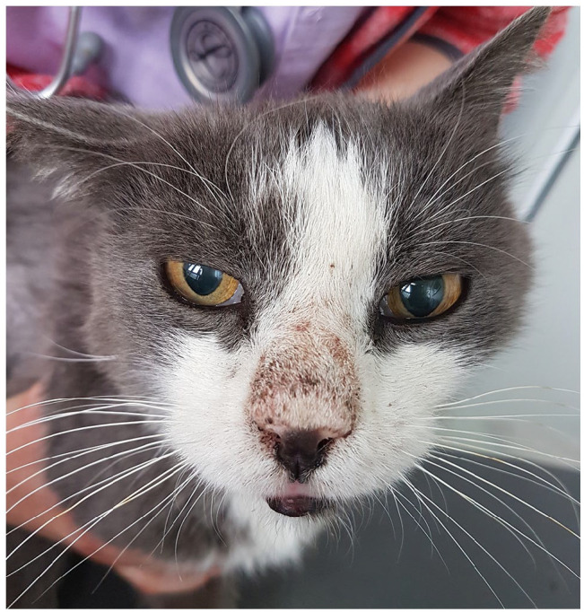
At the time of presentation to the dermatology service, the cat was clinically well, and the DM was controlled with glargine insulin (0.25 IU/kg q12h SC; Lantus, Sanofi-Aventis Deutschland) and dietary therapy (Feline Diabetic Canned Cat Food; Royal Canin). Dermatological examination revealed erythema, hypotrichosis, crusts and a brownish oily exudate on the dorsal nasal region (Figure 1). Ceruminous otic exudate was present in both ear canals and the haircoat was dry and dull. Initial dermatological differential diagnoses included dermatophytosis, demodicosis and solar (actinic) dermatitis.
Figure 1.

Clinical presentation of erythema, hypotrichosis, crusts and a brownish oily a exudate on the dorsal nasal region
A single deep skin scraping and trichograms from the lesional area revealed four live adult D cati mites. No dermatophyte hyphae or arthroconidia were found from the trichograms and on hair shafts obtained from the skin scraping of the lesional area. The parasitological examination of the otic exudate was also positive for D cati. Two adult live mites were seen. Few Malassezia yeasts were retrieved with ear canal cytology. The owner declined further diagnostic tests. The final diagnosis was feline demodicosis due to D cati.
A single dose of 250 mg (55.5 mg/kg) fluralaner spot-on for medium-sized cats (Bravecto; MSD Animal Health) was applied to the base of the cat’s neck. Four weeks later, the ceruminous otitis had resolved completely and the nasal lesions were markedly reduced. One dead adult D cati mite was found in the deep skin scrapings, while adhesive tape strips, trichograms and the parasitological examination of both ear canals were negative for mites. No further treatment was administered.
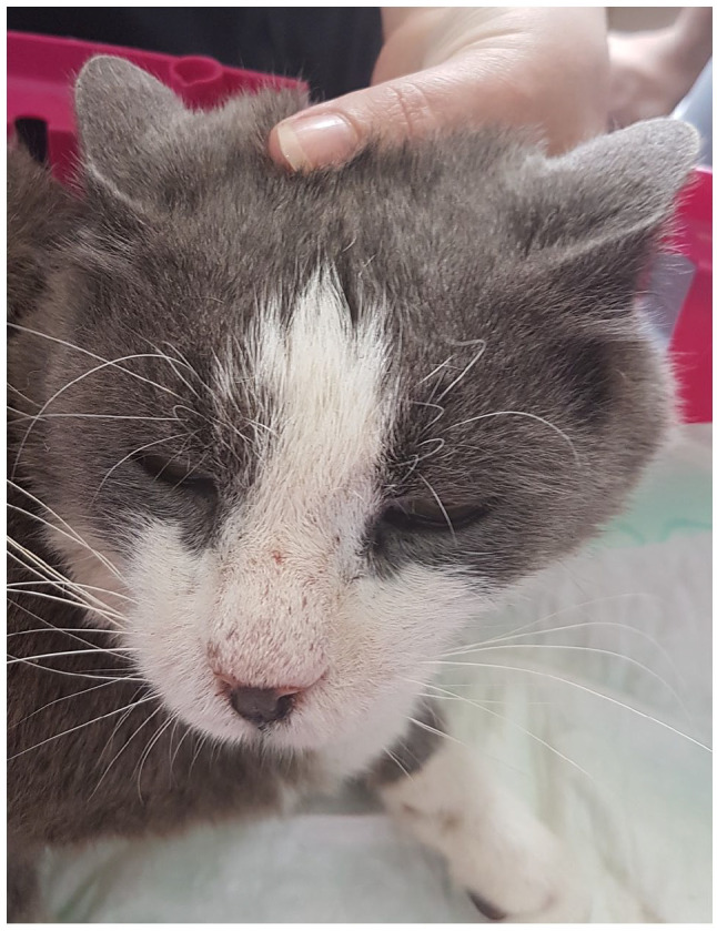
After 2 further weeks, while the cat was normoglyceamic and DM was well controlled, only a mild nasal hypotrichosis persisted. Deep skin scrapings, trichograms and parasitological examination of both ear canals were negative for mites. Eight weeks after the initial examination, the skin lesions had almost clinically resolved (Figure 2) and all parasitological samples were again negative for mites. A second fluralaner spot-on was applied 12 weeks after the first for prevention of relapse. No recurrence was noted at the 6-month follow-up.
Figure 2.

Signs of clinical improvement were evident 8 weeks post-treatment
Discussion
D cati is thought to be part of the normal microflora of the feline skin.15–18 It is believed that kittens become parasitised from the queen during lactation. 19 Unlike Demodex canis in the dog,3,20 there is no report of D cati found in skin scrapings from normal feline skin. Additionally, in one study PCR for D cati DNA was found to be negative in healthy cats and the authors speculated that this relates to the very small number of mites present. 16 However, asymptomatic and healthy cats may be found to be positive for D gatoi on skin scrapings or PCR.1,16
Generalised demodicosis owing to D cati is associated with systemic disease and long-term glucocorticoid use. In many cases, glucocorticoids have preceded clinical signs of demodicosis,2,4,21,22 thus demonstrating their role in the pathogenesis of this dermatitis. But there are also cats that have received glucocorticoids to control pruritus due to demodicosis. The most common underlying systemic conditions are infection with FIV and FeLV, as well as DM,4,11,15,18,21,22 as reported in the present case. However, some cats with D cati have no underlying disease or history of predisposing drug use.1,3 D cati mites have also been identified at lesional sites of Bowenoid in situ carcinoma (BISC).23,24 Although crusting nasal dermatitis can be a sign of BISC, in this case, the absence of hyperkeratotic, hyperpigmented, crusted plaques or papules on the skin did not support such a diagnosis.23,25 The lack of pigment at the lesional site in this cat, along with the history of outdoor activity, initially raised the suspicion of solar-induced lesions. Skin lesions in this case could have been compatible with incipient lesions of solar keratosis;26,27 however, skin biopsy, which would indicate any histological changes due to chronic sun exposure, was not performed. According to the literature, solar-induced lesions become progressively severe with each passing summer and early lesions can regress with photoprotection and sun avoidance.26,27 In this case, the lesions did not regress during the winter months and clinical improvement was seen after antiparasitic therapy, and thus a diagnosis of newly emerging solar keratosis was unlikely.
There is no consensus as to what extent or number of lesions describe localised vs generalised feline demodicosis, as has been reported for dogs. Localised demodicosis usually presents as facial lesions and the generalised form can involve the trunk and limbs.1,15,18 Otodemodicosis can be associated with dermatitis or present as a single finding. In a study of 1407 feline dermatology cases, demodicosis was diagnosed in only nine cases (3.4%, 9/266 parasitic diseases). Demodicosis was associated with D cati in 7/9 cases and in six of these cases mites were confined to the ear canals. 6
Pruritus is usually associated with contagious D gatoi, although asymptomatic infection with D gatoi has been reported.16,28 The host response is attributed to a hypersensitivity reaction, which may explain why some animals do not exhibit pruritus, despite the presence of the otherwise pruritic D gatoi. In the case of D cati, the pruritus can be absent or mild to moderate. 1 Considering that there are mixed infestations with D cati, D gatoi and the third unnamed Demodex species, it may be that the actual number of pruritic D cati cases are even fewer.14,15,18,21,29 In our case, the cat was not pruritic at all, a finding consistent with previous case reports.2,4,18
Localised demodicosis has been reported to be self-limiting.19,30 However, remission of generalised demodicosis owing to D cati is thought to happen by therapeutically targeting the underlying condition or by stopping immunosuppressive drugs. 2 This is in agreement with our case, because both the methylprednisolone had been stopped and the underlying condition (DM) was controlled. There is no time frame as to when clinical and parasitological cure occurs. Analysing published cases, for dips and rinses at least 1–2 months was required for clinical cure and relapses may be seen. In one case of otodemodicosis treated with topical solution of sarolaner/selamectin, complete mite resolution was achieved after four monthly treatments. 12 In a study, a single dose of topical fluralaner spot-on achieved parasitic clearance within 14 days and clinical cure within 1 month in seven cases with generalised dermatitis owing to D cati. 11 The use of oral fluralaner for D cati generalised demodicosis was successful in 1 and 2 months for parasitological and clinical cure, respectively, in another case report. 7 In the present case, clinical and parasitological cure of otodemodicosis was achieved in 4 weeks. For dermatitis, parasitological cure and almost complete disappearance of skin lesions plus hair regrowth occurred in 6 and 8 weeks, respectively.
Isoxazolines seem to be well tolerated and safe. The most common adverse effects reported in cats are vomiting, pruritus, diarrhoea, loss of appetite and alopecia at the site of application.31–35 These are sporadic in nature and usually self-limiting. Potential exists for neurological adverse effects including tremors, ataxia and seizures, even in cats without a history of neurological disorders.31,32 Information on the safety of isoxazolines in diseased animals is scarce. In particular, there is limited information on the safety of antiparasitic treatment for FIV-positive demodectic cats, since these cats died or were euthanased.15,18,22 The first report of an FIV-positive demodectic cat that received therapy was reported in 2005. The cat was treated with ivermectin and later with doramectin, but it developed ataxia and became lethargic, and finally was euthanased. 21 Generalised demodicosis in seven cats, three of them FIV positive, was successfully treated with spot-on fluralaner with no adverse effects reported. 11 In another report, an FeLV-positive demodectic cat received oral sarolaner, also with no adverse effects. 14 Cats with pre-existing chronic disease should be in good health at the time of isoxazoline administration and the underlying condition should be well controlled. The same was true for our case and no side effects were noted.
Isoxazolines have a broad spectrum of ectoparasitic activity in both dogs and cats. 31 In the cat in particular, studies have proved fluralaner’s efficacy against Lynxacarus radovskyi and Otodectes cynotis.36–38 In the first study, 36 fluralaner was administered orally, whereas in the other two studies a spot-on formulation was used,37,38 either alone or in combination with moxidectin. One application was successful in eradicating the infection, although in the case of L radovskyi, fluralaner appeared to have a shorter duration of action than the reported 12 weeks. Oral afoxolaner was administered in cats with otodectic mange and the therapeutic effect lasted for 65 days, and no re-infestation was observed. 39 The use of oral sarolaner was evaluated against lynxacariosis and otodectic mange and was also effective.40,41 A spot-on formulation combining selamectin and sarolaner was successful for the therapy of otodectic mange after a single application. 42 In another study, the efficacy of the above formulation against ear mites was assessed and it reached 94.4% following 1 month of administration. 43 Further studies are required to establish the efficacy of different fluralaner formulations or other isoxazolines against mites and insects.
Conclusions
In this case, spot-on fluralaner successfully treated, with no relapse and no obvious adverse effects, a cat with localised demodicosis and otodemodicosis due to D cati. This would suggest that the topical application of fluralaner is a long-term effective, easy to use, practical and less time-consuming alternative treatment for feline demodicosis.
Footnotes
Accepted: 1 December 2021
Conflict of interest: The authors declared no potential conflicts of interest with respect to the research, authorship, and/or publication of this article.
Funding: The authors received no financial support for the research, authorship, and/or publication of this article.
Ethical approval: The work described in this manuscript involved the use of non-experimental (owned or unowned) animals. Established internationally recognised high standards (‘best practice’) of veterinary clinical care for the individual patient were always followed and/or this work involved the use of cadavers. Ethical approval from a committee was therefore not specifically required for publication in JFMS Open Reports. Although not required, where ethical approval was still obtained, it is stated in the manuscript.
Informed consent: Informed consent (verbal or written) was obtained from the owner or legal custodian of all animal(s) described in this work (experimental or non-experimental animals, including cadavers) for all procedure(s) undertaken (prospective or retrospective studies). No animals or people are identifiable within this publication, and therefore additional informed consent for publication was not required.
ORCID iD: Pavlina Bouza-Rapti  https://orcid.org/0000-0002-4140-5683
Rania Farmaki  https://orcid.org/0000-0002-3748-223X
References
- 1. Beale K. Feline demodicosis. A consideration in the itchy or overgrooming cat. J Feline Med Surg 2012; 14: 209–213. [DOI] [PMC free article] [PubMed] [Google Scholar]
- 2. Bizikova P. Localized demodicosis due to Demodex cati on the muzzle of two cats treated with inhalant glucocorticoids. Vet Dermatol 2014; 25: 222–225. [DOI] [PubMed] [Google Scholar]
- 3. Mueller RS, Rosenkrantz W, Bensignor E, et al. Diagnosis and treatment of demodicosis in dogs and cats: clinical consensus guidelines of the World Association for Veterinary Dermatology. Vet Dermatol 2020; 31: 4–26. [DOI] [PubMed] [Google Scholar]
- 4. Guaguere E, Muller A, Degorce-Rubiales F. Feline demodicosis: a retrospective study of 12 cases [abstract]. Vet Dermatol 2004; 15: S34. [Google Scholar]
- 5. Poucke SV. Ceruminous otitis externa due to Demodex cati in a cat. Vet Rec 2001; 149: 651–652. [DOI] [PubMed] [Google Scholar]
- 6. Scott DW, Miller WH, Erb HN. Feline dermatology at Cornell University: 1407 cases (1988–2003). J Feline Med Surg 2012; 15: 307–316. [DOI] [PMC free article] [PubMed] [Google Scholar]
- 7. Matricoti I, Maina E. The use of oral fluralaner for the treatment of feline generalized demodicosis: a case report. J Small Anim Pract 2017; 58: 476–479. [DOI] [PubMed] [Google Scholar]
- 8. Newbury S, Moriello K, Verbrugge M, et al. Use of lime sulphur and itraconazole to treat shelter cats naturally infected with Microsporum canis in an annex facility: an open field trial. Vet Dermatol 2007; 18: 324–331. [DOI] [PubMed] [Google Scholar]
- 9. Cowan LA, Campbell K. Generalized demodicosis in a cat responsive to amitraz. J Am Vet Med Assoc 1988; 192: 1442–1444. [PubMed] [Google Scholar]
- 10. Duangkaew L, Hoffman H. Efficacy of oral fluralaner for the treatment of Demodex gatoi in two shelter cats. Vet Dermatol 2018; 29: 262. DOI: 10.1111/vde.12520. [DOI] [PubMed] [Google Scholar]
- 11. Beccati MB, Pandolfi PP, DiPalma AD. Efficacy of fluralaner spot-on in cats affected by generalized demodicosis: seven cases [abstract]. Vet Dermatol 2019; 30: 454. [Google Scholar]
- 12. Simpson AC. Successful treatment of otodemodicosis due to Demodex cati with sarolaner/selamectin topical solution in a cat. JFMS Open Rep 2021; 7. DOI: 10.1177/2055116920984386. [DOI] [PMC free article] [PubMed] [Google Scholar]
- 13. Walker C. Treatment of Demodex gatoi mange in two sibling Bengal cats with a combination of selamectin and sarolaner. Compan Anim 2019; 24: 127–131. [Google Scholar]
- 14. De Almeida GPS, Campos DR, Scott FB, et al. Successful treatment of feline demodicosis with oral sarolaner: case report. Braz J Vet Med 2020; 42: e113420. DOI: 10.29374/2527- 2179.bjvm113420. [Google Scholar]
- 15. Neel J, Tarigo J, Tater KC, et al. Deep and superficial skin scrapings from a feline immunodeficiency virus-positive cat. Vet Clin Pathol 2007; 36: 101–104. [DOI] [PubMed] [Google Scholar]
- 16. Frank LA, Kania SA, Chung K, et al. A molecular technique for the detection and differentiation of Demodex mites on cat. Vet Dermatol 2013; 24: 367–369. [DOI] [PubMed] [Google Scholar]
- 17. Silbermayr K, Horvath-Ungerboeck C, Eigner B, et al. Phylogenetic relationships and new genetic tools for the detection and discrimination of the three feline Demodex mites. Parasitol Res 2015; 114: 747–752. [DOI] [PubMed] [Google Scholar]
- 18. Taffin ER, Casaert S, Claerebout E, et al. Morphological variability of Demodex cati in a feline immunodeficiency virus-positive cat. J Am Vet Med Assoc 2016; 249: 1308–1312. [DOI] [PubMed] [Google Scholar]
- 19. Chesney CJ. Demodicosis in the cat. J Small Anim Pract 1989; 30: 689–695. [Google Scholar]
- 20. Ravera I, Altet L, Francino O, et al. Small Demodex populations colonize most parts of the skin of healthy dogs. Vet Dermatol 2013; 24: 168–172. [DOI] [PubMed] [Google Scholar]
- 21. Lowenstein C, Beck W, Bessmann K, et al. Feline demodicosis caused by concurrent infection with Demodex cati and an unnamed species of mite. Vet Rec 2005; 157: 290–292. [DOI] [PubMed] [Google Scholar]
- 22. Iliev PT, Zhelev G, Ivanov A, et al. Demodex cati and feline immunodeficiency virus co-infection in a cat. Bulgarian J Vet Med 2019; 22: 237–242. [Google Scholar]
- 23. Guaguere E, Olivry T, Delverdier-Poujade E. Demodex cati infestation in association with feline cutaneous squamous cell carcinoma in situ: a report of 5 cases. Vet Dermatol 1999; 10: 61–67. [DOI] [PubMed] [Google Scholar]
- 24. Conceição LG, Camargo LP, Costa PRS, et al. Squamous cell carcinoma (Bowen’s disease) in situ in three cats. Arq Bras Med Vet Zootec 2007; 59: 816–820. [Google Scholar]
- 25. Wilhelm S, Degorce-Rubiales F, Godson D, et al. Clinical, histological and immunohistochemical study of feline viral plaques and Bowenoid in situ carcinomas. Vet Dermatol 2006; 17: 424–431. [DOI] [PubMed] [Google Scholar]
- 26. Almeida EM, Caraça RA, Adam RL, et al. Photodamage in feline skin: clinical and histomorphometric analysis. Vet Pathol 2008; 45: 327–335. [DOI] [PubMed] [Google Scholar]
- 27. Peters-Kennedy J, Scott DW, Miller WH. Apparent clinical resolution of pinnal actinic keratoses and squamous cell carcinoma in a cat using topical imiquimod 5% cream. J Feline Med Surg 2008; 10: 593–599. [DOI] [PMC free article] [PubMed] [Google Scholar]
- 28. Silbermayr K, Joachim A, Litschauer B, et al. The first case of Demodex gatoi in Austria, detected with fecal flotation. Parasitol Res 2013; 112: 2805–2810. [DOI] [PubMed] [Google Scholar]
- 29. Moriello K, Newbury S, Steinberg H. Five observations of a third morphologically distinct feline Demodex mite. Vet Dermatol 2013; 24: 460–462. [DOI] [PubMed] [Google Scholar]
- 30. Miller WH, Griffin CE, Campbell KL. Parasitic skin disease. In: Miller WH, Griffin CE, Campbell KL. (eds). Small animal dermatology. 7th ed. St Louis, MO: Elsevier Mosby, 2013, pp 284–342. [Google Scholar]
- 31. Zhou X, Hohman A, Hsu WH. Review of extralabel use of isoxazolines for treatment of demodicosis in dogs and cats. J Am Vet Med Assoc 2020; 256: 1342–1346. [DOI] [PubMed] [Google Scholar]
- 32. Zhou X, Hohman A, Hsu WH. Current review of isoxazoline ectoparasiticides used in veterinary medicine. J Vet Pharmacol Ther 2021. DOI: 10.1111/jvp.12959. [DOI] [PubMed] [Google Scholar]
- 33. Chappell K, Paarlberg T, Seewald W, et al. A randomized, controlled field study to assess the efficacy and safety of lotilaner flavored chewable tablets (Credelio™ CAT) in eliminating fleas in client-owned cats in the USA. Parasit Vectors 2021; 14: 127. DOI: 10.1186/s13071-021-04617-5. [DOI] [PMC free article] [PubMed] [Google Scholar]
- 34. Meadows C, Guerino F, Sun F. A randomized, blinded, controlled USA field study to assess the use of fluralaner topical solution in controlling feline flea infestations. Parasit Vectors 2017; 10: 37. DOI: 10.1186/s13071-017-1972-4. [DOI] [PMC free article] [PubMed] [Google Scholar]
- 35. Packianathan R, Pittorino M, Hodge A, et al. Safety and efficacy of a new spot-on formulation of selamectin plus sarolaner in the treatment and control of naturally occurring flea infestations in cats presented as veterinary patients in Australia. Parasit Vectors 2020; 13: 227. DOI: 10.1186/s13071-020-04099-x. [DOI] [PMC free article] [PubMed] [Google Scholar]
- 36. Han H, Noli C, Cena T. Efficacy and duration of action of oral fluralaner and spot-on moxidectin/imidacloprid in cats infested with Lynxacarus radovskyi. Vet Dermatol 2016; 27: 474–477. [DOI] [PubMed] [Google Scholar]
- 37. Taenzler J, de Vos C, Roepke R, et al. Efficacy of fluralaner against Otodectes cynotis infestations in dogs and cats. Parasit Vectors 2017; 10: 30. DOI: 10.1186/s13071-016-1954-y. [DOI] [PMC free article] [PubMed] [Google Scholar]
- 38. Taenzler J, de Vos C, Roepke R, et al. Efficacy of fluralaner plus moxidectin (Bravecto® Plus spot-on solution for cats) against Otodectes cynotis infestations in cats. Parasit Vectors 2018; 11: 595. DOI: 10.1186/s13071-018-3167-z. [DOI] [PMC free article] [PubMed] [Google Scholar]
- 39. Machado MA, Campos DR, Lopes NL, et al. Efficacy of afoxolaner in the treatment of otodectic mange in naturally infested cats. Vet Parasitol 2018; 256: 29–31. [DOI] [PubMed] [Google Scholar]
- 40. Campos DR, Chaves JKO, Assis RCP, et al. Efficacy of oral sarolaner against Lynxacarus radovskyi in naturally infested cats. Vet Dermatol 2020; 31: 355–358. [DOI] [PubMed] [Google Scholar]
- 41. Campos DR, Chaves JKO, Guimaraes BC, et al. Efficacy of oral sarolaner for the treatment of feline otodectic mange. Pathogens 2021; 10: 341. DOI: 10.3390/pathogens10030341. [DOI] [PMC free article] [PubMed] [Google Scholar]
- 42. Becskei C, Reinemeyer C, King V, et al. Efficacy of a new spot-on formulation of selamectin plus sarolaner in the treatment of Otodectes cynotis in cats. Vet Parasitol 2017; 238: S27–S30. [DOI] [PubMed] [Google Scholar]
- 43. Vatta A, Myers M, Rugg J, et al. Efficacy and safety of a combination of selamectin plus sarolaner for the treatment and prevention of flea infestations and the treatment of ear mites in cats presented as veterinary patients in the United States. Vet Parasitol 2019; 270: S3–S11. [DOI] [PubMed] [Google Scholar]


