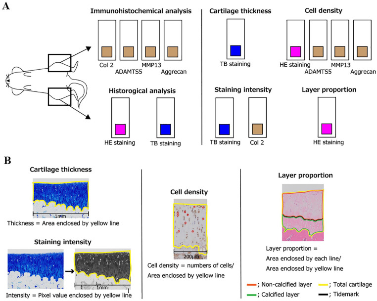
Figure 1.
Schematic of histological analysis for articular cartilage: (A) staining method and analysis parameters and (B) overview of each analysis method. The right knee was used for histological analysis, and the left knee was used in the immunohistochemical analysis. For each staining, cartilage thickness, cell density, staining intensity, and layer proportion were evaluated. For details on these parameters, please refer to the text for evaluation methods. ADAMTS-5 = A disintegrin and metalloproteinase with thrombospondin motifs 5.
HE, hematoxylin–eosin; TB, toluidine blue.

