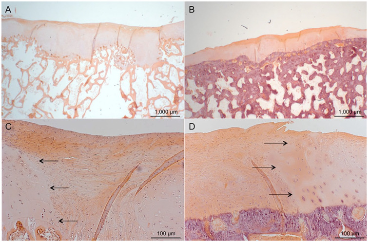
Figure 3.
Hematoxylin and eosin staining; scale bars 1000 μm (A and B) and 100 μm (C and D). (A and C) Histological section of defect treated with CC and PRP. The arrows mark the border between native cartilage and repair tissue. (B and D) A defect treated with CC alone. The defect is sufficiently filled, and the repair tissue is a mixture of fibrous tissue and fibrocartilage. Samples represent mean repair tissue quality. CC, autologous cartilage chips transplantation; PRP, platelet-rich plasma.

