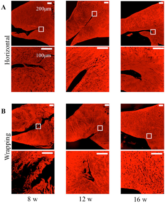
Figure 6.
Picrosirius red staining for collagen fibers of meniscus tear. White square in the upper figure shows the peripheral meniscal tear in the lower figure. (A) Horizontal group at 8, 12, and 16 weeks. (B) Wrapping group at 8, 12, and 16 weeks. In the upper figures, the bar indicates 200 µm. In the lower figures, the bar indicates 100 µm.

