
Figure 1.
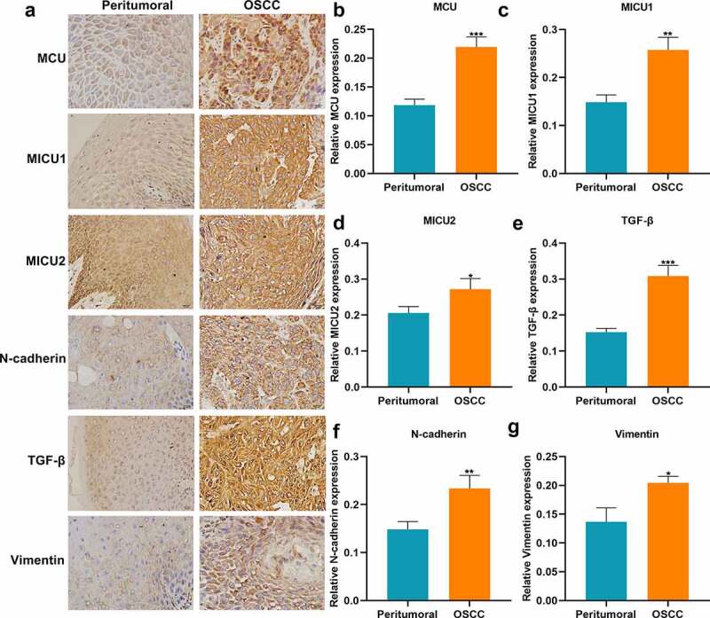
Immunohistochemistry for the expression of MCU, MICU1, MICU2, TGF-β, N-cadherin and vimentin proteins in OSCC and peritumoral tissues. (a) Representative images of immunohistochemistry of MCU, MICU1, MICU2, TGF-β, N-cadherin and vimentin proteins in OSCC and peritumoral tissues. Bar = 20 μm. (b–g) Quantification results of the expressions of (b) MCU; (c) MICU1; (d) MICU2; (e) TGF-β; (f) N-cadherin and (g) vimentin in OSCC and peritumoral tissues. *p < 0.05; **p < 0.01; ***p < 0.001.