

Abstract
Introduction and importance
Refractory diabetic foot ulcers (DFUs) do not easily respond to standard therapeutic approaches and the prevention of DFU-related amputation is one of the most important aspects of treatment in patients with DFU.
Case presentation
The present case report is 51-year old male patient with a history of 5-years type 2 diabetes who has had DFU on the first distal phalanx foot of his right foot with size of 2 × 2 cm. The patient was repeatedly hospitalized for receiving DFU treatment, he did not recover using routine wound treatment. The patient was transferred to our wound care team. After ten sessions (one session every 48 h) of maggot debridement therapy (MDT) using sterile Lucilia sericata, the patients' DFU had completely healed.
Clinical discussion
DFUs can change patient's quality of life and lead to infection, amputation, sepsis, and finally death. Thus, efficient therapeutic methods are necessary for management of DFUs.
Conclusion
This case report was revealed that the maggot therapy is an affordable and highly efficacious treatment method to enhance the recuperation of DFUs. Therefore, it is recommended that wound care teams use this approach to speed up the healing process.
Keywords: Diabetic foot, Larva, Debridement, Amputation, Case report, Iran
Highlights
-
•
DFU is one of the most significant complications of diabetes mellitus.
-
•
Affordable and efficacious treatment is necessary for the management of DFUs.
-
•
Maggot debridement therapy is effective approach for healing DFUs.
1. Introduction
Diabetes mellitus (DM) is a group of metabolic disorders characterized by hyperglycemia due to defects in insulin secretion, insulin action, or both [1]. DM is also associated with an increased occurrence of macrovascular diseases, including coronary artery disease (myocardial infarction), cerebrovascular disease (stroke), and peripheral vascular disease [2]. Complications related to DM are classified as acute and chronic [3]. Diabetic Foot Ulcer (DFU), as a chronic complication of DM, is one of the most significant and debilitating complications [2]. Approximately over 25% of individuals with DM develop DFU during their lifetime [4], and one-fifth of moderate to severe DFUs cause amputation [5].
Many patients with diabetic ulcers have underlying conditions that make it very difficult for them to heal [6]. Therefore, many chronic non-healing wounds need targeted approaches instead of the conventional therapies [7]. In fact refractory diabetic ulcers do not easily respond to standard therapeutic approaches, and the prevention of DFU-related amputation is one of the most important aspects of treatment in these patients [6].
Larval therapy or maggot debridement therapy (MDT) is a method used to treat chronic wounds, including patients with DFUs or bed sores [8]. In this method, the larvae of a type of fly called Lucilia sericata, which are cultured sterile in the laboratory, are used to treat patients [9]. The chief function of maggot therapy is to decrease the microorganism load at the infection site through the digestion of bacteria, production of antibacterial secretions, and elimination of biofilms [8]. As a result of the flourishing use of maggot therapy in numerous countries, particularly the U. S. and Europe, the FDA has approved MDT [10]. The combination of these features and previous reports led us to trial MDT in a case of DFU. This case report was reported according to the SCARE 2020 Guidelines to ensure the quality of reporting [11].
2. Case presentation
This case is a 51-year old male patient with a history of 5-years type 2 diabetes. He was living in Urmia city, Iran. Moreover, he was from a family with low socioeconomic status with a primary education level. He was working in a carpet weaving workshop and had a sedentary lifestyle. During history-taking and physical assessment, the patient mentioned a family history of diabetes mellitus and hypertension (HTN). In order to regulate his blood glucose level, he had undergone an oral medications therapy with metformin 500 mg tablet two times a day (BID). Moreover, Captopril 25 mg tablet had been prescribed for his HTN twice a day (BID). In addition, during taking the patient's history, we discovered that he did not take his drugs regularly and that he did not follow his adequate diet and even his blood sugar control was inappropriate. Therefore, he had uncontrolled DM. This condition led to the formation of DFU on the first distal phalanx foot of his right foot with size of 2 × 2 cm (Fig. 1). He smoked 10 cigarettes per day for 15 years, but he denied any history of drug or alcohol abuse. On his neurological assessment no pathological findings were revealed. Moreover, the culture antibiogram was obtained and it revealed drug resistance to Staphylococcus aureus. The patient received intravenous antibiotics such as Amp Meropenem 1 g three times a day, and Amp Vancomycin 1 g two times a day. Although the patient was repeatedly hospitalized for receiving DFU treatment, he did not recover using routine wound treatment. Therefore, he was referred to our wound management team on 1 July 2020.
Fig. 1.

Diabetic foot ulcer before beginning the maggot debridement therapy.
The vital signs of patient on admission were as follows: Pulse Rate: 110 beat per minute (bpm); Temperature: 38.1 °C; Blood Pressure: 150/95 mmHg; Respiration Rate: 18 bpm. The patient suffered from symptoms of infection and sepsis (such as fever). In addition, some of the patient's laboratory information during admission is provided as follows (Table 1):
Table 1.
The patient's laboratory information during admission.
| Cell blood count (CBC) | Biochemistry |
|---|---|
| WBC: 24000 μl | BUN: 14.9 mg/dl |
| RBC: 5390000 μl | Creatinine: 1.2 mg/dl |
| HGB: 11.5 g/dl | Urea: 23.2 mg/dl |
| HCT: 38% | Calcium: 8.65 mg/dl |
| MCV: 65.5 fl | Phosphorous: 4.5 mg/dl |
| MCH: 19.7 pg | Sodium: 145 meq/dl |
| MCHC: 30.44 g/dl | Potassium: 3.9 meq/dl |
| RDW-CV: 15.7% | SGOT(AST): 65 u/l |
| RDW-SD: 42.3 fl | SGPT(ALT): 81 u/l |
| PLT: 259000 μl | Al/phosphatase: 457 u/l |
| PDW: 16.0 | Bilirubin total: 0.9 mg/dl |
| MPV: 8 fl | Bilirubin direct: 0.4 mg/dl |
| PCT: 0.199% | Blood sugar: 349 mg/dl |
| Serology | LDL: 87 mg/dl |
| CRP: positive(+2) | HDL: 36 mg/dl |
| Thyroid function | Cholesterol: 198 mg/dl |
| TSH: 12 mlu/l | Triglycerides: 128 mg/dl |
| Free T4: 10 pmol/l | HemoglobinA1C: 7.0% |
| Free T3: 3.4 pmol/l | – |
2.1. Management
First, written informed consent was obtained from the patient for maggot debridement therapy. Then the maggots of Lucilia sericata were disinfected with Dakin's solution (0.5% NaOCl), and the patient underwent maggot debridement therapy. The larvae of L. sericata digest dead tissue and microorganisms at the wound site. In fact these larvae release antimicrobial enzymes that improve wound healing. In this case, maggot debridement therapy was carried out in ten sessions (one session every 48 h) (Fig. 2). This method (i.e., DFU preparation, application of the L. sericata to the wound, hydrocolloid dressing and removal of larvae after 48 h) was conducted by a nurse who was trained and licensed in this area. At each stage of the MDT, the patient was asked a question about tolerating the MDT. If the patient's answer was “yes” the MDT was continued, however if the patient's answer was “no,” the MDT was discontinued. In addition, after maggot therapy sessions were completed, the diabetic foot ulcer was stimulated by mechanical debridement as well as normal saline. Therefore, the necrotic tissue was again eliminated and granulation tissue emerged (Fig. 3). The diabetic foot ulcer of the patient had relatively healed on 1 September 2020 and closed three months after the MDT (Fig. 4). Finally, the patient in a good general health condition was discharged (Fig. 5). During discharging stage, the patient was educated to prevent putting excessive pressure on the area and uses a crutch or wheelchair. In fact offloading is an essential stage for DFU healing.
Fig. 2.
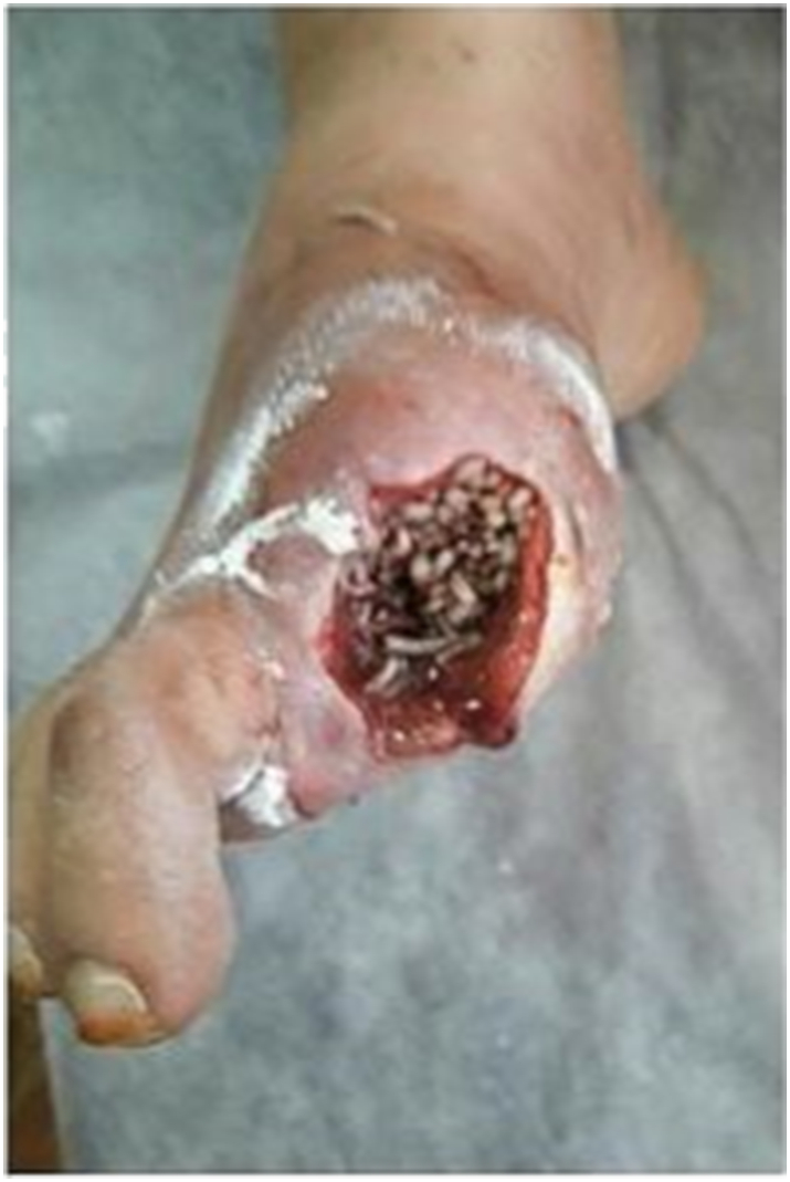
Maggot debridement therapy in diabetic foot ulcer.
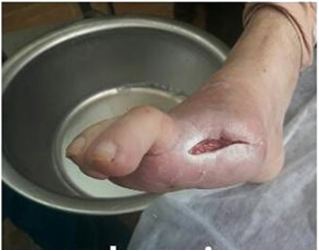
Fig. 3.

Diabetic foot ulcer after maggot debridement therapy.
Fig. 4.
The patient's DFUs three months after maggot debridement therapy.
Fig. 5.

The patient's DFUs five months after maggot debridement therapy.
3. Discussion
Wounds, especially DFUs, are a common complication of DM that can lead to physical disability and stress in patients. DM causes diabetic foot ulcers by several mechanisms. Lack of pain in neuropathy is usually the main cause of ulcers. On the other hand, neuropathy itself makes the skin of the foot dry and brittle and increases the skin's tendency to crack [12]. Moreover, the mechanism of fighting against microorganisms in patients with DM is impaired and the wounds of diabetics are infected [4]. In addition, due to vascular involvement, blood supply to the damaged tissues is disrupted and this tissue repair is impaired [2]. Diabetic foot ulcers can change quality of life and lead to infection, sepsis, amputation, and finally death. Thus, efficient therapeutic methods are necessary for the management of DFUs [7]. Nowadays, concerning the appearance of antibiotic-resistant bacteria, many professionals health care have turned their attention to the use of MDT [9]. According to Food and Drug Administration, MDT will be very effective for treating wounds that do not heal easily, as well as for treating open wounds with dead tissue [10]. The larvae feed on dead tissues and thus destroy these tissues completely. The saliva of these larvae can digest dead wound tissue well and can also remove the microorganisms at the wound site infection [13].
Maggot therapy can be easily conducted by trained healthcare professionals such as nurse, even without the need for hospitalization. It is also an affordable treatment method. MDT in contrast to antibiotic therapy, causes no hazardous complication [9]. However, MDT may has led to pain and anxiety and stress in the patient. Of course, these symptoms will rarely be seen in patients undergoing maggot therapy. In addition, patients may experience bleeding and infection [14]. The risk of infection will only increase if the larvae are not well disinfected before being placed on the wound. In this case, the patient may develop infections in his wound [8].
In line with our study results, Malekian et al. revealed that maggot therapy is a very efficient in treating refractory DFUs [15]. Moreover, consistent with our results, Parizad et al. showed that the combination of surgical debridement, maggot therapy, negative pressure wound therapy, and silver foam dressing is an appropriate treatment method for DFU [16]. Siavash et al. also showed that maggot therapy is efficient in treating DFUs that are resistant to routine methods [17]. In addition, consistent with our results, Mirabzadeh et al. indicated that the maggot therapy is an appropriate and easy-to-use methodology for the treatment of sophisticated and in depth DFUs [18]. Hajimohammadi et al. in a case report study showed that surgical debridement and maggot therapy is an effective approach for improving DFUs and preventing amputation [14].
Many research in this field have revealed the effectiveness of Maggot therapy in improving DFU and the result of this case report showed that maggot therapy could be used in patients who have refractory DFUs.
4. Conclusion
Diabetic foot ulcers remain a problem of concern, causing increased morbidity and mortality in patients with DM. Therefore, efficient therapeutic approaches are essential to prevent foot amputation and death. This case report was revealed that the use of maggot therapy is an affordable treatment method to enhance the recuperation of DFUs and prevent foot amputation. Therefore, it is recommended that wound care teams use this approach to speed up the healing process.
Sources of funding
This case report did not receive any specific grant from funding agencies in the public, commercial, or not-for-profit sectors.
Ethical approval
All ethical principles were considered in conducting this case report. All patient information kept confidential.
Consent
Written informed consent was obtained from the patient for publication of this case report and accompanying images. A copy of the written consent is available for review by the Editor-in-Chief of this journal on request.
Research registration
Not applicable.
Guarantor
Rasoul Goli.
Provenance and peer review
Not commissioned, externally peer-reviewed.
CRediT authorship contribution statement
Rasoul Goli: Study concept, data collection, writing the paper and making the revision of the manuscript following the reviewer's instructions. Babak Choobianzali: Study concept, reviewing and validating the manuscript's credibility. Amireh Hassanpour: reviewing and validating the manuscript's credibility.
Declaration of competing interest
None.
References
- 1.Nanayakkara N., Curtis A.J., Heritier S., Gadowski A.M., Pavkov M.E., Kenealy T., Owens D.R., Thomas R.L., Song S., Wong J., Chan J.C. Impact of age at type 2 diabetes mellitus diagnosis on mortality and vascular complications: systematic review and meta-analyses. Diabetologia. 2021 Feb;64(2):275–287. doi: 10.1007/s00125-020-05319-w. [DOI] [PMC free article] [PubMed] [Google Scholar]
- 2.Jameson J.L. McGraw-Hill Education; 2018. Harrison's Principles of Internal Medicine. [Google Scholar]
- 3.Hinkle J.L., Cheever K.H. Wolters Kluwer India Pvt Ltd; 2018 Aug 30. Brunner and Suddarth's Textbook of Medical-surgical Nursing. [Google Scholar]
- 4.Hurlow J.J., Humphreys G.J., Bowling F.L., McBain A.J. Diabetic foot infection: a critical complication. Int. Wound J. 2018 Oct;15(5):814–821. doi: 10.1111/iwj.12932. [DOI] [PMC free article] [PubMed] [Google Scholar]
- 5.Lipsky B.A., Berendt A.R., Cornia P.B., Pile J.C., Peters E.J., Armstrong D.G., Deery H.G., et al. 2012 Infectious Diseases Society of America clinical practice guideline for the diagnosis and treatment of diabetic foot infections. Clin. Infect. Dis. 2012 Jun 15;54(12):e132–e173. doi: 10.1093/cid/cis346. [DOI] [PubMed] [Google Scholar]
- 6.Armstrong D.G., Boulton A.J., Bus S.A. Diabetic foot ulcers and their recurrence. N. Engl. J. Med. 2017 Jun 15;376(24):2367–2375. doi: 10.1056/NEJMra1615439. [DOI] [PubMed] [Google Scholar]
- 7.Faraji N., Goli R., Choobianzali B., Bahrami S., Sadeghian A., Sepehrnia N., Ghalandari M. Ozone therapy as an alternative method for the treatment of diabetic foot ulcer: a case report. J. Med. Case Rep. 2021 Dec;15(1):1–8. doi: 10.1186/s13256-021-02829-y. [DOI] [PMC free article] [PubMed] [Google Scholar]
- 8.Nishijima A., Yamamoto N., Yoshida R., Yanagibayashi S., Takikawa M., Hayasaka R., et al. Maggot debridement therapy with a direct dressing can cause compression injuries in patients with chronic limb ischemia. Case Rep. Plast. Surg. Hand Surg. 2017 Jan 1;4(1):84–88. doi: 10.1080/23320885.2017.1373596. [DOI] [PMC free article] [PubMed] [Google Scholar]
- 9.Stadler F. The maggot therapy supply chain: a review of the literature and practice. Med. Vet. Entomol. 2020;34(1):1–9. doi: 10.1111/mve.12397. [DOI] [PubMed] [Google Scholar]
- 10.Food and Drug Administration . Monarch Labs LLC. FDA; 2007. 510(k) Summary.https://www.accessdata.fda.gov/cdrh_docs/pdf7/K072438.pdf [accessed on d Month 2019] [Google Scholar]
- 11.Agha R.A., Franchi T., Sohrabi C., Mathew G., Kerwan A., Thoma A., et al. The SCARE 2020 guideline: updating consensus Surgical CAse REport (SCARE) guidelines. Int. J. Surg. 2020 Dec 1;84:226–230. doi: 10.1016/j.ijsu.2020.10.034. [DOI] [PubMed] [Google Scholar]
- 12.Alsanawi Y., Alismail H., Alabd Rabalnabi M., Alturki H., Alsuhaibani A., Mahbub M., et al. Pathogenesis and management of diabetic foot ulcers. Int. J. Commun. Med. Public Health. 2018 Nov;5(11):4953. [Google Scholar]
- 13.Mohd Zubir M.Z., Holloway S., Mohd Noor N. Maggot therapy in wound healing: a systematic review. Int. J. Environ. Res. Public Health. 2020 Jan;17(17):6103. doi: 10.3390/ijerph17176103. [DOI] [PMC free article] [PubMed] [Google Scholar]
- 14.Hajimohammadi K., Parizad N., Hassanpour A., Goli R. Saving diabetic foot ulcers from amputation by surgical debridement and maggot therapy: a case report. Int. J. Surg. Case Rep. 2021 Sep 1;86 doi: 10.1016/j.ijscr.2021.106334. [DOI] [PMC free article] [PubMed] [Google Scholar]
- 15.Malekian A., Djavid G.E., Akbarzadeh K., Soltandallal M., Rassi Y., Rafinejad J., et al. Efficacy of maggot therapy on Staphylococcus aureus and Pseudomonas aeruginosa in diabetic foot ulcers: a randomized controlled trial. J. Wound Ostomy Cont. Nurs. 2019;46(1):25–29. doi: 10.1097/WON.0000000000000496. [DOI] [PubMed] [Google Scholar]
- 16.Parizad N., Hajimohammadi K., Goli R. Surgical debridement, maggot therapy, negative pressure wound therapy, and silver foam dressing revive hope for patients with diabetic foot ulcer: a case report. Int. J. Surg. Case Rep. 2021 Apr 29 doi: 10.1016/j.ijscr.2021.105931. [DOI] [PMC free article] [PubMed] [Google Scholar]
- 17.Siavash M., Najjarnezhad A., Mohseni N., Abtahi S.M., Karimy A., Sabzevari M.H. Efficacy of maggot debridement therapy on refractory atypical diabetic foot ulcers: an open-label study. Int. J. Low. Extrem. Wounds. 2020 May 5 doi: 10.1177/1534734620920403. 1534734620920403. [DOI] [PubMed] [Google Scholar]
- 18.Mirabzadeh A., Ladani M.J., Imani B., Rosen S.A., Sherman R.A. Maggot therapy for wound care in Iran: a case series of the first 28 patients. J. Wound Care. 2017 Mar 2;26(3):137–143. doi: 10.12968/jowc.2017.26.3.137. [DOI] [PubMed] [Google Scholar]