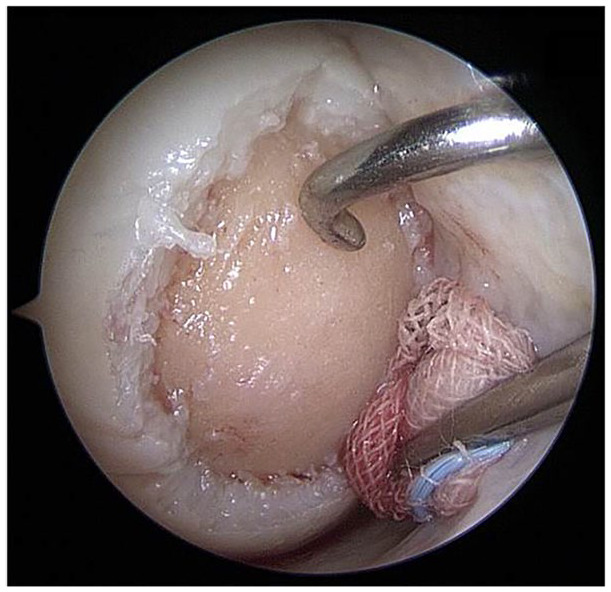
Figure 2.

Arthroscopically prepared cartilage defect (medial femoral condyle, right knee joint, dry conditions). Using the proximal medial portal, a probing hook is placed for defect size measurement (application devices can be introduced into the joint via the same portal). In the distal medial portal, a swab is placed to safely keep the joint dry and to tense the joint capsule and Hoffa fat, to distant them from the defect.
