Version Changes
Revised. Amendments from Version 1
This article has improved by including the following developments:
Improvements in the ContactJ macro code ( https://doi.org/10.5281/zenodo.5810874) performance (GUI, Batch Mode...).
Extended information about the different steps throughout ContactJ macro has been included.
Comparison between Manual and Automatic Analysis with ContactJ.
Implementation of a straightforward core code of ContactJ, ContactJ4All, that facilitates the applicability of ContactJ in 3D or time series images, and its combination with 3D objects analysis and objects tracking methods. ContactJ4All is ready to be included and adapted to any workflow. It is available on GitHub and Zenodo ( https://doi.org/10.5281/zenodo.5810614).
Abstract
Lipid droplets (LDs) are the major lipid storage organelles of eukaryotic cells and together with mitochondria key regulators of cell bioenergetics. LDs communicate with mitochondria and other organelles forming “metabolic synapse” contacts to ensure that lipid supply occurs where and when necessary. Although transmission electron microscopy analysis allows an accurate and precise analysis of contacts, the characterization of a large number of cells and conditions can become a long-term process. In order to extend contact analysis to hundreds of cells and multiple conditions, we have combined confocal fluorescence microscopy with advanced image analysis methods. In this work, we have developed the ImageJ macro script ContactJ, a novel and straight image analysis method to identify and quantify contacts between LD and mitochondria in fluorescence microscopy images allowing the automatic analysis. This image analysis workflow combines colocalization and skeletonization methods, enabling the quantification of LD-mitochondria contacts together with a complete characterization of organelles and cellular parameters. The correlation and normalization of these parameters contribute to the complex description of cell behavior under different experimental energetic states. ContactJ is available here: https://doi.org/10.5281/zenodo.5810874
Keywords: Contact sites, Lipid Droplets, Mitochondria, Image Processing and Analysis, ImageJ, Fluorescence Microscopy, Bioimaging, Interactome
Introduction
Lipid droplets (LDs) are the major lipid storage organelles of eukaryotic cells and together with mitochondria key regulators of cell’s bioenergetics. They supply essential lipids to produce signalling molecules, membrane building blocks, and the metabolic energy needed to survive during nutrient poor periods. 1
In order to achieve their functions, LDs communicate with mitochondria and other organelles (endoplasmic reticulum, endosomes, peroxisomes and vacuoles) forming membrane contact sites, 2 “metabolic synapses”, to ensure that lipid provision occurs where and when necessary. 1 , 3 , 4 Contact sites between these organelles have been described and characterized by transmission electron microscopy (TEM) as it resolves at the membrane scale where these contacts take place. 2 , 5 Whereas TEM allows accurate and precise characterization of contacts, their analysis on a large number of cells and conditions can become a long-term process. On the other hand, confocal fluorescence microscopy combined with advanced image analysis methods enable to extend contact analysis to hundreds of cells and multiple conditions.
Typically, in fluorescence microscopy, the contacts between cellular organelles, the organelle interactome, have been studied by colocalization or overlapping regions of the organelles masks 6 or measuring the fraction of intensity of other organelles near to LD. 7
In the present work, we describe a novel and straight image analysis method (ContactJ) to identify and quantify contact regions between LD and mitochondria in fluorescence microscopy images allowing the automatic analysis of hundreds of cells and multiple conditions. This image analysis workflow combines colocalization and skeletonization methods, enabling the detection of LD-mitochondria contacts together with a complete characterization of organelles and cellular parameters (morphometry and distribution). The correlation and normalization of these parameters contribute to the complex description of cells response under different experimental conditions such as metabolic or pathogenic states.
Methods
Sample preparation and imaging
Sample preparation and imaging have been previously described in detail. 4 Briefly, HEK293 cells were grown in fibronectin coated glass coverslips. Cells were fixed for 60 min in 4% paraformaldehyde, permeabilized in 0.15% Triton X-100 for 10 min, followed by blocking with 1% BSA (A7906, Sigma-Aldrich), 0.1% Tween in PBS for 15 min. Labeling was achieved by incubating cells for 1 hour at room temperature with rabbit polyclonal anti-TOM20 (1:500; ab186734, Abcam) diluted in blocking solution. Primary antibody was detected with donkey anti-rabbit IgG AlexaFluor 555 (A321094) from ThermoFisher Scientific, diluted 1:250 in blocking solution. Finally, cells were labeled with DAPI (1: 4000; ThermoFisher) and LDs were stained with BODIPY 493/503 (1:1000; Molecular Probes) for 10min at room temperature, washed twice with PBS and coverslips were mounted with Mowiol (475904; Calbiochem, Merck).
The present work has been adapted to the particular conditions of this experiment dataset where membrane or cell mask labelling couldn’t be implemented. For this reason, a smart strategy for the cell segmentation has been used: the addition of the available cytoplasmic labellings (LD plus Mitochondria) and the Nucleus staining to define the cell region. Whenever it is possible, it is recommended to use a cell mask labelling to facilitate cell segmentation and individualization. In such case, the workflow for cell segmentation should be modified.
Imaging of LDs, mitochondria and nuclei was performed using a LSM880 laser scanning spectral confocal microscope equipped with an AxioObserver Z1 inverted microscope. DAPI, BODIPY 493/503, and Alexa Fluor 555 images were acquired sequentially using 405, 488 and 561 nm lasers, dichroic beam splitters, emission detection ranges of 415-480 nm, 500-550 nm and 571-625 nm, respectively, and the confocal pinhole was set at 1 Airy Unit (AU). Spectral detection was performed using 2 photomultipliers and 1 central GaAsP. Images were acquired in a 1024 × 1024 format, pixel size at 93 × 93 nm, and integration time of 0.51 microseconds. Sample preparation and image acquisition of TEM image from Figure 3a has been previously described in detail. 5
Implementation
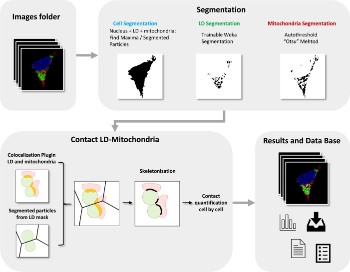
We have developed ContactJ, a macro script for the open-source image analysis software ImageJ. 8 , 9 This macro automatically and rapidly quantifies confocal images (1 section, 8-12-16 bit depth) that are saved in a folder and returns the table of the resulting measurements, images and Regions of Interests (ROIs) in a “Results” folder. Thus, inexperienced users with no prior image analysis experience will find it easy to use. As can be seen in Figure 1, the flowchart illustrates how the macro automatically detects and measures LD-mitochondria linear contacts by combining standard and machine learning segmentation processes and the novel use of colocalization together with skeletonization methods from a large number of fluorescence images.
Figure 1. Flowchart of ContactJ Macro that automatically segments and measures the LD-mitochondria contacts cell by cell by combining standard and machine learning segmentation processes and the novel use of colocalization together with skeletonization methods from a large number of fluorescence images.
LD, Mitochondria, Colocalization Area and final Contact Site are represented in green, red, yellow and black respectively.
First, ContactJ macro performs the segmentation of the cells, LD and mitochondria separately. Cell segmentation was adapted to the particular conditions of this experiment dataset where a membrane or a cell mask labelling couldn’t be implemented. Briefly, cell segmentation was achieved by adding the available cytoplasmic labelling (LD plus Mitochondria) and the Nucleus staining. To obtain similar intensity contributions from cytoplasmic labelling and nucleus, LD and mitochondria channels were multiplied each by 0.2 to reduce its intensity contribution and make it more similar to the nucleus intensity. As these factors are dependent on the different experimental conditions, they can be modified at the graphical user interface (GUI) at the beginning of the macro (referred at the GUI as Compensation).
Intensity compensated channels from mitochondria and LD were added to a binary mask from nuclei.
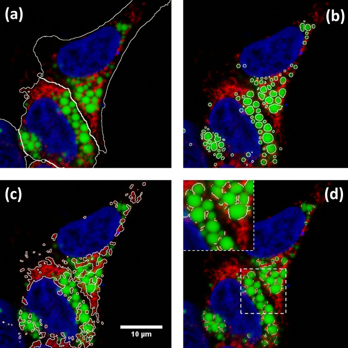
The resulting image was used to find local maxima (with prominence of 100) and to obtain subsequently the segmented particles binary image. Segmented particles limits were encoded as 0 value on the binarized image from the three added channels. The original 3 channel image was sequentially converted to RGB and to 8bit, median filtered (radius 2) and thresholded (Huang). After performing the minimum operation between the processed and thrsesholded original image and the segmented particles limits image, cells were individualized and stored as ROIs (see Figure 2a), by particle analysis. Cell ROIs were saved as zip file in the results folder.
LD segmentation was achieved through a Trainable Weka Segmentation classifier 10 on LD channel image (see Figure 2b) using the classification algorithm Fast Random Forest and the following Training features: Gaussian Blur, Hessian, Membrane projections, Sobel filtre, Difference of Gaussians. Mitochondria were segmented by intensity thresholding (autothreshold method “Otsu”) (see Figure 2c).
Figure 2. Results of ContactJ macro.
Hek293 cells were labelled with anti TOM20 antibody (mitochondria) in red, Bodipy493/505 (LDs) in green and DAPI (nuclei) in blue. The different regions of interest resulting from segmentation are highlighted in white (a) Cell Segmentation, (b) LD segmentation, (c) mitochondria segmentation and (d) LD-mitochondria contacts. Insert in d) shows a detail of how contact regions found by ContactJ are accurate and individualized per LD.
Once cells and LD were segmented, contact regions between mitochondria and LD were first obtained using the Colocalization plugin. 11 This plugin highlights the colocalized “contact” points between mitochondria and LD. The plugin generates an 8 bit binary image with only the colocalized points (Display value = 255). It analyses Mitochondria and LD channels, pixel by pixel, and considers a colocalized point if its respective intensities are strictly higher than the threshold determined for its channels (autothreshold methods “Yen” for LD channel and “Otsu” for mitochondria channel), and if its ratio (of intensity) is strictly higher than the ratio setting value (set to 50%: ratio (0-100%)). Secondly, the regions of individualized LD were obtained using the Find Maxima tool with Segmented particles result from the LD mask. Finally, individualized contact regions were converted to a contour line section by performing the skeletonization of the minimum image calculation from the colocalization mask and the segmented particles result from LD. Contact perimeter and contact counts (a contact is defined as a continuous contact line) were quantified, obtaining the linear LD-mitochondria contact of each cell (see Figure 2d).
Throughout the execution of the macro, all the data is stored in arrays (cell, LD and mitochondria areas and perimeters, contact perimeter, number of contacts, etc). Moreover, this data is stored in a.txt file allowing the traceability of the results for each cell and each image.
Operation
ImageJ/Fiji with the Colocalization 11 and WEKA 10 plugins should be installed and ContactJ run from ImageJ macro editor. The software can be tested with the sample data provided (in Underlying data 12 ). First, the user should prepare a set of images and organize them into a folder. In this images folder the user should create a subfolder named “Model” with the data and model files obtained specifically for the segmentation of LD channel using the machine learning WEKA plugin. Once ContactJ runs, macro asks to the user the folder to analyse. Automatically, ContactJ opens the images one by one analysing them, cell by cell, and saving ROIs and all the measurements data obtained (areas, intensity, contact, perimeter …) in a .txt file as a table.
Use cases
ContactJ has been developed and tested for the contact analysis of hundreds of HEK293 cells treated or untreated with lipopolysaccharide (LPS) and expressing or not a protein of interest PLIN5. 4
Taking advantage of fluorescence multilabelling, the cells have been segmented and all parameters can be expressed per cell individually. The macro segments the nucleus, LDs and mitochondria from each cell and it obtains the following parameters that are stored in a data base table: Cell Area, number of LDs and Mitochondria, LDs and Mitochondria Total Area, Mean LD Area per cell, Standard deviation of the LD mean Area, Mean LD Perimeter and Total Mitochondria and LD Perimeter
The main novelty and distinctive feature from ContactJ are that it creates a contact line corresponding to each contact site between mitochondrion and LD. In order to obtain the contact site, ContactJ first generates a colocalization region corresponding to the overlapping fluorescence from both organelles, using the Colocalization plugin. Then, this shape is skeletonized generating a line of equidistant points to its boundaries representing the contact site. The macro stores in the data base file also the total length of the contact sites, the mean length of each contact and the number of contacts detected per cell. In the mentioned work, the results were expressed as Total Contact Length/Cell and compared between cell populations and treatments. 4
Discussion
One of the innovative and distinctive features of ContactJ is that it creates a linear contact region on the mid plane of the LD in close proximity to Mitochondrion, representing the most probable contact site between both organelles.
Although light microscopy resolution limit prevents assertion of true interaction, the analysis of inter organelle contacts by fluorescence microscopy is accepted as an indicator of possible communication between these two organelles, bringing many advantages when performing contact analysis at a high scale of samples and conditions. TEM is used to measure contacts between organelles as it resolves at the membrane scale where these contacts take place, as can be seen in Figure 3(a). ContactJ measurements of contact perimeter between LD and mitochondria are 2-3 times bigger compared to TEM measurements 4 ( Figure 3). The main reasons for this difference are, first, that 2D confocal microscopy image represents a projection of approximately 500nm sample thickness, compared to the 70nm of the ultra-thin TEM lamella and consequently, it is collecting a higher proportion of membrane and contacts. Secondly, the intrinsic difference in resolution would affect more directly the measurements of small contacts by light microscopy overestimating them. Therefore, the contacts obtained with ContactJ can be considered reliable compared to those observed by TEM.
Figure 3. (a) TEM image showing LD-mitochondria contacts indicated by arrows, (b) result of the LD-mitochondria contacts (in white) detected by ContactJ in fluorescence microscopy image in Hek293 cells labelled with anti TOM20 antibody (mitochondria) in red, Bodipy493/505 (LDs) in green and DAPI (nuclei) in blue; and (c) magnification of the square region of (b) showing the contacts in detail.
In order to test quality and performance of ContactJ, a comparison between contact manual tracing analysis compared to ContactJ analysis has been performed on 20 cells. Mean and Standard Deviation (microns) from total contact length from Manual quantification were: 44.9220 and 30.1140, respectively, and from ContactJ analysis were: 30.8055 and 18.30792, respectively. ANOVA test was performed and showed no significant difference between both groups means (F = 3.209 and p = 0.08) B). Correlation Analysis of the values obtained with both methods showed a correlation index of 0.899 significant at the 0.01 level (see extended data Manual_vs_ContactJ_methods.pdf).
The comparison between ContactJ analysis vs Manual analysis shows that although both methods correlate, manual analysis-increases significantly standard deviation reducing reproducibility of the method and overestimating contact length.
Moreover, while comparing both methods we have experienced that manual quantification doesn’t allow precise contact tracing around organelles, secondly, that it’s difficult to stablish visual criteria of proximity and finally, that manual tracing is not only time consuming but, after defining tens of contacts per cell, it results in quick user fatigue and loss of detection and precision. All these results reinforce the idea that ContactJ is an objective and reproducible method valid to define and quantify contacts between organelles automatically.
Furthermore, robustness of ContactJ has been tested and validated also in contact analysis between Endoplasmic Reticulum and Lipid Droplets in U2OS cells in live cell imaging experiments. 13
Obtaining contact regions, together with multiple morphological parameters of organelles, allows the calculation of descriptive statistics that would help describing cellular response. In front a metabolic or pathogenic event, cells need to regulate the transfer and communication between organelles. Among many possible cell reactions, they may change the contact surface between organelles together with the organelle size, number and distribution. For example, in this case, the quantification with ContactJ of the contact length and the LD perimeter would allow the calculation of the LD-Mitochondria “transfer or communication” efficiency for each cell (Contact length/LD Perimeter Length) helping in the comparison between cells response.
ContactJ application can be extended to the contact analysis between other organelles, in 2D, 3D or time series. In fact, this code has been already adapted for the contact tracking analysis between LD and Endoplasmic Reticulum in time lapse experiments. 13
To facilitate the applicability of ContactJ in 3D or time series images, and its combination with 3D objects analysis and objects tracking methods, a straightforward core code of ContactJ, named ContactJ4All, has been developed. 14 ContactJ4All is ready to be included and adapted to any workflow. It is available on github 14 and example datasets from xyz and xyt images are included to test its performance. 14
In conclusion, the described image analysis workflow unveils a wide range of possibilities in the automatic quantification of LD and mitochondria contacts and it also has been tested, and it is applicable, to the study of other organelles in 2D, 3D images or time series. Obtaining contact regions together with multiple cell and organelles parameters allow building descriptive statistics of the cells response. Moreover, its application in a large number of images enables the use of High Content Screening and Analysis, highly increasing the quality and statistical confidence of the results.
Data availability
Underlying data
Zenodo: UB-BioMedMicroscopy/ContactJ: ContactJ, https://doi.org/10.5281/zenodo.5810874 12
This project contains the trained model and data for Weka plugin and example images.
Extended data
Zenodo: UB-BioMedMicroscopy/ContactJ: ContactJ, https://doi.org/10.5281/zenodo.5810874 12
This project contains the comparison between manual and ContactJ Methods.
Zenodo: UB-BioMedMicroscopy/ContactJ4All: ContactJ4All, https://doi.org/10.5281/zenodo.5810614 14
This project contains the following extended data:
-
•
ContactJ4All xyz dataset
-
•
ContactJ4All xyt dataset
-
•
Processed movie of LD and Mitochondria contact analysis with ContactJ4All
Data are available under the terms of the Creative Commons Zero “No rights reserved” data waiver (CC0 1.0 Public domain dedication).
Software availability
Contact J source code available from: https://github.com/UB-BioMedMicroscopy/ContactJ
Archived source code as at time of publication: https://doi.org/10.5281/zenodo.5810874 12
License: CC0
ContactJ4All source code available from: https://github.com/UB-BioMedMicroscopy/ContactJ4All
Archived source code as at time of publication: https://doi.org/10.5281/zenodo.5810614 14
License: CC0
Acknowledgements
This publication was supported by COST Action NEUBIAS (CA15124), funded by COST (European Cooperation in Science and Technology).
We acknowledge NEUBIAS Cost Action NEUBIAS - COST ActionCA15124 www.neubias.org for Image Analysis courses, tutorials and repositories and their important task in training Image Analysts.
Data presented in this work is part of the published article. 4
This paper is dedicated to the living memory of Anna Bosch.
Funding Statement
This work was supported by the European Cooperation in Science and Technology [CA15124].
[version 2; peer review: 2 approved]
References
- 1. Bosch M, Parton RG, Pol A: Lipid Droplets, Bioenergetic Fluxes, and Metabolic Flexibility. Semin Cell Dev Biol. Elsevier Ltd;December 2020;33–46. 10.1016/j.semcdb.2020.02.010 [DOI] [PubMed] [Google Scholar]
- 2. Parton RG, Bosch M, Steiner B, et al. : Novel Contact Sites between Lipid Droplets, Early Endosomes, and the Endoplasmic Reticulum. J Lipid Res. American Society for Biochemistry and Molecular Biology Inc;November 1, 2020; p1364. 10.1194/jlr.ILR120000876 [DOI] [PMC free article] [PubMed] [Google Scholar]
- 3. Schuldiner M, Bohnert M: A Different Kind of Love – Lipid Droplet Contact Sites. Biochim Biophys Acta Mol Cell Biol Lipids. Elsevier B.V;October 2017;1188–1196. 10.1016/j.bbalip.2017.06.005 [DOI] [PubMed] [Google Scholar]
- 4. Bosch M, Sánchez-Álvarez M, Fajardo A, et al. : Mammalian Lipid Droplets Are Innate Immune Hubs Integrating Cell Metabolism and Host Defense. Science (80-.). 2020;370(6514). 10.1126/science.aay8085 [DOI] [PubMed] [Google Scholar]
- 5. Herms A, Bosch M, Reddy BJN, et al. : Activation Promotes Lipid Droplet Dispersion on Detyrosinated Microtubules to Increase Mitochondrial Fatty Acid Oxidation. Nat. Commun. 2015;6. 10.1038/ncomms8176 [DOI] [PMC free article] [PubMed] [Google Scholar]
- 6. Valm AM, Cohen S, Legant WR, et al. : Applying Systems-Level Spectral Imaging and Analysis to Reveal the Organelle Interactome. Nature. Nature Publishing Group;June 1, 2017;162–167. 10.1038/nature22369 [DOI] [PMC free article] [PubMed] [Google Scholar]
- 7. Freyre CAC, Rauher PC, Ejsing CS, et al. : MIGA2 Links Mitochondria, the ER, and Lipid Droplets and Promotes De Novo Lipogenesis in Adipocytes. Mol. Cell. 2019;76:811–825. e14. 10.1016/j.molcel.2019.09.011 [DOI] [PubMed] [Google Scholar]
- 8. Schindelin J, Arganda-Carreras I, Frise E, et al. : Fiji: An Open-Source Platform for Biological-Image Analysis. Nat Methods. July 2012;676–682. 10.1038/nmeth.2019 [DOI] [PMC free article] [PubMed] [Google Scholar]
- 9. Schneider CA, Rasband WS, Eliceiri KW: NIH Image to ImageJ: 25 Years of Image Analysis. Nat Methods. July 2012;671–675. 10.1038/nmeth.2089 [DOI] [PMC free article] [PubMed] [Google Scholar]
- 10. Arganda-Carreras I, Kaynig V, Rueden C, et al. : Trainable Weka Segmentation: A Machine Learning Tool for Microscopy Pixel Classification. Bioinformatics. 2017;33(15):2424–2426. 10.1093/bioinformatics/btx180 [DOI] [PubMed] [Google Scholar]
- 11. Pierre Bourdoncle (Institut Jacques Monod, Service Imagerie, P: Colocalization Plugin. Reference Source
- 12. MariaCalvo& GemmaMar: UB-BioMedMicroscopy/ContactJ: ContactJ (v5.0.0). Zenodo. 2021. 10.5281/zenodo.5810874 [DOI]
- 13. Lu A, Hsieh F, Sharma BR, et al. : CRISPR screens for lipid regulators reveal a role for ER-bound SNX13 in lysosomal cholesterol export. J Cell Biol. 2022;221(2):e202105060. 10.1083/jcb.202105060 [DOI] [PMC free article] [PubMed] [Google Scholar]
- 14. MariaCalvo: UB-BioMedMicroscopy/ContactJ4All: ContactJ4All (v1.1.0). Zenodo. 2021. 10.5281/zenodo.5810614 [DOI]


