

Abstract
Objective:
To investigate condylar symmetry and condyle fossa relationships in subjects with functional posterior crossbite comparing findings before and after rapid maxillary expansion (RME) treatment through low-dose computed tomography (CT).
Materials and Methods:
Twenty-six patients (14 girls and 12 boys, mean age 9.6 ± 1.4 years) with functional posterior crossbite (FPXB) diagnosis underwent rapid palatal expansion with a Hyrax appliance. Patients' temporomandibular joints (TMJ) underwent multislice CT scans before rapid palatal expansion (T0) and after (T1). Joint spaces were compared with those of a control sample of 13 subjects (7 girls and 5 boys, mean age 11 ± 0.6 years).
Results:
Anterior space (AS), superior space (SS), and posterior space (PS) joint space measurements at T0 between the FPXB side and contralateral side demonstrated no statistically significant differences. After RME treatment (T1), all three joint spaces increased on both the FPXB side and the non-crossbite side. However, differences were statistically significant only for the SS when comparing the two sides at T1. SS increased more than AS and PS in the non-crossbite condyle (0.28 mm) and FPXB condyle (0.37 mm), and PS increased only on the FPXB side (0.34 mm).
Conclusions:
There were no statistically significant differences in condyle position within the glenoid fossa between the FPXB and non-crossbite side before treatment. Increases in joint spaces were observed after treatment with RME on both sides. These changes were, however, of small amounts.
Keywords: Computed tomography, Crossbite, Condyle fossa, TMJ
INTRODUCTION
Posterior crossbite is one of the most frequently occurring malocclusions in deciduous and mixed dentitions, with a reported prevalence of 7% to 23%.1–9 The most common form of posterior crossbite is a unilateral presentation, with a functional shift of the mandible toward the crossbite side.10 This condition is known as functional posterior crossbite (FPXB).11 Because crossbite develops early and has a low rate of spontaneous correction,10 an early treatment12–21 of this malocclusion has been recommended.
Interest in literature in FPXB has grown not only with regard to the choice of timing for treatment but also for an eventual association with an asymmetrical mandible growth alteration and for the presence of an asymmetric condyle position within the glenoid fossa. In this respect, it has been suggested that FPXB may result in right-to-left-side differences in the condyle fossa relationship, which are responsible for temporomandibular joint (TMJ) disc displacement and internal derangement.22 According to some authors,11,21,23 this different relationship seemed to be corrected after treatment of FPXB with an establishment of a more symmetric position of both condyles. On the contrary, some other studies reported no differences in condylar position between the crossbite and non-crossbite sides and before and after treatment.8,19
Presumably, the main reasons for such controversies may be ascribed to the radiographic technique used to investigate the condyle fossa relationship, which have been mainly based on transcranial radiographs or tomograms. An earlier developed imaging technology for the evaluation of the condyle fossa relationship is computed tomography (CT). This method overcomes the methodological drawbacks of previous techniques; in fact, data obtained from CT have been shown to delineate the joint structures with higher accuracy.24,25
Accordingly, the purposes of this prospective study were to investigate via low-dose computed tomography the condyle fossa relationships in subjects with FPXB before (T0) and after treatment (T1) with rapid maxillary expansion (RME) and to compare findings at T0 and T1.
MATERIALS AND METHODS
The sample consisted of 39 subjects aged 8.2 to 11.6 years in mixed dentition from the Department of Orthodontics, Faculty of Dentistry, Catania University, Italy, and the Department of Orthodontics, Faculty of Dentistry, La Sapienza, Rome University, Italy. The patient group (n = 26 patients) included 14 girls and 12 boys (mean age, 9.6 ± 1.4 years) diagnosed with FPXB and Angle Class I malocclusion.
To be included in this study, all patients had to have at the start of the treatment a transverse maxillary deficiency and a crossbite on one side only, with the mandibular midline shifted toward the crossbite site at the maximum intercuspal position and not at mouth opening, indicating an FPXB. The mandibular midline shift was 2 ± 1 mm. Exclusion criteria included (1) developmental or acquired craniofacial deformities involving condyles and/or mandible; (2) no systemic diseases; (3) no history of orthodontic treatment; (4) no anterior crossbite; (5) no signs or symptoms of temporomandibular disorder, according to the research diagnostic criteria for TMJ disorders26; (6) no missing teeth, excluding the third molars; and (7) no carious lesions, extensive restorations, or pathologic periodontal status.
The control group included 13 subjects, 7 girls and 5 boys (mean age, 11 ± 0.6 years), with an Angle Class I relationship and one palatally displaced canine that needed surgical exposure. The exclusion criteria were the same as the patient group, plus the absence of FPXB. All procedures were explained to the patients and their parents. Informed consent forms were obtained, and institutional approval was granted to conduct the study.
A Hyrax palatal expander was used for each subject of the patient group, and the activation protocol required the screw to be turned three times per day (0.25 mm per turn) for an average of 18 days, for all subjects.
Multislice CT scans were performed before rapid palatal expansion (time T0) and again at the end of the active expansion phase (time T1) without removing the expander. The CT scans were carried out by a trained radiographer at the same scanner console with the primary indication of evaluating buccal bone of maxillary posterior teeth in the FPXB group and to assess the canine displacement in the control group.
A low-dose CT scan protocol was used, as previously described.27,28 Briefly, patients were examined with a multidetector helical CT scanner (Lightspeed Ultra, GE Medical Systems, Giles, UK). The scanning parameters were 80 kV, 10 mA (low dose), 0.625 mm collimation, pitch 1, and gantry tilt 0°. Multiplanar reformation and 3D postprocessing were performed on a workstation (Advantage Windows 4.1, GE Medical Systems).
The CT images were obtained with the patients in maximum dental intercuspation. Patients were scanned in the supine position with shoulder rests, as well as having their head positioned with a Camper's plane perpendicular to the ground.
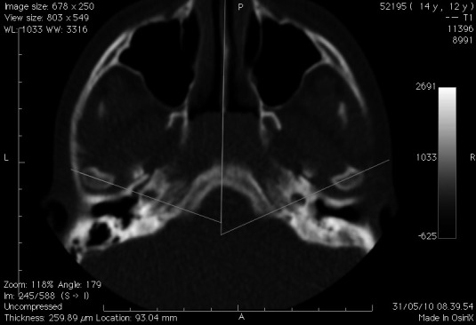
The data of each patient were reconstructed with 0.5-mm slice thickness and saved as DICOM (Digital Imaging and COmmunications in Medicine) files. The data were then transferred to a workstation (Mac Pro Quad 2.66 GHz, Apple, Cupertino, Calif) and visualized by using the OsiriX medical imaging software program, which allowed the reorientation of the CT images and then performance of measurements (Open-Source, OsiriX Medical Imaging Software, www.osirix-viewer.com). In fact, to minimize eventual measurement errors due to the absence of a cephalostat, the 3D images were reoriented according to two references planes: NFZ (a frontal plane passing through the two frontozygomatic sutures at the inner rim of the orbit and nasion) and the Frankfurt plane, as described recently by Cho.29 Afterward, the long axis of the condyle was determined on axial section (Figure 1a), and a vertical plane bisecting the long axis was defined as the sagittal section (Figure 1b).24 Linear measurements were made of the anterior joint space (AS), superior joint space (SS), and posterior joint space (PS) using landmarks and variables shown in Figure 2. Furthermore, the angle between the long axis of the mandibular condylar process and the midsagittal plane (MSP) was assessed on the axial plane (Figure 3). Linear and angular measurements were taken to the nearest 0.01 mm and 0.01°.
Figure 1.

The long axis of the condyle is determined on the axial section (a), and a vertical plane bisecting the long axis was defined as the sagittal section (b).
Figure 2.

Landmarks and linear measurements of the space between the condyle and glenoid fossa. A horizontal line parallel to FH and drawn through the most superior point of the fossa is used as a reference plane. Lines tangent to the most prominent anterior and posterior aspect of the condyle are drawn. The distance from the most superior condyle point is measured as superior joint space (SS). Distances from the anterior and posterior tangent points to the glenoid fossa were measured as the anterior joint space (AS) and posterior joint space (PS).
Figure 3.
Angular measurement to evaluate the mandibular condyle relative to the cranium. The midsagittal plane is constructed by using the best-fit line connecting opistion, basion, and odontoid. The angle between the long axis of the mandibular condyle process and the midsagittal plane is measured.
Statistical Analysis
Every measurement was made twice by the same blinded observer with a 2-week interval between the first and second reading, thus eliminating interobserver error. The average value of the first and second readings was used, as recommended by Baumrind and Frantz.30
One-way group analysis of variance (ANOVA), consisting of a between main factor group with three levels (FPXB sides vs non-FPXB sides vs control), was used to compare AS, SS, and PS between groups FPXB, non-FPXB, and controls.
Descriptive statistics (mean, standard deviation) were calculated separately on the FPXB side condyle, non-crossbite side condyle, and control condyles. In the patient group, calculations were made for each period, T0 (before the treatment) and T1 (after the treatment), and for each side.
Student's paired t-test was carried out to evaluate the differences between the two sides before (T0) and after (T1) treatment with RME and within the same side (both for crossbite and non crossbite). Moreover, the 95% confidence interval was evaluated to describe the best possible interval at which the true values of the measurements could be expected to be found.
Three levels of statistical significance were considered: P < .05 (*), P < .01 (**), and P < .001 (***). Data were analyzed using SPSS for EX Windows, version 16.0 (SPSS Inc, Chicago, Ill).
RESULTS
There was an even distribution of crossbite sides in the patient group: 12 patients had left FPXB and 14 had right FPXB. Sex differences regarding condyle fossa relationship were not statistically significant (P > .05); therefore, data from girls and boys were grouped together. A total of 52 low-dose TMJ CTs were studied in the patient group: 26 TMJs for the FPXB side and 26 TMJs for the non-crossbite side.
In TMJ CT scans of the control group, there were no statistically significant differences (P > .05) for joint spaces between the two sides; accordingly, data from the two joints were tabulated together. Thus, a total of 26 TMJs comprised the control group.
Before treatment (T0), when comparing condyle joint spaces from the control group, FPXB side and non-FPXB contralateral condyles (Tables 1–3), no statistically significant difference was observed for SS and PS among FPXB, non-crossbite, and control TMJs before maxillary expansion (Tables 2 and 3), whereas ANOVA values for AS were statistically significant (P < .01; Table 1). On the other hand, Student's paired t-test values between these recordings were not statistically significant (Table 4).
Table 1.
Analysis of Variance for Anterior Space Measurements Between the Groups Functional Posterior Crossbite, Non–functional Posterior Crossbite, and Controls at Time T0

Table 2.
Analysis of Variance for Superior Space Measurements Between the Groups Functional Posterior Crossbite, Non–functional Posterior Crossbite, and Controls at Time T0

Table 3.
Analysis of Variance for Posterior Space Measurements Between the Groups Functional Posterior Crossbite, Non–functional Posterior Crossbite, and Controls at Time T0

Table 4.
Statistical Analysis for Crossbite vs Non-crossbite Before Treatment (T0)a

After RME treatment (T1), all three joint spaces increased both in the FPXB side and in the non-FPXB side. However, differences between the two sides, also in this instance, were of small amount and were statistically significant only for SS (Tables 5 and 6).
Table 5.
Statistical Analysis for Crossbite vs Non–crossbite After Treatment (T1)a

Table 6.
Statistical Analysis for Non-crossbite Side Before (T0) and After Treatment (T1)a

The increased three joint spaces observed after RME treatment (T1) both on the FPXB and non-FPXB sides (Tables 6 and 7) were of a different extent. In fact, when comparing measurements at T0 and T1, SS increased more than AS and PS, both in non-crossbite condyle (0.28 mm; Table 6) and in FPXB condyle (0.37 mm; Table 7), and PS increased just in the FPXB side (0.34 mm). Almost all of these differences were statistically significant (P < .01 and P < .001). It is interesting to note that the standard deviations of the relative condylar position at both T0 and T1 were also slight, indicating narrow variation in condylar positions in both sides of the study group.
Table 7.
Statistical Analysis for Crossbite Side Before (T0) and After Treatment (T1)a

No statistically significant differences were observed when comparing MSP∧ between the FPXB sides to the non-crossbite side both at T0 and at T1 and T1 versus T0 (Tables 4–7).
DISCUSSION
Actually, CT imaging has been shown to be the ideal tool for TMJ assessment. Here, for the first time, low-dose CT was used to evaluate condylar position in patients affected by FPXB, before and after treatment. The axial slice was used to assess the symmetry between the condyles in the anteroposterior and mediolateral aspects because it shows both condyles in the same image and allows the determination of reference planes such as the median sagittal plane. In fact, all attempts were made to select pretreatment and posttreatment CT scan slices of similar cut depth to minimize bias due to the documented variation in condylar position between adjacent CT sections. On the other hand, the sagittal slice was used for assessing the condyle fossa relationship, allowing the analysis of condylar concentricity.31 Furthermore, we also focused on the central cuts of condyles because landmark identification is difficult outside this area because of the anatomy of the glenoid fossa.24
The present study demonstrated that there were no statistically significant differences in condyle position within the glenoid fossa between the FPXB and non-FPXB side before treatment. Any positional asymmetry of the mandible was found in relation to the cranial floor (based on axial scan), as there was no significant difference between the MSP∧ values for the right and left sides.
Measurements of the joint spaces (AS, SS, and PS) in our sample of patients affected by FPXB were very close to the control sample and to values reported for healthy subjects obtained with limited cone-beam CT.24 The SS distance was greatest both in the patients and control group, followed by PS and AS; this is in agreement with data on the concentric position of the mandibular condyle in the glenoid fossa. All three joint spaces increased, however, after treatment with RME on both sides; these changes, even if statistically significant, were of small amounts (at most 0.37 mm). This symmetric condyle–glenoid fossa relationship, recorded in both condyles before treatment, could be explained by compensatory condyle fossa remodeling and/or variation in thickness of the articular TMJ disc.23,32 In fact, it has been suggested that the TMJ disc has the ability to adapt to any alteration caused by occlusal changes occurring in the space between the condyle and fossa.32 The very slight asymmetric condylar position recorded in our sample following treatment, on the other hand, may be explained by the rapid changes of occlusal contacts between the mandibular and maxillary arch after RME therapy. In fact, our study describes early treatment changes in the condyle position within the glenoid fossa, with CT scans obtained after the active phase of maxillary expansion and not following the fixed orthodontic appliance.
Therefore, the beliefs that children with functional unilateral posterior crossbite are characterized by an asymmetric pretreatment condylar position (with the non-crossbite side condyle in an anterior and inferior position in the glenoid fossa) and that a more symmetric condylar relationship is established after maxillary expansion are not confirmed in the present study.
For the sake of clarity, it should be underlined that these beliefs were based on studies that documented condylar position with transcranial radiographs11,21 and tomography,23 but in this latter instance, not every patient but rather few patients had a submental vertex radiograph to determine which angulation and depth of the linear section would pass through the center of the condyle. On the other hand, we used, as in some other studies, a multidetector helical CT scanner with patients in the supine position, which somehow could have affected results. In this respect, the use of a maximum intercuspation wax bite is advised for future studies. Furthermore, ours is a short-term study, and therefore, any information on the stability of mandible positional changes should be obtained in a longer-term study.
All in all, our results corroborate a previous study19 that showed, with horizontal corrected tomograms, no differences in condylar position between the crossbite and non-crossbite side before and after treatment with a quad helix appliance.
CONCLUSIONS
Young patients with FPXB are characterized by a pretreatment symmetric condylar position.
After RME treatment, the three joint spaces somehow increased, but the symmetric condyle–glenoid fossa relationship persisted.
REFERENCES
- 1.Borzabadi-Farahani A, Eslamipour F. Malocclusion and occlusal traits in an urban Iranian population: an epidemiological study of 11- to 14-year-old children. Eur J Orthod. 2009;31:477–484. doi: 10.1093/ejo/cjp031. [DOI] [PubMed] [Google Scholar]
- 2.da Silva Filho O. G, Santamaria M, Jr, Capelozza Filho L. Epidemiology of posterior crossbite in the primary dentition. J Clin Pediatr Dent. 2007;32:73–78. doi: 10.17796/jcpd.32.1.h53g027713432102. [DOI] [PubMed] [Google Scholar]
- 3.Poveda Roda R, Bagan J. V, Diaz Fernandez J. M, Hernandez Bazan S, Jimenez Soriano Y. Review of temporomandibular joint pathology. Part I: classification, epidemiology and risk factors. Med Oral Patol Oral Cir Bucal. 2007;12:E292–E298. [PubMed] [Google Scholar]
- 4.Scavone H, Jr, Ferreira R. I, Mendes T. E, Ferreira F. V. Prevalence of posterior crossbite among pacifier users: a study in the deciduous dentition. Braz Oral Res. 2007;21:153–158. doi: 10.1590/s1806-83242007000200010. [DOI] [PubMed] [Google Scholar]
- 5.daCosta O. O, Orenuga O. O. Dentofacial anomalies related to the digit sucking habit. Afr J Med Med Sci. 2002;31:239–242. [PubMed] [Google Scholar]
- 6.Thilander B, Pena L, Infante C, Parada S. S, de Mayorga C. Prevalence of malocclusion and orthodontic treatment need in children and adolescents in Bogota, Colombia: an epidemiological study related to different stages of dental development. Eur J Orthod. 2001;23:153–167. doi: 10.1093/ejo/23.2.153. [DOI] [PubMed] [Google Scholar]
- 7.Sonnesen L, Bakke M, Solow B. Malocclusion traits and symptoms and signs of temporomandibular disorders in children with severe malocclusion. Eur J Orthod. 1998;20:543–559. doi: 10.1093/ejo/20.5.543. [DOI] [PubMed] [Google Scholar]
- 8.Brin I, Ben-Bassat Y, Blustein Y, et al. Skeletal and functional effects of treatment for unilateral posterior crossbite. Am J Orthod Dentofacial Orthop. 1996;109:173–179. doi: 10.1016/s0889-5406(96)70178-6. [DOI] [PubMed] [Google Scholar]
- 9.Keeling S. D, McGorray S, Wheeler T. T, King G. J. Risk factors associated with temporomandibular joint sounds in children 6 to 12 years of age. Am J Orthod Dentofacial Orthop. 1994;105:279–287. doi: 10.1016/S0889-5406(94)70122-9. [DOI] [PubMed] [Google Scholar]
- 10.Kennedy D. B, Osepchook M. Unilateral posterior crossbite with mandibular shift: a review. J Can Dent Assoc. 2005;71:569–573. [PubMed] [Google Scholar]
- 11.Kecik D, Kocadereli I, Saatci I. Evaluation of the treatment changes of functional posterior crossbite in the mixed dentition. Am J Orthod Dentofacial Orthop. 2007;131:202–215. doi: 10.1016/j.ajodo.2005.03.030. [DOI] [PubMed] [Google Scholar]
- 12.Petren S, Bondemark L. Correction of unilateral posterior crossbite in the mixed dentition: a randomized controlled trial. Am J Orthod Dentofacial Orthop. 2008;133:790.e7–790e13. doi: 10.1016/j.ajodo.2007.11.021. [DOI] [PubMed] [Google Scholar]
- 13.Vitral R. W, Fraga M. R, de Oliveira R. S, de Andrade Vitral J. C. Temporomandibular joint alterations after correction of a unilateral posterior crossbite in a mixed-dentition patient: a computed tomography study. Am J Orthod Dentofacial Orthop. 2007;132:395–399. doi: 10.1016/j.ajodo.2005.12.033. [DOI] [PubMed] [Google Scholar]
- 14.Enoki C, Valera F. C, Lessa F. C, et al. Effect of rapid maxillary expansion on the dimension of the nasal cavity and on nasal air resistance. Int J Pediatr Otorhinolaryngol. 2006;70:1225–1230. doi: 10.1016/j.ijporl.2005.12.019. [DOI] [PubMed] [Google Scholar]
- 15.Gu Y. Prediction of treatment outcome following correction of anterior crossbites in the mixed dentition: orthodontic versus orthopaedic methods. Aust Orthod J. 2005;21:25–30. [PubMed] [Google Scholar]
- 16.Flores-Mir C. Grinding is effective in early orthodontic treatment of unilateral posterior crossbite. Evid Based Dent. 2005;6:24. doi: 10.1038/sj.ebd.6400315. [DOI] [PubMed] [Google Scholar]
- 17.Petren S, Bondemark L, Soderfeldt B. A systematic review concerning early orthodontic treatment of unilateral posterior crossbite. Angle Orthod. 2003;73:588–596. doi: 10.1043/0003-3219(2003)073<0588:ASRCEO>2.0.CO;2. [DOI] [PubMed] [Google Scholar]
- 18.Harrison J. E, Ashby D. Orthodontic treatment for posterior crossbites. Cochrane Database Syst Rev. 2001:CD000979. doi: 10.1002/14651858.CD000979. [DOI] [PubMed] [Google Scholar]
- 19.Lam P. H, Sadowsky C, Omerza F. Mandibular asymmetry and condylar position in children with unilateral posterior crossbite. Am J Orthod Dentofacial Orthop. 1999;115:569–575. doi: 10.1016/s0889-5406(99)70282-9. [DOI] [PubMed] [Google Scholar]
- 20.de Boer M, Steenks M. H. Functional unilateral posterior crossbite: orthodontic and functional aspects. J Oral Rehabil. 1997;24:614–623. doi: 10.1046/j.1365-2842.1997.00633.x. [DOI] [PubMed] [Google Scholar]
- 21.Myers D. R, Barenie J. T, Bell R. A, Williamson E. H. Condylar position in children with functional posterior crossbites: before and after crossbite correction. Pediatr Dent. 1980;2:190–194. [PubMed] [Google Scholar]
- 22.Egermark I, Magnusson T, Carlsson G. E. A 20-year follow-up of signs and symptoms of temporomandibular disorders and malocclusions in subjects with and without orthodontic treatment in childhood. Angle Orthod. 2003;73:109–115. doi: 10.1043/0003-3219(2003)73<109:AYFOSA>2.0.CO;2. [DOI] [PubMed] [Google Scholar]
- 23.Hesse K. L, Artun J, Joondeph D. R, Kennedy D. B. Changes in condylar postition and occlusion associated with maxillary expansion for correction of functional unilateral posterior crossbite. Am J Orthod Dentofacial Orthop. 1997;111:410–418. doi: 10.1016/s0889-5406(97)80023-6. [DOI] [PubMed] [Google Scholar]
- 24.Ikeda K, Kawamura A. Assessment of optimal condylar position with limited cone-beam computed tomography. Am J Orthod Dentofacial Orthop. 2009;135:495–501. doi: 10.1016/j.ajodo.2007.05.021. [DOI] [PubMed] [Google Scholar]
- 25.Rodrigues A. F, Fraga M. R, Vitral R. W. Computed tomography evaluation of the temporomandibular joint in Class II Division 1 and Class III malocclusion patients: condylar symmetry and condyle-fossa relationship. Am J Orthod Dentofacial Orthop. 2009;136:199–206. doi: 10.1016/j.ajodo.2007.07.033. [DOI] [PubMed] [Google Scholar]
- 26.Dworkin S. F, Huggins K. H, Wilson L, et al. A randomized clinical trial using research diagnostic criteria for temporomandibular disorders-axis II to target clinic cases for a tailored self-care TMD treatment program. J Orofac Pain. 2002;16:48–63. [PubMed] [Google Scholar]
- 27.Leonardi R, Cutrera A, Barbato E. Rapid maxillary expansion affects the spheno-occipital synchondrosis in youngsters: a study with low-dose computed tomography. Angle Orthod. 2010;80:106–110. doi: 10.2319/012709-56.1. [DOI] [PMC free article] [PubMed] [Google Scholar]
- 28.Leonardi R, Sicurezza E, Cutrera A, Barbato E. Early post-treatment changes of circumaxillary sutures in young patients treated with rapid maxillary expansion. Angle Orthod. 2011;81:38–43. doi: 10.2319/050910-250.1. [DOI] [PMC free article] [PubMed] [Google Scholar]
- 29.Cho H. J. A three-dimensional cephalometric analysis. J Clin Orthod. 2009;43:235–252. [PubMed] [Google Scholar]
- 30.Baumrind S, Frantz R. C. The reliability of head film measurements. 2. Conventional angular and linear measures. Am J Orthod. 1971;60:505–517. doi: 10.1016/0002-9416(71)90116-3. [DOI] [PubMed] [Google Scholar]
- 31.Rodrigues A. F, Fraga M. R, Vitral R. W. Computed tomography evaluation of the temporomandibular joint in Class I malocclusion patients: condylar symmetry and condyle-fossa relationship. Am J Orthod Dentofacial Orthop. 2009;136:192–198. doi: 10.1016/j.ajodo.2007.07.032. [DOI] [PubMed] [Google Scholar]
- 32.Wang M. Q, He J. J, Li G, Widmalm S. E. The effect of physiological nonbalanced occlusion on the thickness of the temporomandibular joint disc: a pilot autopsy study. J Prosthet Dent. 2008;99:148–152. doi: 10.1016/S0022-3913(08)60031-1. [DOI] [PubMed] [Google Scholar]