Abstract
Objective:
To identify and review the literature regarding the reliability of cervical vertebrae maturation (CVM) staging to predict the pubertal spurt.
Materials and Methods:
The selection criteria included cross-sectional and longitudinal descriptive studies in humans that evaluated qualitatively or quantitatively the accuracy and reproducibility of the CVM method on lateral cephalometric radiographs, as well as the correlation with a standard method established by hand-wrist radiographs.
Results:
The searches retrieved 343 unique citations. Twenty-three studies met the inclusion criteria. Six articles had moderate to high scores, while 17 of 23 had low scores. Analysis also showed a moderate to high statistically significant correlation between CVM and hand-wrist maturation methods. There was a moderate to high reproducibility of the CVM method, and only one specific study investigated the accuracy of the CVM index in detecting peak pubertal growth.
Conclusions:
This systematic review has shown that the studies on CVM method for radiographic assessment of skeletal maturation stages suffer from serious methodological failures. Better-designed studies with adequate accuracy, reproducibility, and correlation analysis, including studies with appropriate sensitivity-specificity analysis, should be performed.
Keywords: Skeletal maturation, Cervical vertebrae
INTRODUCTION
The issue of optimal timing for orthodontic treatment is linked intimately to the identification of periods of craniofacial growth that can contribute significantly to the treatment of patients with skeletal discrepancies.1,2 The use of radiographic analysis to estimate skeletal maturation stage is a widely used method for predicting the timing of pubertal growth and for estimating growth velocity and the proportion of growth remaining.3
The hand-wrist radiograph is considered4–7 to be the most standardized method of skeletal maturation assessment based upon time and the sequence of appearance of the carpal bones and certain ossification events. Skeletal maturity is generally determined using stages in the ossification of bones of the hand-wrist, because of the relationship between the overall horizontal and vertical facial growth velocity and skeletal maturity determined by hand-wrist methods3 or because of the quantity of different types of bone available,8–11 or by evaluating the ossification onset of the sesamoid.9 The usual means by which to assess the hand-wrist radiograph are the comparison atlas of Greulich and Pyle8 and Tanner et al.9 and the processes that use specific indicators that relate skeletal maturation to the pubertal growth curve, such as the methods described by Bowden10 and Fishman.11
There are some limitations in the interpretation of skeletal maturity from hand-wrist radiographs.3 The ossification sequence and timing of skeletal maturity within the hand-wrist area show polymorphism and sexual dimorphism, which can limit the clinical predictive use of this method.12,13 Moreover, there are concerns about the extra radiation exposure resulting from use of this method,14 and its use must be questioned if other comparable methods of assessment are available. Finally, events in the hand and wrist are indicators of the peak and the end of the pubertal growth spurt, but these events do not signal the onset of the pubertal growth spurt.15
The cervical vertebrae maturation (CVM) method was introduced by Lamparski16 for use in growth assessment, allowing skeletal age evaluation and eliminating the need for additional radiographic exposure since the vertebrae are already recorded in the lateral cephalogram taken as a pretreatment record.1,5,14,17,18 However, the reproducibility of the CVM method has been questioned.19
In spite of extensive research about CVM methods applied to skeletal maturity assessment during the past years, statistical and biological details about the relationship between vertebral shape and skeletal maturation are still missing.20
Systematic reviews are useful tools with which to obtain evidence-based clinical information.21 The literature is controversial regarding the CVM method in terms of its utility for determining skeletal maturity. The aim of this systematic review was to identify and qualify the evidence and methodology of those reports and to evaluate the following question: How reliable is CVM staging in terms of predicting the pubertal spurt?
MATERIALS AND METHODS
To identify potentially relevant studies reporting data related to maturational indexes, specifically CVM methods, detailed search strategies were developed and executed. The search also included appropriate changes in the vocabulary and followed each database's syntax rules. Citations to potentially relevant studies in journals, dissertations, and conference proceedings were located by searching the appropriate electronic database in an effort to minimize publication bias.
Table 1 shows the database search and outlines our search strategy. This electronic investigation was conducted through July 2010. We also searched by hand by checking the references of the retrieved articles to identify all possible articles to be included in this review. No language restriction was applied.
Table 1.
Electronic Database Searched and Search Strategy
The selection criteria for considering studies included in this systematic review (SR) were the following: (1) cross-sectional or longitudinal descriptive studies in humans that evaluated qualitatively or quantitatively an established CVM method on lateral cephalometric radiographs to determine skeletal maturation; (2) studies that used hand-wrist radiographs as the standard method when evaluating correlation or comparison with the CVM method; and (3) studies that evaluated the reproducibility of the CVM method. Studies with inadequate sample sizes (ie, cleft lip/palate patients or repeated samples) that introduced a new or additionally modified version of the CVM method, editor's summaries, reference-only works, patents, meeting abstracts, opinion articles, and reviews were excluded.
Initially, the titles and abstracts identified were reviewed. Duplicate articles appearing in more than one database search were considered only once. Each abstract was checked to determine whether it presented data related to patients' skeletal maturation stage, as assessed by the CVM method. Any investigation not fulfilling this criterion was excluded from further evaluation. If the reviewer could not decide on a study's eligibility by examining the title and abstract, its full text was retrieved.
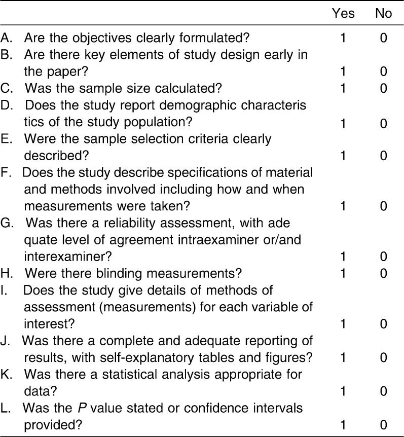
The full texts of remaining articles were retrieved for further evaluation in duplicate by two reviewers. In addition, to document the methodological soundness of each article, a quality assessment modified from the Strengthening the Reporting of Observational Studies in Epidemiology (STROBE),22 Standards for the Reporting of Diagnostic Accuracy Studies (SATRD),23 and Lagravere et al.24 was performed (Table 2). When two reviewers disagreed, a third investigator was called in, and consensus was reached.
Table 2.
Criteria for Assessing Quality Components in the Studies Included
One point was given to each criterion, if fulfilled. Quality assessment scores ranged from 0 to 12. The studies were classified as “low” (score 0 to 6), “moderate” (score 7 to 10), or “high” (score 11 to 12) according to this assessment. Studies with a low score (<7 points) were considered to be of poor methodological quality and were not considered at all in terms of the SR conclusions. Disagreements between the reviewers were resolved by reexamining the article in question, with discussion until both researchers were satisfied with the decision.
RESULTS
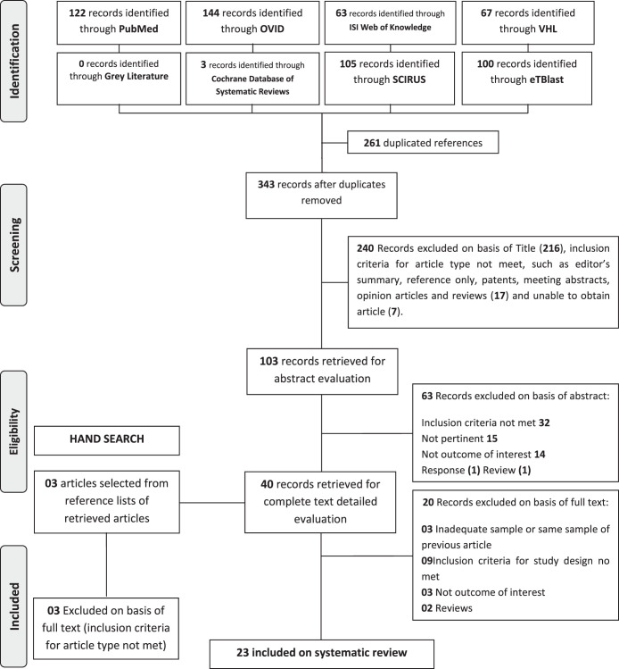
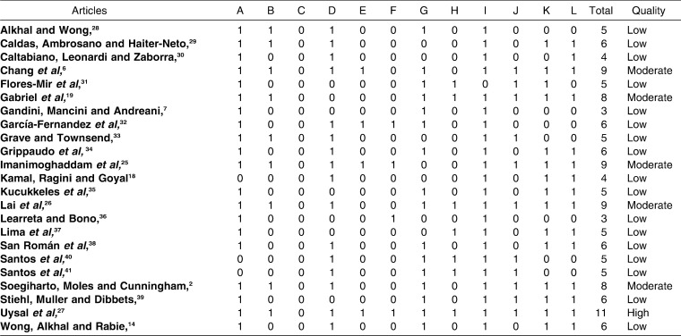
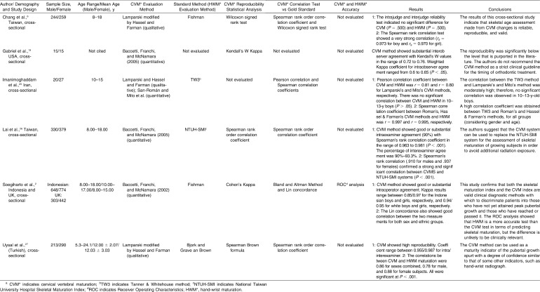
The electronic searches identified 343 records after duplicated references were removed. From these, 109 were selected for abstract evaluation. A total of 39 abstracts were retrieved for complete text detailed evaluation, including two articles added from hand searching. Ultimately, our search yielded 23 articles that met the inclusion criteria (Figure 1). Once the articles were selected, we systematically assigned a methodological score to each study in order to characterize them as useful or not (Table 3). Based on quality assessment, we found that six studies2,6,19,25–27 were particularly useful, with a moderate to high score. As stated above, 17 studies4,7,14,28–41 with a low score (<6.5 points) were considered to be of low methodological quality and were not particularly useful. The characteristics of moderate- and high-quality studies are presented in Table 4.
Figure 1.
Flow diagram of literature search.
Table 3.
Quality Assessment of Selected Full Text Article
Table 4.
Characteristics of Moderate- and High-Quality Included Studiesa
DISCUSSION
One objective of orthodontic treatment in adolescents is to take advantage of potential skeletal growth to treat skeletal discrepancies. Sexual maturation characteristics, chronologic age, dental development, height, weight, and skeletal development are some of the more common methods that have been used to identify stages of growth. Peak growth velocity in standing height is the most valid representation of rate of overall skeletal growth. It forms a useful historic longitudinal measure of an individual's growth pattern but has little predictive value in terms of future growth rate or percentage of total growth remaining. On the other hand, skeletal maturation staging from radiographic analysis is a widely used approach to predict timing of pubertal growth, to estimate growth velocity, and to estimate the proportion of growth remaining.3
According to a previous study,15 skeletal age offers no value over chronological age, either in assessing or predicting the time of pubertal growth. On the other hand, the hand-wrist radiograph is considered to be the most standardized method of skeletal assessment.4,7,18,27 Although some studies4,12,13 stated that the use of hand-wrist radiographs to predict growth spurt is not sufficiently accurate to be of value in clinical orthodontics, the validity of skeletal maturity assessment using the hand-wrist radiographs has been observed.2,3 However, to avoid taking additional radiographs, it is relevant to relate maturational stages to skeletal features other than the bones in the hand and wrist.
The cervical vertebrae are already shown on the lateral cephalogram film taken as a pretreatment record, and it is well known that the lateral view of the cervical vertebrae bodies changes with growth. In recent years, evaluation of the cervical vertebrae has been increasingly used to determine skeletal maturity.1,5,14,17,18
None of the studies included in this SR could be used for a meta-analysis because of the different methods of CVM and hand-wrist maturation (HWM) evaluation applied. The study of Imanimoghaddam et al.25 showed different correlation levels between four different CVM methods and the same HWM (TW3 method) in a unique sample. Thus, accuracy, correlation, and reproducibility may be influenced by the method.
There are a great variety of CVM methods, including simple qualitative analysis of the vertebral shape and size5,16 to quantitative measurements of vertebral shape (some of which are limited to height and width distances and ratios) and the depth of the inferior concavity1,42 and to other, more specific measurements, rendered through geometric morphometric analysis20 or linear regression formulae.40,43
Caution should be taken in the application of the results presented in this SR. Most of the studies investigating the use of cervical vertebral radiographic analysis of skeletal maturation did not report any kind of randomization, blinding, and sample size calculation. Articles that presented methodological deficiencies, which would likely compromise the interpretation of reported results, were excluded from this study. We found 23 articles that met the inclusion criteria,2,6,7,14,18,19,25–42 but only six2,6,19,25–27 were considered to be of moderate to high methodological quality.
Initially, the research on the direct relationship between cervical vertebral anatomical alterations and facial growth was limited because of the problems of longitudinal radiographic recordings, although there are a few longitudinal databases that can be accessed that have hand-wrist and lateral cephalometric radiographs taken simultaneously.17 All of the moderate- to high-ranked studies selected in this review were based on cross-sectional data, and these studies' designs have inherent limitations in terms of analyzing growth. Cross-sectional sampling is relatively insensitive to the individual variability seen easily in a longitudinal sample. In the study of growth and development, longitudinal research is an essential method for the detailed study of craniofacial growth, which can determine a patient's unique development type and make possible continuous comparison.
According to Soegiharto et al.,2 studies of this type should be longitudinal, but the difficulties of obtaining a large sample size and the associated increase in the number of radiographic exposures tend to preclude this methodology. With regard to randomization, only the studies of Chang et al.6 and Gabriel et al.19 superficially mentioned that the sample records analyzed were randomly selected. However, the specific criteria for randomization were not described in their articles.
Only the article of Uysal et al.27 established rigorous sample selection criteria, taking into account confounding factors such as lack of relative medical history, ethnicity, systematic diseases, medical syndromes, and hormonal disorders. Considering the influence of these co-factors on general growth and development, we judge that their rigorous criterion is a pleasant surprise that should be mimicked in future related studies.
The validity of CVM in the evaluation of craniofacial growth has been questioned. According to Gabriel et al.,19 most of the studies that cited high-reproducibility results for the CVM method (>90%) used tracings of the cervical vertebrae instead of the actual radiographs during the CVM stage process, which may introduce bias in the staging results, and some studies used few rigorous measures of association for measuring agreement between judges. In a sample of 30 untreated subjects randomly selected, Gabriel et al.19 concluded that CVM was a poorly reproducible method. We do agree that the study of Gabriel et al.19 was the first specifically designed to eliminate the methodological errors observed in other studies of CVM reproducibility. However, the intraexaminer reproducibility outcome they found was interpreted as “low,” while a widely accepted scale of reproducibility44 would score it from “moderate” to “substantial.” Furthermore, during frequency analysis of interobserver agreement and disagreement at the second time point in the study, the percentage of agreement increased 10% (607 to 665), while disagreements decreased (743 to 685). At the second time point, 3 weeks after the first observation, the observers were retrained in the CVM method. Finally, the interexaminer reproducibility (Kendall's W) was moderate at the initial and 3-week time periods.
Soegiharto et al.,2 Chang et al.,6 Lai et al.,26 and Uysal et al.27 cited reproducibility results ranging from 85% to 98% using patients' actual radiographs. To determine values of reproducibility, these studies used the Spearman rank correlation test or Cohen's Kappa statistic. These are adequate measures of association used for ordinal data, recommended for measuring agreement between judges. However, two of these four studies6,27 used the authors themselves as observers in interobserver and intraobserver agreement. According to Gabriel et al.,19 authors who serve as observers have a “research-level” understanding of the CVM method, and, because of this, reproducibility results might be overstated. We concluded that in studies in which the authors themselves serve as observers, both the discussion and conclusion sections should report clearly that the results were obtained by observers with a high level of expertise in the CVM method.
Three studies6,26,27 included in this review described significant correlation (variable correlation strength) between hand-wrist skeletal maturation and CVM. Uysal et al.27 reported moderate to high correlation between CVM and HWM for boys (r = 0.78) and girls (r = 0.88). Chang et al.6 and Lai et al.26 found a high degree of correlation between CVM and HWM for boys (r = 0.97/0.91) and girls (r = 0.97/0.94), respectively.
This difference may be attributed to the variety of CVM methods and different sample sizes. The study of Imanimoghaddam et al.25 reported a different correlation between specific CVM methods and the same HWM (TW3 method) in a unique sample. This finding is consistent with those publications6,26,27 that identified correlations of varying strengths between CVM and HWM using different CVM methods.
Wong et al.14 stated that the CVM method is not sensitive for detecting maturity except in the growth-spurt period and that studies with wide age range, such as 5 to 18 years, might affect the correlation coefficient obtained because of the inclusion of subjects with skeletal maturity far from the pubertal growth. They used an age range from 10 to 17 years in a previous study. In four articles2,6,25,26 from our sample of five selected studies the age ranged from 8 to 18 years, but in two of the studies6,26 the correlation coefficient seemed not to have been affected by age variety.
According to Soegiharto et al.,2 correlation values are not able to demonstrate that one method is better than the other. They evaluated the effectiveness of the skeletal maturation index and the CVM index through receiver operating characteristics analysis. We judge that their results provided the unique and best-documented evidence that really introduced a reliable analysis of the effectiveness of the CVM method in detecting peak pubertal growth. They also determined significant differences in the ability of the CVM and HWM methods to predict pubertal growth. Although statistically significant differences were observed between the two methods, their findings indicate that both the skeletal maturation index and the CVM index are valid clinical diagnostic indices for the prediction of peak growth of the maxilla and the mandible.
Finally, the prediction of skeletal maturation methods improves as the time of the growth spurt is approached. As stated by Houston et al.,4 the use of individual ossification events is of limited use during pubertal growth-spurt prediction, and analysis that includes bone stages as well as ossification events is recommended.3
CONCLUSION
Although some studies indicate that the CVM method shows good correlation with the HWM method, with considerable levels of reproducibility, these parameters are not good enough for determining the validity of the CVM method. Furthermore, these conclusions were based on six articles, which points to a low level of evidence, and the biggest question remains unanswered: How reliable is CVM staging for predicting the pubertal spurt?
REFERENCES
- 1.Baccetti T, Franchi L, McNamara J. A., Jr The cervical vertebral maturation (CVM) method for the assessment of optimal treatment timing in dentofacial orthopedics. Semin Orthod. 2005;11:119–129. [Google Scholar]
- 2.Soegiharto B. M, Moles D. R, Cunningham S. J. Discriminatory ability of a skeletal maturation index and the cervical vertebrae maturation index in detecting peak pubertal growth in Indonesian and white subjects with receiver operating characteristics analysis. Am J Orthod Dentofacial Orthop. 2008;134:227–237. doi: 10.1016/j.ajodo.2006.09.062. [DOI] [PubMed] [Google Scholar]
- 3.Flores-Mir C, Nebbe B, Major P. W. Use of skeletal maturation based on hand-wrist radiographic analysis as a predictor of facial growth: a systematic review. Angle Orthod. 2004;74:118–124. doi: 10.1043/0003-3219(2004)074<0118:UOSMBO>2.0.CO;2. [DOI] [PubMed] [Google Scholar]
- 4.Houston W. J. B, Miller J. C, Tanner J. M. Prediction of the timing of the adolescent growth spurt from ossification events in hand-wrist films. Br J Orthod. 1979;6:145–152. doi: 10.1179/bjo.6.3.145. [DOI] [PubMed] [Google Scholar]
- 5.Hassel B, Farman A. G. Skeletal maturation evaluation using cervical vertebrae. Am J Orthod Dentofacial Orthop. 1995;107:58–66. doi: 10.1016/s0889-5406(95)70157-5. [DOI] [PubMed] [Google Scholar]
- 6.Chang H. P, Liao C. H, Yang Y. H, Chang H. F, Chen K. C. Correlation of cervical vertebral maturation with hand-wrist maturation in children. Kaohsiung J Med Sci. 2001;17:29–35. [PubMed] [Google Scholar]
- 7.Gandini P, Mancini M, Andreani F. A comparison of hand-wrist bone and cervical vertebral analysis in measuring skeletal maturation. Angle Orthod. 2006;76:984–989. doi: 10.2319/070605-217. [DOI] [PubMed] [Google Scholar]
- 8.Greulich W. W, Pyle S. I. Radiographic Atlas of Skeletal Development of Hand and Wrist 2nd ed. Stanford, Calif: Stanford University Press; 1959. [Google Scholar]
- 9.Tanner J. M, Whitehouse R. H, Cameron N, Marshall W. A, Healy M. J. R, Goldstein H. Assessment of Skeletal Maturity and Prediction of a Adult Height (TW2 Method) 2nd ed. London, UK: Academic Press; 1983. [Google Scholar]
- 10.Bowden B. D. Epiphysial changes in the hand/wrist area as indicators of adolescent stage. Aust Orthod J. 1976;4:87–104. [PubMed] [Google Scholar]
- 11.Fishman L. S. Radiographic evaluation of skeletal maturation. A clinically oriented method based on hand-wrist films. Angle Orthod. 1982;52:88–112. doi: 10.1043/0003-3219(1982)052<0088:REOSM>2.0.CO;2. [DOI] [PubMed] [Google Scholar]
- 12.Houston W. J. Relationships between skeletal maturity estimated from hand-wrist radiographs and the timing of the adolescent growth spurt. Eur J Orthod. 1980;2:81–93. doi: 10.1093/ejo/2.2.81. [DOI] [PubMed] [Google Scholar]
- 13.Smith R. J. Misuse of hand-wrist radiographs. Am J Orthod. 1980;77:75–78. doi: 10.1016/0002-9416(80)90225-0. [DOI] [PubMed] [Google Scholar]
- 14.Wong R. W, Alkhal H. A, Rabie B. M. Use of cervical vertebral maturation to determine skeletal age. Am J Orthod Dentofacial Orthop. 2009;136:484.e1–484.e6. doi: 10.1016/j.ajodo.2007.08.033. [DOI] [PubMed] [Google Scholar]
- 15.Mellion Z. J. The Pattern of Facial Skeletal Growth and its Relationship to Various Common Indices of Maturation [Thesis] St Louis, Mo: St Louis University; 2007. [Google Scholar]
- 16.Lamparski D. G. Skeletal Age Assessment Utilizing Cervical Vertebrae [thesis] Pittsburgh, Pa: University of Pittsburgh; 1972. [Google Scholar]
- 17.Chen L. L, Xu T. M, Jiang H. J, Zhang X. Z, Lin J. X. Quantitative cervical vertebral maturation assessment in adolescents with normal occlusion: a mixed longitudinal study. Am J Orthod Dentofacial Orthop. 2008;134:720.e1–720.e7. doi: 10.1016/j.ajodo.2008.03.014. [DOI] [PubMed] [Google Scholar]
- 18.Kamal M, Ragini and Goyal S. Comparative evaluation of hand wrist radiographs with cervical vertebrae for skeletal maturation in 10–12 years old children. J Indian Soc Pedod Prev Dent. 2006;24:127–135. doi: 10.4103/0970-4388.27901. [DOI] [PubMed] [Google Scholar]
- 19.Gabriel D. B, Southard K. A, Qian F, et al. Cervical vertebrae maturation method: poor reproducibility. Am J Orthod Dentofacial Orthop. 2009;136:478.e1–478.e7. doi: 10.1016/j.ajodo.2007.08.028. [DOI] [PubMed] [Google Scholar]
- 20.Chatzigianni A, Halazonetis D. J. Geometric morphometric evaluation of cervical vertebrae shape and its relationship to skeletal maturation. Am J Orthod Dentofacial Orthop. 2009;136:481.e1–481.e9. doi: 10.1016/j.ajodo.2009.04.017. [DOI] [PubMed] [Google Scholar]
- 21.Papadopoulos M. A. Meta-analysis in evidence-based orthodontics. Orthod Craniofac Res. 2003;6:112–126. [PubMed] [Google Scholar]
- 22.Elm E. V, Altman D. G, Egger M, et al. The strengthening the reporting of observational studies in epidemiology (STROBE) statement: guidelines for reporting observational studies. PLoS Med. 2007;4:1623–1627. doi: 10.1371/journal.pmed.0040296. [DOI] [PMC free article] [PubMed] [Google Scholar]
- 23.Bossuyt P. M, Reitsma J. B, Bruns D. E, et al. The STARD Statement for Reporting Studies of Diagnostic Accuracy: explanation and elaboration. Clin Chem. 2003;49:7–18. doi: 10.1373/49.1.7. [DOI] [PubMed] [Google Scholar]
- 24.Lagravere M. O, Major P. W, Flores-Mir C. Long-term dental arch changes after rapid maxillary expansion treatment: a systematic review. Angle Orthod. 2005;75:155–161. doi: 10.1043/0003-3219(2005)075<0151:LDACAR>2.0.CO;2. [DOI] [PubMed] [Google Scholar]
- 25.Imanimoghaddam M, Heravi F, Khalaji M, Esmaily H. Evaluation of the correlation of different methods in determining skeletal maturation utilizing cervical vertebrae in lateral cephalogram. J Mashad Dent School. 2008;32:95–102. [Google Scholar]
- 26.Lai E. H. H, Liu J. P, Chang J. Z. C, et al. Radiographic assessment of skeletal maturation stages for orthodontic patients: hand-wrist bones or cervical vertebrae. J Formos Med Assoc. 2008;107:316–325. doi: 10.1016/S0929-6646(08)60093-5. [DOI] [PubMed] [Google Scholar]
- 27.Uysal T, Ramoglu S. I, Basciftci F. A, Sari Z. Chronologic age and skeletal maturation of the cervical vertebrae and hand-wrist: is there a relationship. Am J Orthod Dentofac Orthop. 2006;130:622–628. doi: 10.1016/j.ajodo.2005.01.031. [DOI] [PubMed] [Google Scholar]
- 28.Al Khal H. A, Ricky W, Wong K. Elimination of hand-wrist radiographs for maturity assessment in children needing orthodontic therapy. Skeletal Radiol. 2008;37:195–200. doi: 10.1007/s00256-007-0369-4. [DOI] [PubMed] [Google Scholar]
- 29.Caldas M. P, Ambrosano G. M. B, Haiter-Neto F. Computer-assisted analysis of cervical vertebral bone age using cephalometric radiographs in Brazilian subjects. Braz Oral Res. 2010;24:120–126. doi: 10.1590/s1806-83242010000100020. [DOI] [PubMed] [Google Scholar]
- 30.Caltabiano M, Leonardi R, Zaborra G. Valutazione delle vertebre cervicali per la determinazione Dell'éstà scheletrica. Riv Ital Odontoiatr Infant. 1990;21:15–20. [PubMed] [Google Scholar]
- 31.Flores-Mir C, Burgess C. A, Champney M, et al. Correlation of skeletal maturation stages determined by cervical vertebrae and hand-wrist evaluations. Angle Orthod. 2006;76:1–5. doi: 10.1043/0003-3219(2006)076[0001:COSMSD]2.0.CO;2. [DOI] [PubMed] [Google Scholar]
- 32.Garcia-Fernadez P, Torre H, Flores L, Rea J. The cervical vertebrae as maturational indicators. J Clin Orthod. 1998;32:221–225. [PubMed] [Google Scholar]
- 33.Grave T, Townsend G. Hand-wrist and cervical vertebral maturation indicators: how can these events be used to time Class II treatments. Aust Orthod J. 2003;19:33–45. [PubMed] [Google Scholar]
- 34.Grippaudo C, Garcovich D, Volpe G, Lajolo C. Comparative evaluation between cervical vertebral morphology and hand-wrist morphology for skeletal maturation assessment. Minerva Stomatol. 2006;55:271–280. [PubMed] [Google Scholar]
- 35.Kucukkeles N, Acar A, Biren S, Arun T. Comparisons between cervical vertebrae and hand-wrist maturation for the assessment of skeletal maturity. J Clin Pediatr Dent. 1999;24:47–52. [PubMed] [Google Scholar]
- 36.Learreta J. A, Bono A. E. Correlación existente entre la determinación de la edad ósea proveniente de la mano/muñeca y La edad ósea de lãs vértebras cervicales. Soc Argent Ortod. 1998;62:19–28. [Google Scholar]
- 37.Lima K. T. F, Sales R. D, Soares E. A, Cruz H. N, Soares R. F. P. Comparação entre três métodos para a determinação da maturação esquelética. Odontologia. Clín Cient. 2006;5:49–55. [Google Scholar]
- 38.San Román P, Palma J. C, Oteo M. D, Nevado E. Skeletal maturation determined by cervical vertebrae development. Eur J Orthod. 2002;24:303–311. doi: 10.1093/ejo/24.3.303. [DOI] [PubMed] [Google Scholar]
- 39.Stiehl J, Muller B, Dibbets J. The development of the cervical vertebrae as an indicator of skeletal maturity: comparison with the classic method of hand-wrist radiograph. J Orofac Orthop. 2009;70:327–335. doi: 10.1007/s00056-009-9918-x. [DOI] [PubMed] [Google Scholar]
- 40.Santos E. C. A, Bertoz F. A, Arantes F. M, Reis P. M, Bertoz A. P. M. Skeletal maturation analysis by morphological evaluation of the cervical vertebrae. J Clin Pediatr Dent. 2006;30:265–270. doi: 10.17796/jcpd.30.3.f53857h02n3t1022. [DOI] [PubMed] [Google Scholar]
- 41.Santos E. C. A, Bertoz F. A, Arantes F. M, Reis P. M. P. Avaliação da reprodutibilidade do método de determinação da maturação esquelética por meio das vértebras cervicais. R Dent Press Ortodont Ortop Facial. 2005;10:62–68. [Google Scholar]
- 42.Baccetti T, Franchi L, McNamara J. A. An improved version of the cervical vertebral maturation (CVM) method for the assessment of mandibular growth. Angle Orthod. 2002;72:316–323. doi: 10.1043/0003-3219(2002)072<0316:AIVOTC>2.0.CO;2. [DOI] [PubMed] [Google Scholar]
- 43.Caldas M. P, Ambrosano G. M. B, Haiter-Neto F. New formula to objectively evaluate skeletal maturation using lateral cephalometric radiographs. Braz Oral Res. 2007;21:330–335. doi: 10.1590/s1806-83242007000400009. [DOI] [PubMed] [Google Scholar]
- 44.Landis JR, Koch GG The measurement of observer agreement for categorical data. Biometrics. 1977;33:159–174. [PubMed] [Google Scholar]


