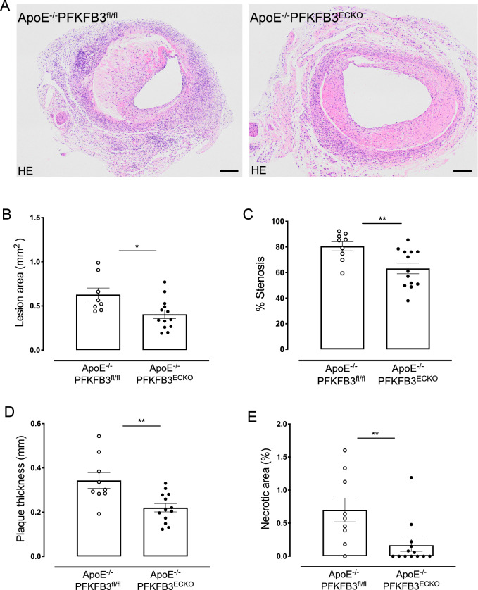
Fig. 7.
PFKFB3 deficiency in endothelial cells reduces the area, percentage stenosis, thickness, and necrotic area of vein graft lesions. A Representative cross sections of hematoxylin & eosin-stained vein grafts from ApoE−/−PFKFB3fl/fl and ApoE−/−PFKFB3ECKO mice. Scale bar = 200 μm. B–E Quantification of lesion area, percentage stenosis, thickness, and necrotic area of vein graft lesions. *P = 0.01, **P = 0.0095 versus ApoE−/−PFKFB3fl/fl. Independent samples t test; n = 8 (ApoE−/−PFKFB3fl/fl, panel B), n = 9 (ApoE−/−PFKFB3fl/fl, panel C–E), or n = 13 (ApoE−/−PFKFB3ECKO)

