Int J Mol Med 42: 322-330, 2018; DOI: 10.3892/ijmm.2018.3592
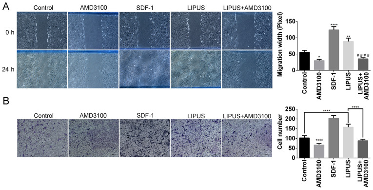
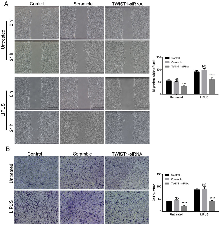
Subsequently to the publication of the above paper, an interested reader drew to the authors' attention that the 'Control' and 'AMD3100' panels in Fig. 3B on p. 326 appeared to show strikingly similar data, such that they may have been derived from the same original source; likewise, the 'Control' and 'Scramble' data panels in the 'Untreated' row of data panels in Fig. 5B on p. 327 also appeared to share some of the same data.
Figure 3.
LIPUS treatment promotes PDLSCs migration. (A) Representative images and quantification from three separate experiments of wound healing assays. (B) Representative images and quantification of transwell migration assays. PDLSCs that penetrated to the lower surface of the membrane were fixed, stained with 0.1% crystal violet, and counted per group. PDLSCs penetrating the membrane were fixed and stained with 0.1% crystal violet after 24 h. Quantification of PDLSCs invasion determined by cell counting. *P<0.05, **P<0.01 and ****P<0.001 vs. control group; ####P<0.001 vs. LIPUS group. LIPUS, low-intensity pulsed ultrasound; PDLSCs, periodontal ligament stem cells; SDF-1, stromal cell-derived factor-1.
Figure 5.
Effect of TWIST1 knockdown on LIPUS-promoted migration of PDLSCs. (A) Representative images and quantification from three separate experi- ments of wound healing assays. (B) Representative images and quantification of transwell migration assays. PDLSCs that penetrated to the lower surface of the membrane were fixed, stained with 0.1% crystal violet, and counted per group. Crystal violet staining of the penetrated cells after 24 h. Numbers of PDLSCs that crossed the upper transwell chamber. ***P<0.005 and ****P<0.001 vs. the control group. TWIST1, twist family bHLH transcription factor 1; LIPUS, low- intensity pulsed ultrasound; PDLSCs, periodontal ligament stem cells; si, small interfering; ns, not significant.
The authors have re-examined their data, and realized that a pair of the data panels included in these figures were inadvertently selected incorrectly. The corrected versions of Figs. 3 and 5, containing the correct data for the 'Control' panel in Fig. 3B and the 'Control' panel for the 'Untreated' experiments in Fig. 5B, are shown on the next page. These errors did not affect the major conclusions reported in the paper. All the authors have agreed to this Corrigendum, and thank the Editor of International Journal of Molecular Medicine for allowing them the opportunity to publish this. The authors regret these errors went unnoticed during the compilation of the figures in question, and apologize to the readership for any confusion that this may have caused.


