Abstract
The blood brain barrier (BBB) forms a stringent barrier that protects the brain from components in the circulation that could interfere with neuronal function. At the same time, the BBB enables selective transport of critical nutrients and other chemicals to the brain. Beyond these functions, another recently recognized function is even less characterized, specifically the role of the BBB in modulating behavior by affecting neuronal function in a sex-dependent manner. Notably, signaling in the adult Drosophila BBB is required for normal male courtship behavior. Courtship regulation also relies on male-specific molecules in the BBB. Our previous studies have demonstrated that adult feminization of these cells in males significantly lowered courtship. Here, we conducted microarray analysis of BBB cells isolated from males and females. Findings revealed that these cells contain male- and female-enriched transcripts, respectively. Among these transcripts, nuclear receptor Hr46/Hr3 was identified as a male-enriched BBB transcript. Hr46/Hr3 is best known for its essential roles in the ecdysone response during development and metamorphosis. In this study, we demonstrate that Hr46/Hr3 is specifically required in the BBB cells for courtship behavior in mature males. The protein is localized in the nuclei of sub-perineurial glial cells (SPG), indicating that it might act as a transcriptional regulator. These data provide a catalogue of sexually dimorphic BBB transcripts and demonstrate a physiological adult role for the nuclear receptor Hr46/Hr3 in the regulation of male courtship, a novel function that is independent of its developmental role.
Author summary
The blood brain barrier very tightly regulates which molecules can enter the brain. This is an important protection for the brain, however, it also complicates communication between molecules in the circulating fluid and the brain. In fly courtship, for example, circulating male-specific products are crucially required for normal courtship. But the neuronal circuits that ultimately control the behavior are inside the brain, separated from these molecules by the blood brain barrier. The mechanisms of this communication are not known. Here we show that the blood brain barrier itself contains sex-specific RNAs and we show that one of them, a nuclear receptor called Hr46 or Hr3, is required in adult males for normal courtship. These findings promise new insight into the communication between blood brain barrier and the brain.
Introduction
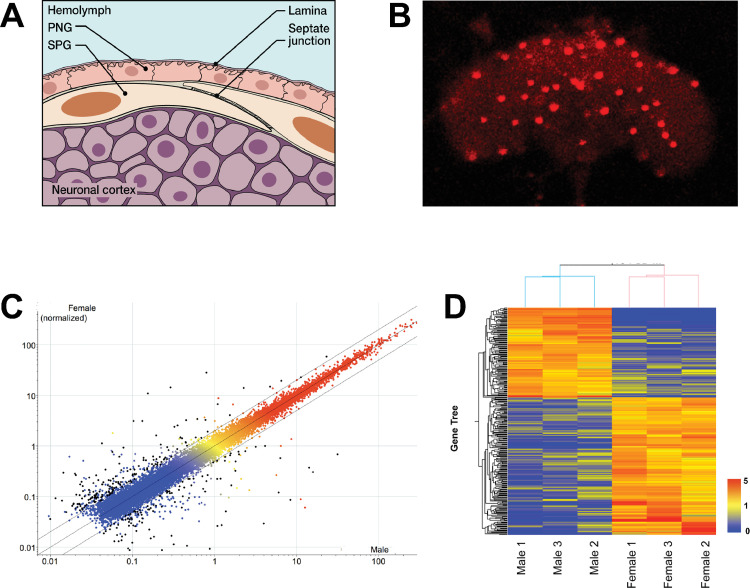
It is well established that the two layers of glial cells that tightly surround the nervous system form the Drosophila blood brain barrier (BBB) [1,2]. Flies have a non-vascular open circulatory system that distributes the hemolymph. The BBB forms the tight exclusion barrier that is essential to protecting neurons from hemolymph components that could interfere with neuronal function [1,3]. At the same time, the barrier needs to allow selective uptake of nutrients and other molecules needed for brain function. The Drosophila blood brain barrier (BBB) surrounds the brain like a tight cap and is made up of two layers of specialized glial cells (for recent reviews on Drosophila glial cells see [4,5]). The outer perineurial glia cells (PG cells) are thought to function as a barrier for large-molecular weight molecules. The inner layer, the subperineurial glia (SPG), is adjacent to the neuronal cell bodies and contains the tight junctions that form the physical barrier (Fig 1A). It has been shown in a number of genetic and functional studies that the barriers in flies and vertebrates share not only structure and function, but also many homologous proteins that ensure their function, as shown in [6]. A recent microarray study of isolated BBB cells has expanded on these earlier findings and shown that besides the characteristic barrier proteins, fly and mouse BBB cells share a large number of conserved proteins [7]. That study has also provided the first detailed “inventory” of these cells in Drosophila. While the barrier and selective uptake functions of the BBB are its most obvious essential function, evidence is starting to accumulate that other physiological processes in BBB cells are contributing to brain function. For example, the G-protein-coupled receptor moody is specifically expressed in the subperineurial glial cells (SPG)[8,9]. While the absence of both moody isoforms leads to a leaky BBB [8,9], mutants with only one of the isoforms have intact barriers, but have behavioral defects in their response to cocaine and alcohol [8]. In addition, moody, in a function independent of its function in barrier integrity, is also required in BBB cells for normal male courtship [10]. That active signaling processes in BBB cells regulate neuronal output was further indicated by the finding that BBB-specific reductions in the G protein Galpha(o) cause courtship defects, while leaving the barrier integrity intact [10]. It has been found that the circulating hemolymph contains male-specific factors from the fat body that are needed to ensure normal courtship [11]. It is not clear how these factors interact with the male brain circuits that regulate the behavior. Here we examine whether the BBB expresses sex-specific transcripts that might be part of this communication. This would be in agreement with the finding that feminization of the BBB by expression of the feminizing TRA protein specifically in the BBB of adult males results in a significant reduction in male courtship [10]. In these experiments, the tightness of the barrier was unaffected, suggesting that specific male transcripts are physiologically participating in courtship control. The identity of these factors and their function is unknown. Here we identify sex-preferentially expressed transcripts in the BBB of males and females and demonstrate that the nuclear receptor Hr46/Hr3, best known for its roles in larval development [12–14], is physiologically required in the BBB of adult mature males to ensure normal male courtship behavior.
Fig 1. Microarray analysis of isolated SPG cells of the BBB.
(A) Schematic of the Drosophila Blood Brain Barrier (BBB). The BBB consists of two layers of glial cells, the outer Perineurial Glia (PG) facing the circulating hemolymph, and the inner Subperineurial Glia (SPG) with septate junctions that form the main barrier. The SPG is in contact with the underlying nuclei of the neuronal cortex. (B) Isolated fly brain with SPG cells labeled by nuclear DsRed expression driven by SPG-Gal4. Fluorescently marked cells like these from males and females were hand-isolated for RNA extraction. 20x Magnification. (C) Probes present (above background) in all male or female samples are displayed as normalized to the 75th percentile intensity of each array (19,218 probes). Each spot is the mean of 3 samples from each condition. Black spots = differentially expressed genes (>2Fold, T-test p-value < 0.05, 284 probes). Red/orange = High expression, Yellow = Medium expression, Blue = Low expression. (D) Differentially expressed genes (>2 fold, T-test p-value < 0.05) in Male vs. Female are displayed as normalized to the median value of each probe across six samples (284 probes). The heat map color scale is shown on the right.
Results
A microarray screen identifies male- and female-enriched transcripts in the BBB
In our previous experiments, there was a strong reduction in male courtship when we conditionally feminized adult BBB cells [10]. This suggests that feminization disrupts male-specific transcripts that are physiologically required for normal mating behavior. In order to identify these transcripts, we isolated BBB cells from males and females and characterized their transcripts. The Gal4/UAS system was used to mark these cells [15]. We expressed the fluorescent protein DsRed in the nuclei of SPG cells, using the moody-Gal4 driver that drives expression in SPG cells (SPG-Gal4) [8]. As seen in Fig 1B, the large nuclei of the SPG cells were specifically marked. We dissected fly brains and manually removed and collected fluorescent cells. The RNA was subsequently used for microarray analysis by GenUs Biosystems (http://www.genusbiosystems.com/). The results confirmed the presence of sex-preferentially expressed transcripts in the BBB of males and females, respectively. 284 transcripts were identified that were enriched > 2 fold in either males or females (Fig 1C and 1D). Of those, 112 were male-preferentially expressed (S1 Table). As expected, the male-specifically expressed rox RNAs that are required for dosage compensation were highly specific to males. Furthermore, sex-specific dsx transcripts were identified because male and female transcripts use different polyA-sites and can thus be identified by microarray [16]. An analysis of the GeneOntology of the enriched transcripts is shown in S2 Table. Sex specific categories such as sex determination, sex-specific behavior and dosage compensation are well represented, further confirming that the isolated cells are sexually determined. About half of the genes with identified GO associations fall into one of these categories. The rest of the genes could not be assigned to a specific category. In addition to identifying sex-preferentially expressed RNAs, the experiment also provided an inventory of RNAs present in the BBB cells. The vast majority of BBB transcripts is equally expressed in males and females. Among them, as predicted for SPG cells, were RNAs that are characteristic of BBB cells [6,7]: RNAs for the junction proteins sinu and neurexin, for example, and the previously characterized SPG transcripts for moody and Mdr65. The most likely contaminating cells from the dissections would be fat body cells which are in close proximity to the BBB, and neuronal cells. We found very small amounts of the fat body transcript Lsp-2, or of the neuronal marker elav. They were not preferentially present in males or females, indicating that low amounts of these cells are unlikely to affect the identification of sex-specific transcripts in the BBB.
The nuclear receptor Hr46/Hr3 is required in the BBB for courtship
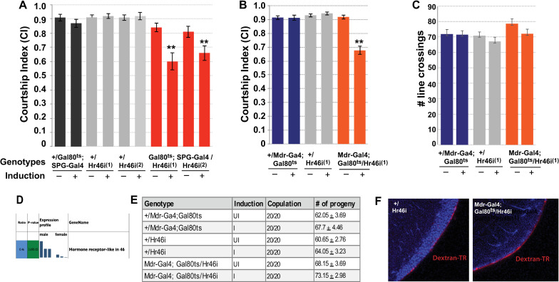
One of the male-enriched BBB RNAs is the transcript for the nuclear receptor Hr46/Hr3. Hr46/Hr3 is an orphan nuclear receptor that is most related to the mammalian ROR receptor (Retinoic acid related orphan receptor) [17]. Hr46/Hr3 is a well-described transcriptional regulator of larval developmental processes in response to Ecdysone, but no adult functions have been described so far. We have recently found an adult role for Juvenile Hormone, the other major insect hormone, in courtship [18], and we were intrigued by a possible link to Ecdysone signaling. To examine whether Hr46/Hr3 is required in the BBB for courtship we conditionally expressed two independent Hr46/Hr3-RNAi constructs specifically in the BBB of mature adult males and examined their courtship. We used the Gal4/Gal80ts system to restrict knockdown to mature males [19]. Two different BBB-Gal4 drivers were used to direct expression, the previously described SPG-Gal4, and a SPG-specific Mdr65-Gal4 driver that was generated in our lab (Fig 2A–2C). The ATP binding cassette (ABC) transporter Mdr65 has previously been shown to be specifically expressed in the SPG cells of the BBB [20,21], a pattern of expression shared by our Mdr-Gal4 line. For the courtship experiments, control flies containing a copy of just one of the two respective transgenes were grown, treated and tested in parallel to the knockdown flies as controls. Courtship results are shown in Fig 3A and 3B. The Courtship Index (CI) is calculated as the fraction of time the male spends displaying any element of courtship behavior (orienting, following, wing extension, licking, attempted copulation, copulation) within a 10 minute observation period [22]. At 18°C, Gal4 is inhibited by Gal80ts, and Hr46/Hr3-RNAi is not expressed. At this temperature, all genotypes exhibited normal courtship. In contrast, following induction at 32°C, males expressing Hr46/Hr3-RNAi in the BBB had significantly reduced courtship (p≤0.001) (Fig 3A and 3B). Reduction was observed with both drivers in combination with either of two independent UAS-Hr46/Hr3-RNAi constructs. In a short term activity assay [23], induced mutant flies did not differ from control flies in their activity (Fig 3C), suggesting that the observed courtship reduction is not due to general sickness of the males. We conclude that Hr46/Hr3 is required in the BBB of mature males for normal courtship behavior. While courtship was reduced, the males were capable of performing all of the steps of courtship, but they did so with lower probability. As shown in Fig 3E, they were also capable of copulation, and their average number of offspring did not differ from that of uninduced flies or controls.
Fig 2. Mdr-Gal4 driver is expressed in SPG nuclei.
(A, B) Visualization of our newly constructed Mdr-Gal4 driver with a nuclear dsRed reporter reveals expression in SPG nuclei. The nuclei differ in size because of different polyploidy grades [24]. Mdr-Gal4/UAS-dsRed; tubP-GAL80ts males were induced using the same protocol used in courtship assays. 20x magnification, size bar = 50um. (C) Mdr-Gal4>UAS-dsRed expression (red) coincides with expression of Indy-GFP in the SPG, but not the outer PG layer. Indy-GFP is a marker for both PG and SPG cells.
Fig 3. Knockdown of Hr46/Hr3 in the BBB of mature males reduces courtship.
(A-C) Graphs show the courtship index CI (fraction of time males spend courting during the observation period) ± SEM (A, B), or the performance of males in a control activity assay (# of line crossings ± SEM) (C) of the indicated genotypes. N = 20. Data were analyzed by ANOVA followed by Tukey multiple comparisons (p<0.05). Indices that are significantly different from the controls are marked by asterisks. UAS-Hr46i (abbreviated as Hr46i) expression is restricted by the presence of tubP-Gal80ts (abbreviated as “Gal80ts”) at 18°C (induction -). Placement of 5-day-old males at 32°C for 16 hours (induction +) releases the Gal80 inhibition and leads to the expression of RNAi. (A) Conditional expression of two different UAS-Hr46-RNAi transgenes (1-line 27253 and 2-line 27254) using SPG-Gal4 significantly reduces male courtship. (B) Conditional expression of UAS-Hr46i (27254) using Mdr-Gal4 in adult males similarly reduces courtship in comparison to controls. The controls are +/ tubP-GAL80ts;+/SPG-Gal4 and +/Mdr-Gal4; +/tubP-GAL80ts, respectively, as well as +; +/UAS-Hr46 RNAi. (C) The activity of the mutants as measured by number of line crossings is not lower than in control flies. Some variation in activity is likely caused by genetic background. (D) Hr46/Hr3) RNA is preferentially expressed in males. (E) Males with identical genotypes and induction regimen as in A-C were combined with a mature adult Canton-S virgin and scored for copulation. N = 20. All males copulated (20/20) and produced similar numbers of progeny. (F) Blood–brain barrier integrity is not compromised in Mdr-Gal4/ UAS-Hr46-RNAi males. Experimental and controls flies were injected with 10 kDa TR-Dextran (red) and dye penetration into or exclusion from the brain was examined by confocal microscopy. The brain nuclei are stained with DAPI. A tight BBB is indicated by the demarcated red line on the surface of the brain indicating exclusion of TR-dextran from the brain of Mdr-Gal4/UAS-Hr46-RNAi males. Flies were injected and processed in parallel and imaged with identical settings. Magnification = 63x.
To assess whether Hr46/Hr3 knockdown affects the integrity of the BBB, we tested the tightness of the BBB by injecting 10kD Texas-Red (TR)-marked Dextran. It is well documented that in wildtype animals TR-Dextran will be kept out of the brain and accumulate at the BBB, whereas a leaky BBB would allow entry into the brain [8]. As shown in Fig 3F, males expressing Hr46/Hr3 RNAi have normal BBB barrier function with the dye accumulating at the barrier like in the control. These findings indicate that BBB integrity is not compromised in the mutants, giving support to a physiological function for Hr46/Hr3 in the BBB that is required for normal regulation of courtship.
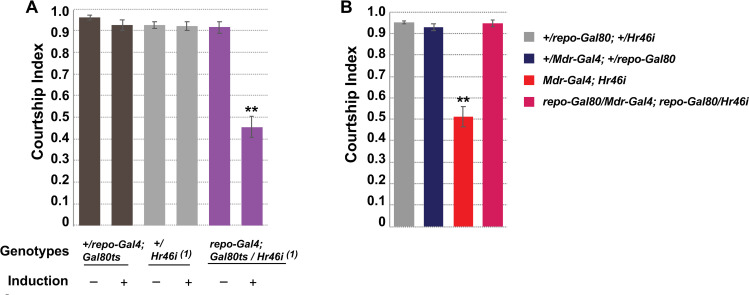
To confirm the glial requirement for Hr46/Hr3 we used the glia-specific driver repo-Gal4 to drive UAS-Hr46/Hr3-RNAi and observed equally reduced courtship (p<0.001) (Fig 4A). As expected, when we expressed Hr46/Hr3-RNAi in the BBB with Mdr-Gal4 in the presence of a glial-expressed Gal80 blocker (repo-Gal80) we observed a reversal of the courtship defects (Fig 4B). Together our findings demonstrate that Hr46/Hr3 is needed in the glial SPG cells for normal courtship.
Fig 4. Hr46/Hr3 is required in the glial SPG cells for courtship.
Graphs show the courtship index CI (fraction of time males spend courting during the observation period) ± SEM. N = 20. Data were analyzed by ANOVA followed by Tukey multiple comparisons (p<0.05). Indices that are significantly different from the controls are marked by asterisks. (A) Conditional glial knockdown of Hr46/Hr3 in mature males using repo-Gal4; tubP-GAL80ts reduces courtship. (B) The courtship reduction of Mdr-Gal4 directed Hr46/Hr3 knockdown can be reversed by Gal80 expression in glial cells by repo-Gal80.
Hr46/Hr3 and its ligand are present in SPG nuclei
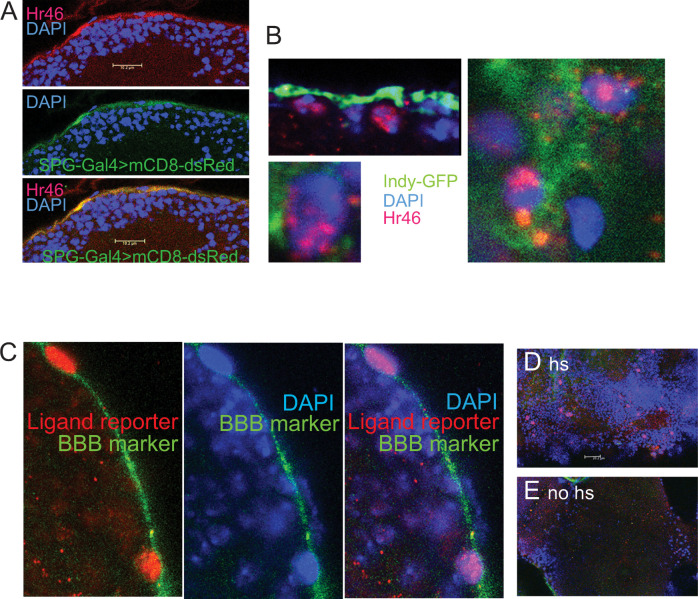
To study Hr46/Hr3 protein distribution in SPG cells of mature animals (Fig 5) we used antibodies generated by Lam et al [13]. As markers of BBB cells we used SPG-Gal4 (moody-Gal4)>UAS-mCD8-dsRed or Indy-GFP. While SPG-Gal4 is SPG-cell specific, Indy-GFP is expressed in both PG and SPG cells [7]. SPG cells are big flat cells with large polyploid nuclei [24]. Anti-Hr46/Hr3 antibody staining was observed in the SPG cells marked by SPG-Gal4>UAS-dsRed (Fig 5A) and in the SPG layer marked by a subset of indy-GFP expressing cells (Fig 5B). The protein was detected in the cytoplasm and the nucleus of SPG cells (Fig 5B). Hr46/Hr3 is a transcriptional activator in larvae, and its presence in the nucleus of BBB cells is consistent with a transcriptional role in these cells.
Fig 5. Hr46/Hr3 protein is located in SPG nuclei, and the Hr46/Hr3 ligand is present in the SPG cells of mature males.
(A) Anti-Hr46 antibody staining (red) co-localizes with SPG-cell specific expression of dsRed in SPG-Gal4 (= moody-Gal4)>UAS-dsRed males (green). Blue: DNA staining (DAPI). Size bar: 10um. (B) Anti-Hr46 antibody staining (Red) shows the presence of Hr46 in the nuclei and cytoplasm of SPG cells. Indy-GFP (green) marks both PG and SPG cells. Blue: DNA staining (DAPI). 63x magnification. (C) Activation of the hs-Gal4LBD HR3 reporter [25] indicates the presence of the Hr46/Hr3 ligand in SPG cells. hs-Gal4LBD HR3/UAS-dsRed/indy-GFP mature males were heat-shocked to express Gal4LBD HR3. Following binding of Hr3 ligand, Gal4 initiates transcription at UAS-dsRed(nuclear). dsRed can be seen expressed in the characteristic large nuclei of SPG cells (red). Indy-GFP expression is used as a BBB marker (green). Blue: DNA staining (DAPI, blue). 63x magnification. (D) 40x magnification showing expression of dsRed in SPG nuclei following induction as described for C. Size bar = 20um. (E) No ds Red expression was observed in no-hs controls of the same genotype that were grown, treated, stained and imaged in an identical fashion.
Hr46/Hr3 belongs to the family of ligand activated nuclear receptors. And while its ligand is unknown, insertion of the putative ligand binding domain into the Gal4 activation domain results in the transcriptional activation of Gal4 in cells containing the putative ligand. Palanker et al. have shown that this construct recapitulates Hr46 activation in cells where Hr46 transcriptional activity has been observed [25]. Binding of the putative ligand activates Gal4LBD(HR3) whose activity can then be visualized by a UAS- reporter. Importantly, in the construct, the Gal4LBD(HR3) reporter is driven by a hsp70 heat shock promoter (hsp70- Gal4LBD(HR3). This makes it possible to interrogate the presence of the ligand at a time of choice. We induced the Gal4LBD(HR3) ligand sensor in mature males by exposing the flies to 37 degrees Celsius for one hour and fixed their brains four hours later. We combined hsp70- Gal4LBD(HR3 with UAS-dsRed to visualize Gal4 activity, and indy-GFP for visualization of the BBB. As shown in Fig 5C and 5D, dsRed staining is observed in the large nuclei of SPG cells, indicating that the ligand for Hr46 is present in these cells in adult mature males. This staining was not observed in non-heatshocked flies (Fig 5E). Together our findings support a scenario in which Hr46/Hr3 is activated and physiologically needed in the SPG cells of the BBB of adult males to support normal male courtship behavior.
Discussion
Our microarray screen revealed that the Drosophila BBB contains male-enriched transcripts in males, as well as female-enriched transcripts in females. We have previously observed a reduction in male courtship when we conditionally feminized the BBB cells of mature males. Together these findings suggest that sex-specifically enriched transcripts contribute to a “male-specific” state of BBB cells that shapes its physiology and its dynamic interaction with the brain to modulate courtship. The feminization experiments were done in mature adult males by conditional induction of the female-specific TRA protein [26]. TRA is a master controller of sex determination by virtue of its direct control of the two major sex specific transcription factors DSX and FRU, which in turn control a multitude of genes [27,28]. Non-induced males were normal, demonstrating that it was the acute adult change in transcripts that resulted in disturbed courtship. In the microarray experiments described here we sampled all transcripts that were present in the BBB cells of mature males and females. These animals were of the same age as the flies in the TRA induction studies. Neither males nor females had mating experience. The sex-specific transcripts we identify here therefore likely include transcripts that were affected in the feminization experiment.
We identified a total of 284 sex-preferentially expressed transcripts. It is likely that a number of them are required in the regulation of sex-specific behaviors and that their disruption will affect courtship. Identifying them holds the promise of insight into the physiological processes that underlies BBB-brain communication required for normal courtship. However, there will likely also be commonly expressed transcripts that participate in these sex-specific processes as they interact with sex-specific partners or regulators, or respond to sex-specific incoming signals. The majority of identified SPG transcripts are equally expressed in males and females, representing an insight into the overall transcriptional “makeup” of SPG cells of mature males and females. Our data specifically represent SPG transcripts in males or females. This is in contrast to the study by deSalvo et al. [7] who characterized both layers of the BBB, PG and SPG cells, without distinguishing between males and females.
Hr46/Hr3 belongs to the nuclear-receptor superfamily that is characterized by the presence of a highly conserved DNA-binding domain (DBD) and a less conserved C-terminal ligand-binding and dimerization domain (LBD). The ligand for Hr46/Hr3 is unknown, but the reporter construct made by Palanker et al. strongly indicates that a ligand exists that binds to the LBD in the receptor [25]. In larvae, Palanker et al. observed fairly widespread, but not ubiquitous, activation, including in the fat body, leading to the speculation that Hr46/Hr3 might have metabolic functions. ROR, the mammalian homologue of Hr46/Hr3, is known to bind cholesterol and play a role in lipid homeostasis. Flies do not produce cholesterol, but take it up from their diet and it is an important precursor for the steroid ecdysone among other roles. Another nuclear receptor, DHR96 has been shown to bind cholesterol in Drosophila and to be essential for cholesterol homeostasis [29], but this does not exclude a role for Hr46/Hr3. Palanker et al. observed strong Gal4HR3LDB reporter expression in tissues of late third instars, with expression dropping in pre-pupariation, but strong activation was observed again in late pupae. We show here activation of the reporter construct in the BBB of mature adult males. In these experiments, the reporter construct is conditionally induced by a heat pulse in mature males. Thus, the observed activation reflects a “snapshot” of the presence of the putative ligand at that time. The observed activity coincides with the time when knockdown of Hr46/Hr3 causes a reduction in courtship.
Hr46/Hr3 is best known for its essential role in development as an Ecdysone effector. It is activated by Ecdysone and is a part of an activation cascade in response to Ecdysone. It induces another nuclear receptor, ftz-F1, among numerous other genes. Eventually, it acts as a negative feedback regulator to turn off Ecdysone-receptor signaling [12–14,30,31]. Hr46/Hr3 has essential functions during embryogenesis, prior to molts, and at the onset of metamorphosis. To our knowledge, this is the first report of an adult non-developmental role for Hr46/Hr3. Our conditional knockdown experiments demonstrate that its presence in the BBB of mature males is needed for normal courtship. Whether this reflects a role for an Ecdysone-induced signaling cascade and transcriptional activation of downstream targets remains to be determined. Data from [32] suggest that Ecdysone and the Ecdysone receptor (EcR) are present in the BBB. We have likewise found in our screen that EcR and ftz-F1 RNAs are present in SPG cells in a non-sex-specific manner. In analogy to its developmental role, Hr46/Hr3 most likely acts as a transcriptional regulator. Our observation that the Hr46 protein is present in SPG nuclei supports this interpretation. However, in an unexpected finding Montagne et al have identified Hr46/Hr3 as a S6K interacting protein in late larvae/prepupae [33]. Intriguingly, this function required a novel form of Hr46/Hr3 that did not contain the DNA binding domain, but did require the ligand binding domain. The presence of this altered form of Hr46/Hr3 increased phosphorylation activity of S6K. This finding, together with the short time scale of the response led the authors to propose an alternative non-genomic role of Hr46/Hr3, possibly as a mediator of the metabolic state of these cells. We do not know whether this isoform plays a role in courtship and whether Hr46/Hr3 might have a role that is independent of EcR in the BBB, conceivably in addition to the transcriptional role that is suggested by its presence in the nucleus.
Taken together, the data presented here demonstrate an adult physiological role for Hr46/Hr3, a nuclear receptor mainly known for its crucial function in development, in the glial cells of the BBB where it is required for the regulation of normal male courtship.
Materials and methods
Fly stocks
SPG-Gal4/TM3 [8] was a gift from Roland Bainton, UCSF. tubP-Gal80ts/CyO and tubP-Gal80ts/TM3,Sb flies were a gift from Gregg Roman (University of Mississippi). Hr46/Hr3 RNAi lines y1 v1; P{TRiP.JF02542}attP2 (BL 27253) and y1 v1; P{TRiP.JF02543}attP2 (BL 27254); w1118; P{w[+mC] = UAS-RedStinger (dsRed)}4/CyO (BL 8546); w*; P{PTT-GC}IndyYC0017/TM6C, Sb1 (Indy-GFP) (BL 50860) were obtained from the Bloomington Drosophila stock center (https://bdsc.indiana.edu/). y, repo-Gal4 on X was a gift from Takeshi Awasaki (University of Massachusetts [34]; The y mutation was removed by recombination. w; Pin, repo-Gal80/CyO flies were a gift from Rob Jackson (Tufts University). Pin was removed by recombination. w; +; repo-Gal80 flies [34] were a gift from Christian Klämbt (University of Münster). w1118; P{w[+mC] = hs-GAL4-HR3.LBD was a gift from Carl Thummel (University of Utah).
Gal80ts experiments
tubP-Gal80ts carrying flies and control flies were raised at 18°C. Virgin males were collected at eclosion and kept in individual vials for 5–8 days at 18°C. Flies were then placed at 32°C for 48 hours for induction. Following induction, induced and uninduced flies were kept at 25°C overnight prior to courtship assays.
Behavioral assays
The courtship assay and activity assay were performed as previously described [35]. Male courtship in Drosophila melanogaster consists of well-defined stereotyped behavioral steps that can easily be quantified in a courtship index (CI) [36–38]. The CI is calculated as the fraction of time the male spends displaying any element of courtship behavior (orienting, following, wing extension, licking, attempted copulation, copulation) within a 10 minute observation period [22]. In short, males were placed in a plexiglass “mating wheel” (diameter 0.8 cm), together with a 2–4 hrs old Canton-S virgin female. Short-term activity assays were performed as previously described [23]. Individual males were placed into the “mating wheel” containing a filter paper with a single line dividing the chamber in half. After 2–3 minutes of acclimation time, the number of times the male crossed the center line within the three-minute observation time was counted.
Each graph represents sets of control and experimental genotypes that were grown, collected and aged in parallel. In each behavioral session, equal numbers of all genotypes were tested.
Copulation/fecundity test
Flies were grown, collected and Gal4/tubP-Gal80ts induction performed in the same way as for courtship assays. Individual males were then aspirated into a fresh vial containing a 4–5-day old virgin Canton-S female. The pairs were observed for 30 minutes, then checked periodically every 10 minutes until all pairs were observed copulating. The parents were kept in the vial for 7 days and then removed. Progeny were counted upon eclosion every 1 to 2 days until the vials produced no progeny for at least 2 days.
Microarray analysis
To isolate blood-brain barrier cells, flies bearing the SPG-Gal4 driver were crossed to flies carrying the fluorescent reporter transgene, UAS-DsRed. This resulted in the expression of the visible fluorescent marker DsRed to mark the nuclei of BBB cells. Prior to the experiment, both the driver SPG-Gal4 and the UAS-DsRed lines were outcrossed with a Cantonized w1118 strain for 10 generation. The flies were grown in a 25°C incubator under a 12hrs light/12hrs dark cycle. Eclosing males and females were collected and kept in separate groups of 10–15 flies of the same sex under the same conditions for 4 days and then dissected between ZT 5 and ZT 7 to control for levels of cycling transcripts. Equal numbers of males and females originating from the same culture were dissected in each sitting. The brains were dissected in ice-cold 1 X PBS between ZT5 and ZT7. The dissected brains were then transferred to a new petri dish containing ice-cold 1X PBS within half an hour. Carefully, under the fluorescent microscope, individual and/or groups of blood-brain barrier cells marked with DsRed were isolated manually by using Dumont # 5 SF superfine forceps (Fine Science Tools, Inc). The cells were then immediately transferred to a frozen droplet of Trizol reagent on dry ice and stored in -80°C until processed further. Cells were isolated from at least 50 brains for each genotype. Cells from approximately 50 flies were pooled for each biological replicate, and the RNA of three biological replicas from separate crosses was prepared for each sex. The approximate total number of cells isolated per brain varied from ~60–120. The forceps were cleaned with RNAZap when moving from one genotype to the other.
The isolated BBB cells of male and female flies were provided to GenUs Biosystems (http://www.genusbiosystems.com/) for microarray analysis. A total of 3 biological replicates for males and females were submitted. Cells were lysed in TRI reagent (Ambion) and Total RNA was isolated using phenol/chloroform extraction followed by purification over RNeasy spin columns (Qiagen). The concentration and purity of Total RNA was measured by spectrophotometry at OD260/280 and the quality of the Total RNA sample was assessed using an Agilent Bioanalyzer with the RNA6000 Nano Lab Chip (Agilent Technologies). Labeled cRNA was prepared by linear amplification of the Poly (A)+ RNA population within the Total RNA sample. Briefly, 1 μg of Total RNA was reverse transcribed after priming with a DNA oligonucleotide containing the T7 RNA polymerase promoter 5’ to a dT(24) sequence. After second-strand cDNA synthesis and purification of double-stranded cDNA, in vitro transcription was performed using T7 RNA polymerase. The quantity and quality of the cRNA was assayed by spectrophotometry and on the Agilent Bioanalyzer. One microgram of purified cRNA was fragmented to uniform size and applied to Drosophila (V2) Gene Expression microarray (Agilent Technologies, Design ID 021791) in hybridization buffer. Arrays were hybridized at 37° C for 18 hrs in a rotating incubator, washed, and scanned with a G2565 Microarray Scanner (Agilent Technologies). Arrays were processed with Agilent Feature Extraction software and data was analyzed with GeneSpring GX software (both Agilent Technologies). To compare individual expression values across arrays, raw intensity data from each gene was normalized to the 75th percentile intensity of each array. Genes with values greater than background intensity in all female or all male replicates were filtered for further analysis. Differentially expressed genes were identified with fold change > 2-fold and Welch Ttest, p-value < 0.05.
Generation of Mdr65-Gal4 transgenic flies
650 bp of sequence upstream of the Mdr65 coding sequence was PCR amplified from CS genomic DNA using the primers 5’cggaattc(EcoRI)TCCATCACTTAGCAAAGCAGACTTCAATC and 5’cgggatcc(BamH1) GGTGATGTTTAGTCGGCACTGACGA and inserted into the Drosophila transformation vector pGATN to create Mdr65-Gal4. In pGATN, expression of the yeast transcription factor Gal4 is driven by the inserted promoter sequences [15]. Transgenic flies were generated by Rainbow Transgenic Flies (https://www.rainbowgene.com/) by P-element mediated insertion. The expression pattern in Mdr65-Gal4 transgenic lines was examined by crosses to UAS-dsRed (nuclear) or UAS-mcD8-dsRed.
Immunohistochemistry
Immunohistochemistry on isolated brains was performed as described in Li et al. [39]. The Hr46/Hr3 antibody was a gift from Carl Thummel, University of Utah [13] and was used at 1:50 dilution. To visualize BBB cells, flies carrying Indy-GFP were used for anti-Hr3/anti-GFP double staining. Indy-GFP marks BBB cells [40].
Antibodies used:
Rabbit anti-Hr3, 1:50 or 1:25 (gift of Carl Thummel, University of Utah [13]); Rabbit anti-RFP (abcam ab62341), 1:200; chicken anti-GFP (abcam ab13970), 1:500; Alexa Fluor 555 goat anti-rabbit (Invitrogen A21429) 1:200; Alexa Fluor 635 goat anti-mouse (Invitrogen A31575) 1:200; Alexa Fluor 488 goat anti-chicken (Thermo Fisher Scientific A-11039).
Injection of 10kd Dextran-TR to assess the integrity of the BBB was performed as described in Hoxha et al. [10]. Flies were subjected to the same experimental regimen as in behavioral experiments and control and experimental flies were assayed in parallel and imaged under identical conditions.
Test for presence of Hr46/Hr3 ligand
hs-Gal4LBD(HR3) flies were crossed to UAS-dsRed(nuclear); indy-GFP flies and grown at 18°C. Progeny were collected at eclosion and kept in small groups of males or females for 4 days. Expression of hs-Gal4LBD(HR3 was induced by placing flies in prewarmed food vials at 37°C for one hour, followed by recovery at room temperature for three hours and brain isolation. dsRed expression as a measure of Gal4 activation by Hr3 ligand and GFP (as BBB marker) were assessed by immunohistochemistry. Induced and uninduced flies were assayed in parallel and imaged with identical settings.
Statistical analysis
Two-way analysis of variance (ANOVA) was used to establish overall significance. Post hoc analysis for multiple comparisons was carried out with Tukey (HSD). P values < 0.05 were considered statistically significant. All statistical calculations were done using XLSTAT (Addinsoft, NY, NY) running on Microsoft Excel for Mac (version 16). All ±error bars are standard error of the mean (SEM).
Supporting information
284 transcripts were identified that differ by >2Fold (p<0.05). Of those 112 were male-preferentially expressed, 172 were female-preferentially expressed.
(XLSX)
GO groups shaded in grey are related to sex-specific processes. The total number of genes and GO groups assigned to this category is indicated.
(XLSX)
(XLSX)
Acknowledgments
We thank Carl Thummel for anti-Hr3 antibodies and fly lines and Takeshi Awasaki (University of Massachusetts), Rob Jackson (Tufts University), Christian Klämbt (University of Münster) and Gregg Roman (University of Mississippi) for fly lines. We thank Miguel de la Flor for artwork and Calvin Do for help with brain dissections. We thank GenUS biosystems for expert help with the microarray experiments.
Data Availability
All microarray data are available at GEO Series accession number GSE 157122 (https://www.ncbi.nlm.nih.gov/geo/query/acc.cgi?acc=GSE157122). All other relevant data are within the manuscript and its Supporting Information files.
Funding Statement
Funding was provided by the National Science Foundation (http://nsf.gov), grants IOS-1755385 (BD) and IOS-0919697 (BD). The funders had no role in study design, data collection and analysis, decision to publish, or preparation of the manuscript.
References
- 1.Hindle SJ, Bainton RJ. Barrier mechanisms in the Drosophila blood-brain barrier. Front Neurosci. 2014;8:414. Epub 2015/01/08. doi: 10.3389/fnins.2014.00414 ; PubMed Central PMCID: PMC4267209. [DOI] [PMC free article] [PubMed] [Google Scholar]
- 2.Stork T, Engelen D, Krudewig A, Silies M, Bainton RJ, Klambt C. Organization and function of the blood-brain barrier in Drosophila. J Neurosci. 2008;28(3):587–97. Epub 2008/01/18. doi: 10.1523/JNEUROSCI.4367-07.2008 . [DOI] [PMC free article] [PubMed] [Google Scholar]
- 3.Schirmeier S, Klambt C. The Drosophila blood-brain barrier as interface between neurons and hemolymph. Mech Dev. 2015. Epub 2015/06/24. doi: 10.1016/j.mod.2015.06.002 . [DOI] [PubMed] [Google Scholar]
- 4.Bittern J, Pogodalla N, Ohm H, Bruser L, Kottmeier R, Schirmeier S, et al. Neuron-glia interaction in the Drosophila nervous system. Dev Neurobiol. 2021;81(5):438–52. Epub 2020/02/26. doi: 10.1002/dneu.22737 . [DOI] [PubMed] [Google Scholar]
- 5.Yildirim K, Petri J, Kottmeier R, Klambt C. Drosophila glia: Few cell types and many conserved functions. Glia. 2019;67(1):5–26. Epub 2018/11/18. doi: 10.1002/glia.23459 . [DOI] [PubMed] [Google Scholar]
- 6.Limmer S, Weiler A, Volkenhoff A, Babatz F, Klambt C. The Drosophila blood-brain barrier: development and function of a glial endothelium. Front Neurosci. 2014;8:365. Epub 2014/12/03. doi: 10.3389/fnins.2014.00365 ; PubMed Central PMCID: PMC4231875. [DOI] [PMC free article] [PubMed] [Google Scholar]
- 7.DeSalvo MK, Hindle SJ, Rusan ZM, Orng S, Eddison M, Halliwill K, et al. The Drosophila surface glia transcriptome: evolutionary conserved blood-brain barrier processes. Front Neurosci. 2014;8:346. Epub 2014/11/27. doi: 10.3389/fnins.2014.00346 ; PubMed Central PMCID: PMC4224204. [DOI] [PMC free article] [PubMed] [Google Scholar]
- 8.Bainton RJ, Tsai LT, Schwabe T, DeSalvo M, Gaul U, Heberlein U. moody encodes two GPCRs that regulate cocaine behaviors and blood-brain barrier permeability in Drosophila. Cell. 2005;123(1):145–56. doi: 10.1016/j.cell.2005.07.029 . [DOI] [PubMed] [Google Scholar]
- 9.Schwabe T, Bainton RJ, Fetter RD, Heberlein U, Gaul U. GPCR signaling is required for blood-brain barrier formation in drosophila. Cell. 2005;123(1):133–44. doi: 10.1016/j.cell.2005.08.037 . [DOI] [PubMed] [Google Scholar]
- 10.Hoxha V, Lama C, Chang PL, Saurabh S, Patel N, Olate N, et al. Sex-specific signaling in the blood-brain barrier is required for male courtship in Drosophila. PLoS genetics. 2013;9(1):e1003217. Epub 2013/01/30. doi: 10.1371/journal.pgen.1003217 . [DOI] [PMC free article] [PubMed] [Google Scholar]
- 11.Lazareva AA, Roman G, Mattox W, Hardin PE, Dauwalder B. A role for the adult fat body in Drosophila male courtship behavior. PLoS Genet. 2007;3(1):e16. doi: 10.1371/journal.pgen.0030016 . [DOI] [PMC free article] [PubMed] [Google Scholar]
- 12.Lam G, Hall BL, Bender M, Thummel CS. DHR3 is required for the prepupal-pupal transition and differentiation of adult structures during Drosophila metamorphosis. Developmental biology. 1999;212(1):204–16. Epub 1999/07/27. doi: 10.1006/dbio.1999.9343 . [DOI] [PubMed] [Google Scholar]
- 13.Lam GT, Jiang C, Thummel CS. Coordination of larval and prepupal gene expression by the DHR3 orphan receptor during Drosophila metamorphosis. Development. 1997;124(9):1757–69. Epub 1997/05/01. [DOI] [PubMed] [Google Scholar]
- 14.Ruaud AF, Lam G, Thummel CS. The Drosophila nuclear receptors DHR3 and betaFTZ-F1 control overlapping developmental responses in late embryos. Development. 2010;137(1):123–31. Epub 2009/12/22. doi: 10.1242/dev.042036 ; PubMed Central PMCID: PMC2796934. [DOI] [PMC free article] [PubMed] [Google Scholar]
- 15.Brand AH, Perrimon N. Targeted gene expression as a means of altering cell fates and generating dominant phenotypes. Development. 1993;118(2):401–15. [DOI] [PubMed] [Google Scholar]
- 16.Baker BS, Wolfner MF. A molecular analysis of doublesex, a bifunctional gene that controls both male and female sexual differentiation in Drosophila melanogaster. Genes Dev. 1988;2(4):477–89. doi: 10.1101/gad.2.4.477 [DOI] [PubMed] [Google Scholar]
- 17.King-Jones K, Thummel CS. Nuclear receptors—a perspective from Drosophila. Nat Rev Genet. 2005;6(4):311–23. Epub 2005/04/02. doi: 10.1038/nrg1581 . [DOI] [PubMed] [Google Scholar]
- 18.Wijesekera TP, Saurabh S, Dauwalder B. Juvenile Hormone Is Required in Adult Males for Drosophila Courtship. PloS one. 2016;11(3):e0151912. Epub 2016/03/24. doi: 10.1371/journal.pone.0151912 ; PubMed Central PMCID: PMC4803231. [DOI] [PMC free article] [PubMed] [Google Scholar]
- 19.McGuire SE, Roman G, Davis RL. Gene expression systems in Drosophila: a synthesis of time and space. Trends Genet. 2004;20(8):384–91. doi: 10.1016/j.tig.2004.06.012 . [DOI] [PubMed] [Google Scholar]
- 20.Kanai MI, Kim MJ, Akiyama T, Takemura M, Wharton K, O’Connor MB, et al. Regulation of neuroblast proliferation by surface glia in the Drosophila larval brain. Sci Rep. 2018;8(1):3730. Epub 2018/03/01. doi: 10.1038/s41598-018-22028-y ; PubMed Central PMCID: PMC5829083. [DOI] [PMC free article] [PubMed] [Google Scholar]
- 21.Mayer F, Mayer N, Chinn L, Pinsonneault RL, Kroetz D, Bainton RJ. Evolutionary conservation of vertebrate blood-brain barrier chemoprotective mechanisms in Drosophila. J Neurosci. 2009;29(11):3538–50. Epub 2009/03/20. doi: 10.1523/JNEUROSCI.5564-08.2009 ; PubMed Central PMCID: PMC3040577. [DOI] [PMC free article] [PubMed] [Google Scholar]
- 22.Taylor BJ, Villella A, Ryner LC, Baker BS, Hall JC. Behavioral and neurobiological implications of sex-determining factors in Drosophila. Dev Genet. 1994;15(3):275–96. doi: 10.1002/dvg.1020150309 [DOI] [PubMed] [Google Scholar]
- 23.Anand A, Villella A, Ryner LC, Carlo T, Goodwin SF, Song HJ, et al. Molecular Genetic Dissection of the Sex-Specific and Vital Functions of the Drosophila melanogaster Sex Determination Gene fruitless. Genetics. 2001;158(4):1569–95. doi: 10.1093/genetics/158.4.1569 [DOI] [PMC free article] [PubMed] [Google Scholar]
- 24.Unhavaithaya Y, Orr-Weaver TL. Polyploidization of glia in neural development links tissue growth to blood-brain barrier integrity. Genes & development. 2012;26(1):31–6. Epub 2012/01/05. doi: 10.1101/gad.177436.111 ; PubMed Central PMCID: PMC3258963. [DOI] [PMC free article] [PubMed] [Google Scholar]
- 25.Palanker L, Necakov AS, Sampson HM, Ni R, Hu C, Thummel CS, et al. Dynamic regulation of Drosophila nuclear receptor activity in vivo. Development. 2006;133(18):3549–62. Epub 2006/08/18. doi: 10.1242/dev.02512 ; PubMed Central PMCID: PMC2100403. [DOI] [PMC free article] [PubMed] [Google Scholar]
- 26.Ferveur JF, Greenspan RJ. Courtship behavior of brain mosaics in Drosophila. J Neurogenet. 1998;12(4):205–26. doi: 10.3109/01677069809108559 [DOI] [PubMed] [Google Scholar]
- 27.Baker BS. Sex in flies: the splice of life. Nature. 1989;340(6234):521–4. doi: 10.1038/340521a0 [DOI] [PubMed] [Google Scholar]
- 28.Dauwalder B. The roles of fruitless and doublesex in the control of male courtship. Int Rev Neurobiol. 2011;99:87–105. Epub 2011/09/13. doi: 10.1016/B978-0-12-387003-2.00004-5 . [DOI] [PubMed] [Google Scholar]
- 29.Horner MA, Pardee K, Liu S, King-Jones K, Lajoie G, Edwards A, et al. The Drosophila DHR96 nuclear receptor binds cholesterol and regulates cholesterol homeostasis. Genes & development. 2009;23(23):2711–6. Epub 2009/12/03. doi: 10.1101/gad.1833609 ; PubMed Central PMCID: PMC2788327. [DOI] [PMC free article] [PubMed] [Google Scholar]
- 30.Carney GE, Wade AA, Sapra R, Goldstein ES, Bender M. DHR3, an ecdysone-inducible early-late gene encoding a Drosophila nuclear receptor, is required for embryogenesis. Proceedings of the National Academy of Sciences of the United States of America. 1997;94(22):12024–9. Epub 1997/10/29. PubMed Central PMCID: PMC23691. doi: 10.1073/pnas.94.22.12024 [DOI] [PMC free article] [PubMed] [Google Scholar]
- 31.Sullivan AA, Thummel CS. Temporal profiles of nuclear receptor gene expression reveal coordinate transcriptional responses during Drosophila development. Mol Endocrinol. 2003;17(11):2125–37. Epub 2003/07/26. doi: 10.1210/me.2002-0430 . [DOI] [PubMed] [Google Scholar]
- 32.Hindle SJ, Munji RN, Dolghih E, Gaskins G, Orng S, Ishimoto H, et al. Evolutionarily Conserved Roles for Blood-Brain Barrier Xenobiotic Transporters in Endogenous Steroid Partitioning and Behavior. Cell Rep. 2017;21(5):1304–16. Epub 2017/11/02. doi: 10.1016/j.celrep.2017.10.026 ; PubMed Central PMCID: PMC5774027. [DOI] [PMC free article] [PubMed] [Google Scholar]
- 33.Montagne J, Lecerf C, Parvy JP, Bennion JM, Radimerski T, Ruhf ML, et al. The nuclear receptor DHR3 modulates dS6 kinase-dependent growth in Drosophila. PLoS genetics. 2010;6(5):e1000937. Epub 2010/05/14. doi: 10.1371/journal.pgen.1000937 ; PubMed Central PMCID: PMC2865512. [DOI] [PMC free article] [PubMed] [Google Scholar]
- 34.Awasaki T, Lai SL, Ito K, Lee T. Organization and postembryonic development of glial cells in the adult central brain of Drosophila. J Neurosci. 2008;28(51):13742–53. Epub 2008/12/19. doi: 10.1523/JNEUROSCI.4844-08.2008 . [DOI] [PMC free article] [PubMed] [Google Scholar]
- 35.Dauwalder B, Tsujimoto S, Moss J, Mattox W. The Drosophila takeout gene is regulated by the somatic sex-determination pathway and affects male courtship behavior. Genes Dev. 2002;16(22):2879–92. doi: 10.1101/gad.1010302 . [DOI] [PMC free article] [PubMed] [Google Scholar]
- 36.Greenspan RJ. Understanding the genetic construction of behavior. Sci Am. 1995;272(4):72–8. doi: 10.1038/scientificamerican0495-72 [DOI] [PubMed] [Google Scholar]
- 37.Hall JC. The mating of a fly. Science. 1994;264(5166):1702–14. doi: 10.1126/science.8209251 [DOI] [PubMed] [Google Scholar]
- 38.Villella A, Hall JC. Neurogenetics of courtship and mating in Drosophila. Adv Genet. 2008;62:67–184. Epub 2008/11/18. doi: 10.1016/S0065-2660(08)00603-2 [pii] 10.1016/S0065-2660(08)00603-2. . [DOI] [PubMed] [Google Scholar]
- 39.Li Y, Hoxha V, Lama C, Dinh BH, Vo CN, Dauwalder B. The hector G-protein coupled receptor is required in a subset of fruitless neurons for male courtship behavior. PloS one. 2011;6(11):e28269. Epub 2011/12/06. doi: 10.1371/journal.pone.0028269 ; PubMed Central PMCID: PMC3227663. [DOI] [PMC free article] [PubMed] [Google Scholar]
- 40.Love CR, Dauwalder B. Drosophila as a Model to Study the Blood-Brain Barrier. In: Barichello T, editor. Blood-Brain Barrier. New York, NY: Springer New York; 2019. p. 175–85. [Google Scholar]
Associated Data
This section collects any data citations, data availability statements, or supplementary materials included in this article.
Supplementary Materials
284 transcripts were identified that differ by >2Fold (p<0.05). Of those 112 were male-preferentially expressed, 172 were female-preferentially expressed.
(XLSX)
GO groups shaded in grey are related to sex-specific processes. The total number of genes and GO groups assigned to this category is indicated.
(XLSX)
(XLSX)
Data Availability Statement
All microarray data are available at GEO Series accession number GSE 157122 (https://www.ncbi.nlm.nih.gov/geo/query/acc.cgi?acc=GSE157122). All other relevant data are within the manuscript and its Supporting Information files.


