Abstract
The relapsing fever spirochete Borrelia hermsii and the Lyme disease spirochete Borrelia burgdorferi sensu stricto each produces an abundant, orthologous, outer membrane protein, Vtp and OspC, respectively, when transmitted by tick bite. Gene inactivation studies have shown that both proteins are essential for spirochete infectivity when transmitted by their respective tick vectors. Therefore, we transformed a vtp-minus mutant of B. hermsii with ospC from B. burgdorferi and examined the behavior of this transgenic spirochete in its soft tick vector Ornithodoros hermsi. IFA staining indicated up to 97.8% of the transgenic B. hermsii upregulated OspC in the ticks’ salivary glands compared to no more than 12.8% in the midgut, similar to our previous findings with wild-type B. hermsii producing Vtp. Transformation with ospC also restored B. hermsii infectivity to mice when fed upon by infected ticks. Previous sequence analysis of Vtp for 79 isolates and DNA samples of B. hermsii in our laboratory showed this protein is highly polymorphic with 9 divergent amino acid types, yet strikingly the signal peptide is identical among all samples and the same for all OspC signal peptides for B. burgdorferi and related species examined to date. Searches in multiple genome sequences for other species of relapsing fever spirochetes failed to find the same signal peptide sequence to help identify potential transmission-associated proteins. However, some candidate signal peptides with highly similar sequences were found and worthy of future efforts with other species. While OspC of B. burgdorferi restored infectivity to a Vtp-minus mutant of B. hermsii, the functions of these proteins are not known. Our results should stimulate investigators to search for orthologous transmission-associated proteins in other tick-borne spirochetes to better understand how this group of pathogens has coevolved with diverse tick vectors.
Keywords: Borrelia hermsi, Borrelia burgdorferi sensu stricto
1. Introduction
Vector-borne pathogens cause significant morbidity and mortality throughout most of the world where environmental factors allow blood-feeding arthropods to coexist with humans (Lederberg et al., 1992; Medicine, 2008). Understanding how these disease-causing agents adapt to specific vectors for their subsequent transmission to susceptible human and other hosts can aid in the development of new vaccines and diagnostic tests. The obligatory changes that microorganisms make as they alternate between diverse mammalian and arthropod hosts also demonstrate how parasites and hosts have co-evolved, as exemplified by tick-borne spirochetes that cause relapsing fever and Lyme disease.
The primary species of spirochetes that cause relapsing fever and Lyme disease in North America are Borrelia hermsii and Borrelia burgdorferi sensu stricto (= Borreliella burgdorferi), respectively. These zoonotic pathogens cause very different diseases when infecting humans (Steere, 2001; Dworkin et al., 2008), and are transmitted by ticks that have strikingly different life cycles and feeding behaviors (Sonenshine, 1991; Piesman and Schwan, 2010). The argasid tick vector of B. hermsii, Ornithodoros hermsi, feeds rapidly in all life stages, engorging fully in only15 to 90 minutes (Herms and Wheeler, 1936; Wheeler, 1943). In contrast, the ixodid tick vectors of B. burgdorferi, such as Ixodes scapularis, attach for approximately three to 11 days depending on which stage (larva, nymph, adult) is feeding (Krinsky, 1979). When O. hermsi acquires B. hermsii in the blood from an infected host, the spirochetes disseminate from the midgut during the next few weeks to colonize multiple organs including the salivary glands (Schwan and Hinnebusch, 1998; Raffel et al., 2014). When I. scapularis acquires B. burgdorferi from its host, the spirochetes remain restricted to the midgut until after the tick has molted and feeds again (Benach et al., 1987; Ribeiro et al., 1987; Burgdorfer et al., 1988; Gilmore and Piesman, 2000). Yet when being transmitted by the bite of their respective tick vectors, both species of spirochetes produce an orthologous outer surface protein (Schwan and Piesman, 2002).
Borrelia hermsii produces the variable tick protein (Vtp) when persistently residing in the salivary glands of O. hermsi (Schwan and Hinnebusch, 1998; Raffel et al., 2014), while B. burgdorferi up-regulates outer surface protein (Osp) C in the midgut of I. scapularis only after the onset of feeding (Schwan et al., 1995; Coleman et al., 1997; Schwan and Piesman, 2000). Thus, the temporal presence of Vtp and OspC is vastly different while these two species of spirochetes reside in their respective tick vectors (Schwan and Piesman, 2002). Yet both species of spirochetes produce these orthologous proteins when the bacteria are transmitted via saliva to a vertebrate host, gene inactivations of B. burgdorferi ospC and B. hermsii vtp render both spirochetes noninfectious by tick bite (Grimm et al., 2004; Pal et al., 2004; Raffel et al., 2014).
The work presented here is an extension of our previous study demonstrating that Vtp is essential for the infectivity of B. hermsii when transmitted by tick bite (Raffel et al., 2014). Given the orthologous relationship of Vtp and OspC and their requirement for infectivity by tick bite, we asked if the ospC gene of B. burgdorferi would restore infectivity to a B. hermsii mutant lacking vtp (Battisti et al., 2008; Raffel et al., 2014). Here we show that B. hermsii lacking vtp but transformed with ospC was again infectious by tick bite. We also show that the temporal regulation of OspC by the transformed B. hermsii spirochetes in O. hermsi was comparable to the synthesis of Vtp by wild-type B. hermsii (Raffel et al., 2014).
2. Material and methods
2.1. Bacterial isolates and cultivation
Borrelia hermsii DAH 2E7 serotype 7 was cloned by limiting dilution in mBSK-c medium from a clinical isolate that originated from eastern Washington (Barbour, 1984; Hinnebusch et al., 1998; Porcella et al., 2005; Battisti et al., 2008). Borrelia burgdorferi B31, the source for the ospC gene used herein, originated from a pool of four I. scapularis ticks collected on Shelter Island, New York (Burgdorfer et al., 1982). The high passage strain of B. burgdorferi that constitutively produces OspC (strain B312) (Sadziene et al., 1993) was used in immunoblots. The methods to create the Δvtp B. hermsii mutant lacking an intact vtp gene, its cultivation and experimental infections in ticks and mice are described elsewhere (Battisti et al., 2008; Raffel et al., 2014).
2.2. Complementation of the B. hermsii vtp-minus mutant with ospC
In the Δvtp B. hermsii, the vtp locus was disrupted with a flgBP-kanamycin-resistance cassette and in this study we replaced the disruption cassette with ospC from B. burgdorferi and a flaBP-gentamicin-resistance cassette yielding Δvtp::ospC+, but retained the upstream and downstream flanking regions for homologous recombination. We retained the native vtp promoter because the synthesis of Vtp and OspC are affected differently with temperature changes during in vitro cultivation. With a decrease in temperature, B. hermsii up-regulates Vtp (Schwan and Hinnebusch, 1998) while B. burgdorferi down-regulates OspC (Schwan et al., 1995). The construction of the Δvtp::ospC+ transgenic B. hermsii was done as follows. The vtp promoter (vtpP) was PCR-amplified with primers ReconF+XmaI and vtpP3’-NdeI (Table 1) from the plasmid pOKvtpRECON (Battisti et al., 2008), that contains the wild-type vtp with approximately 2.3 Kb of flanking DNA on each side of vtp and the flaBP-gentamicin-resistance cassette inserted upstream of vtp, and cloned into the pCR2.1 TOPO vector (Invitrogen, Carlsbad, CA). The ospC coding region was amplified with primers OspC5’-NdeI and OspC3’-BamHI/NcoI (Table 1) from B. burgdorferi B31 genomic DNA, digested with NdeI and BamHI, and ligated in frame with vtpP in the pCR2.1 TOPO vector digested with the same enzymes. The plasmid pOKvtpRECON was amplified by inverse PCR with primers Gent5’+XmaI and pOKvtpF-NcoI (Table 1) with the Expand Long Template PCR kit (Roche Applied Science, Indianapolis, IN) to eliminate vtpP-vtp, and the amplicon was digested with XmaI and NcoI. The vtpP-ospC cassette was digested and gel-purified from pCR2.1::vtpP-ospC with the same enzymes and ligated into the vector, yielding pOKvtpPospC, and confirmed by sequencing. The plasmid was linearized with PvuI and 25 μg of DNA was transformed into B. hermsii Δvtp (Battisti et al., 2008). The B. hermsii Δvtp::ospC+ strain was selected by gentamicin resistance at 40 μg/ml and screened for kanamycin sensitivity, and confirmed by PCR with primer pair Gent5’+XmaI and BBB12+ (Table 1), which is outside of the flanking DNA cloned into pOKvtpPospC.
Table 1.
Primers used for the construction of the OspC complementation plasmid.
| Primer Name | Sequencea | Ref |
|---|---|---|
| ReconF+XmaI | GTGCCCGGGGATTATAAGATTTAACAC | (Battisti et al., 2008) |
| vtpP3’-NdeI | CTTCTTCATATGTATGTGCCTCCTTATTAACATACATTAATAGTGC | This work |
| OspC5’-NdeI | GCACAAACATATGAAAAAGAATACATTAAGTGC | This work |
| OspC3’-BamHI/NcoI | GGATCCATGGTTAAGGTTTTTTTGGACTTTCTGCCACAAC | This work |
| Gent5’+XmaI | ATTCGCCCGGGCCGGCAATTCCTAATCAGAAAAATGTGG | (Battisti et al., 2008) |
| pOKvtpF-NcoI | AACCATGGTATATATAATTTTAAATATTATTATAAG | This work |
| BBB12+ | CTTGTCATTCCTTTACCAACCG | This work |
Bases in bold indicate nuclease restriction sites
Spirochete whole-cell lysates were prepared in Laemmli sample buffer as described (Raffel et al., 2018) and electrophoresed in 4–15% Mini-PROTEAN TGX gels (Bio-Rad Laboratories, Inc., Hercules, CA). Proteins were transferred to nitrocellulose membranes with the Trans-Blot Turbo blotting system (Bio-Rad Laboratories, Inc.), blocked 5 hr in TBS-T (25mM Tris base, 150 mM NaCl, pH 7.4, 0.1% Tween-20) with 5% non-fat dry milk, and incubated overnight with single monoclonal antibodies to FlaB (H9724) (Barbour et al., 1986), Vtp Type 6 (H4825) (Barbour, 1987; Porcella et al., 2005), or OspC (B5) (Mbow et al., 1999), each diluted 1:5000 in 1% milk, washed 4 times, incubated with HRP-rec- protein A (1:5000) (Thermo Fisher Scientific, Waltham, MA) for 30 min in TBS-T plus 1% milk, washed 4 times, and incubated with SuperSignal West Pico Plus Chemiluminescent Substrate (Thermo Fisher Scientific, Inc.) and developed on Amersham Hyperfilm ECL (GE Healthcare, Buckinghamshire, UK).
2.3. Infection and tick transmission experiments
The experimental infectious cycle with ticks and mice was initiated by inoculating a laboratory mouse (Mus musculus; strain RML) intraperitoneally with 100 μl of a mid-exponential phase BSK culture (Barbour, 1984) suspension of the Δvtp::ospC+ transgenic B. hermsii. The mouse was examined daily for infection by collecting a drop of blood from the tail vein while anesthetized by inhalation of isoflurane (Fluriso; Vet ONE, MWI Veterinary Supply, Boise, ID) and viewing the sample with a dark-field microscope. When the spirochetes had reached a density of approximately 107 cells per ml, the mouse was anesthetized by intraperitoneal injection of pentobarbital sodium (0.5 mg / 10 g body weight) (Abbott Laboratories, North Chicago, IL), and hair on the abdomen was clipped. Uninfected second-stage nymphs of O. hermsi ticks (SIS colony at the Rocky Mountain Laboratories [RML]) engorged on the abdomen of the mouse for up to 60 min. Thin smears of blood from the mouse were prepared for identifying the serotype of the spirochetes. Four ticks were examined immediately after feeding by dissecting the midgut in PBS and viewing the wet preparations at 400X with a dark-field microscope. Spirochetes were observed in all four ticks, which demonstrated they acquired B. hermsii while feeding. The remaining engorged ticks were kept at 85% relative humidity, 21°C and ambient photoperiod for subsequent examination and transmission experiments.
Ticks were examined for spirochete infection at 2 to 29 weeks after feeding on the infected mouse. Additional ticks in the same cohort were allowed to feed again on mice at 15 and 26 weeks after acquisition of spirochetes to determine the infectiousness of the transgenic B. hermsii by tick bite. Tick transmission experiments were performed with 12 to 23 ticks confined to the abdomen of one of eight mice that were anesthetized with pentobarbital sodium as described above. After ticks fed, the mice were examined daily for infection for up to 12 days by microscopic examination of the blood collected from the tail vein. When mice became spirochetemic, thin blood smears were prepared on microscope slides for subsequent analysis for serotype. Infected blood samples (four drops from a tuberculin syringe) from a subset of mice were inoculated into tubes containing 5 ml of BSK medium with either no antibiotics, gentamicin (40 μg/ml), or kanamycin (200 μg/ml) to confirm the proper antibiotic resistance (KanS, GentR) for the transformed spirochetes. The mice were kept in two groups of four mice each and were euthanized at 146 and 223 days after infection. Terminal blood samples were collected by intracardiac puncture for serological analysis.
2.4. Indirect fluorescence antibody (IFA) staining
Spirochetes in blood and tick tissues were examined by IFA as described (Schwan and Hinnebusch, 1998; Raffel et al., 2014). Thin smears of infected mouse blood were dried at room temperature, fixed with methanol, and incubated with one of five antibodies: mouse monoclonal antibody H9236 (neat) for Vlp7-positive cells (serotype 7) (Barbour, 1987); mouse monoclonal antibody H4825 (neat) for Vtp Type 6 (Barbour, 1987; Porcella et al., 2005); mouse monoclonal antibody B5 for OspC (Mbow et al., 1999); rabbit anti-OspC polyclonal antibody (RML); and rabbit anti-Vtp antibody (RML). Secondary antibodies used to detect mouse and rabbit immunoglobulins binding to spirochetes were goat anti-mouse IgG-FITC (1:100) and goat anti-rabbit IgG-FITC (1:100), respectively (Kirkegaard & Perry Laboratories, Inc., Gaithersburg, MD).
Spirochetes in tick tissues were examined by double-labeled IFA for the presence of B. hermsii producing OspC. Tick midgut and salivary glands were dissected in PBS and placed on glass microscope slides or coverslips, respectively, dried at room temperature, and fixed with acetone. The samples were incubated sequentially with rabbit anti-B. hermsii antibody (1:50), goat anti-rabbit IgG-FITC (1:100), mouse anti-OspC monoclonal antibody B5 (neat to 1:10), and goat anti-mouse IgG-RITC (1:100). The samples were rinsed with distilled water, dried, mounted with glycerol and examined with a Nikon Eclipse 800 epifluorescence microscope at 400X with fluorescence filter cubes specific for fluorescein (# 96107) and rhodamine (# 96110). Spirochetes were counted and scored as positive or negative for OspC. Additional preparations of midgut and salivary glands from infected ticks were not examined with irrelevant antibodies as negative controls.
2.5. Serological analysis
Serum samples from mice infected by tick bite were tested by IFA for anti-B. hermsii antibodies using whole-cell, methanol-fixed spirochetes (B. hermsii DAH) prepared on glass microscope slides as described (Schwan et al., 1996). These samples were tested at 10 serial, 2-fold dilutions from 1:16 to 1:8,192, incubated next with goat anti-mouse IgG-FITC (1:100) (Kirkegaard & Perry Laboratories, Inc.), and examined with an epifluorescence microscope to determine the highest dilution that produced fluorescence of the spirochetes greater than background.
2.6. Ethics Statement
The RML, NIAID, NIH, Animal Care and Use Committee approved study protocols for the feeding of ticks on mice, spirochete infection in mice, and sampling mouse blood for spirochetemia (#2009–32, 2009–87, 2012–29, 2012–70). All work was done in adherence to the institution’s guidelines for animal husbandry and followed the guidelines and basic principals in the Public Health Service Policy on Humane Care and Use of Laboratory Animals, and the Guide for the Care and Use of Laboratory Animals, United States Institute of Laboratory Animal Resources, National Research Council.
3. Results
3.1. Analysis of the transgenic B. hermsii
SDS-PAGE and immunoblot analysis demonstrated that while the Δvtp B. hermsii mutant produced neither Vtp nor OspC, the Δvtp::ospC+ transgenic B. hermsii produced OspC and not Vtp (Fig. 1). Therefore, we proceeded with this novel construct for our experimental infections in ticks.
Fig. 1.
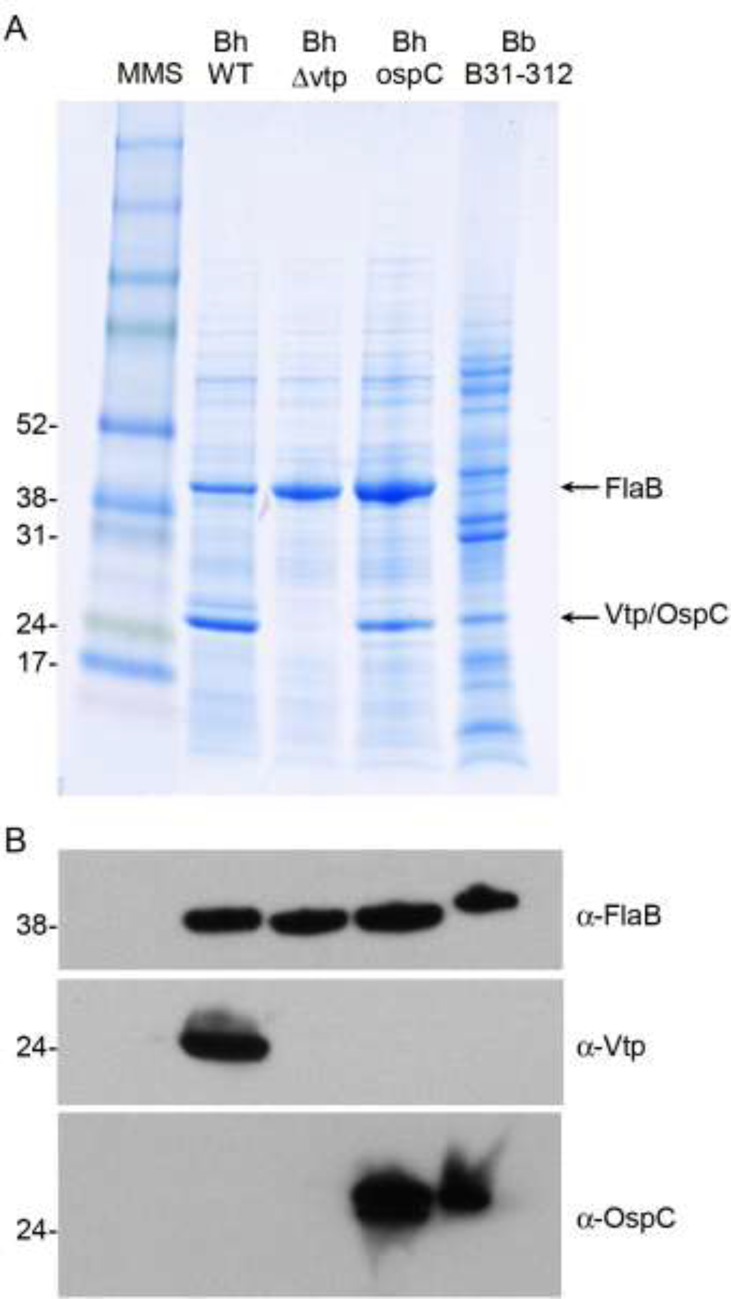
SDS-PAGE and immunoblot analysis of whole-cell lysates of wild-type and genetically manipulated strains. A, stained protein profiles and B, reactivity of FlaB, Vtp and OspC to specific monoclonal antibodies. Bh WT = B. hermsii wild-type; Bh Δvtp = B. hermsii vtp deletion mutant; Bh OspC = B. hermsii Δvtp::ospC+; Bb B31–312 = B. burgdorferi high passage strain. Molecular mass standards (MMS) are shown on the left in kiloDaltons.
3.2. Infection of ticks with the transgenic B. hermsii
We infected a cohort of O. hermsi ticks with the Δvtp::ospC+ transgenic B. hermsii. Spirochetes in blood smears from the mouse used to infect these ticks stained positively by IFA with the mouse anti-Vlp-7 monoclonal antibody (Fig. 2A), which confirmed the identity of the spirochetes acquired by the ticks as serotype 7.
Fig. 2.
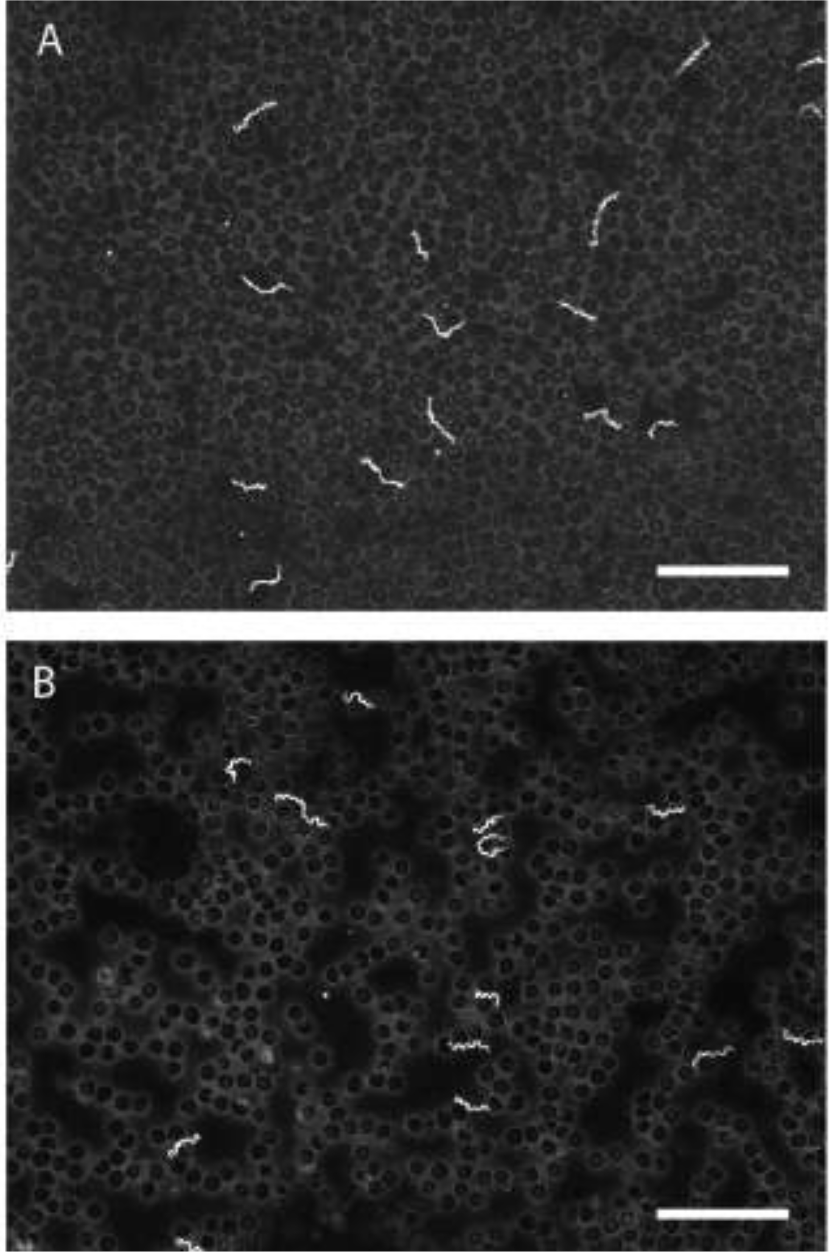
Transgenic Δvtp::ospC+ B. hermsii (A) in blood of mouse used to infect ticks and (B) in a mouse later infected by tick bite. Spirochetes in both smears stained with anti-Vlp7 monoclonal antibody confirming serotype 7. Scale bars = 50 μm.
Ticks were examined after their acquisition blood meal to determine if spirochetes infecting them produced OspC. The midgut and salivary glands from 2 to 5 ticks were examined at 2, 6, 7, 11 and 29 weeks after infection (Table 2). Cumulatively in 16 ticks examined by IFA, OspC was upregulated by a proportion of the spirochetes. OspC was found on significantly more transgenic B. hermsii in the salivary glands (42%; 496 of 1,182) than in the midgut (7.2%; 51 of 713) (Chi Square = 187.1; p <0.0001).
Table 2.
Transgenic Borrelia hermsii Δvtp::ospC+ producing OspC in Ornithodoros hermsi ticks.
| Weeks PIa | No. Ticks | No. and tick stageb | Midgut | Salivary Glands | ||||
|---|---|---|---|---|---|---|---|---|
| Infected | No. OspC+ | % OspC+ | Pairs infected | No. OspC+ | % OspC+ | |||
| 2 | 2 | 1N, 1♂ | 2/2c | 0/123d | 0 | 0/0c | NA | NA |
| 6 | 2 | 1♂, 1♀ | 2/2 | 7/200 | 3.5 | 2/2 | 4/95d | 4.2 |
| 7 | 4 | 2♂, 2♀ | 4/4 | 25/196 | 12.8 | 2/4 | 100/379 | 26.4 |
| 11 | 3 | 3♂ | 2/3 | 10/101 | 9.9 | 2/3 | 210/522 | 40.4 |
| 29 | 5 | 4♂, 1♀ | 4/5 | 9/93 | 9.7 | 3/5 | 182/186 | 97.8 |
| Total | 16 | 1N,11♂,4♀ | 14/16 | 51/713 | 7.2 | 9/16 | 496/1182 | 41.6 |
PI = post infection
N = nymph, ♂ = male, ♀ = female
number examined/number infected
number spirochetes OspC+/number spirochetes examined
The proportion of spirochetes producing OspC increased with time after their acquisition by ticks (Table 2) (Fig. 3). By 29 weeks, 97.8% (182 of 186) of the spirochetes examined in the salivary glands were OspC-positive (Figs. 3 & 4), which was significantly higher than the proportion of spirochetes producing this protein in the samples examined earlier (31.5%; 314 of 996) (Chi Square = 164.5; p < 0.0001). Thus, with a longer residence of the spirochetes in ticks, nearly all the transgenic B. hermsii switched to producing OspC in the salivary glands. This pattern of upregulation of OspC was very similar to wild-type B. hermsii producing Vtp, in which the prevalence of spirochetes producing Vtp in the ticks’ salivary glands increased and peaked at 96.8% at 32 weeks after infection (Raffel et al., 2014).
Fig. 3.
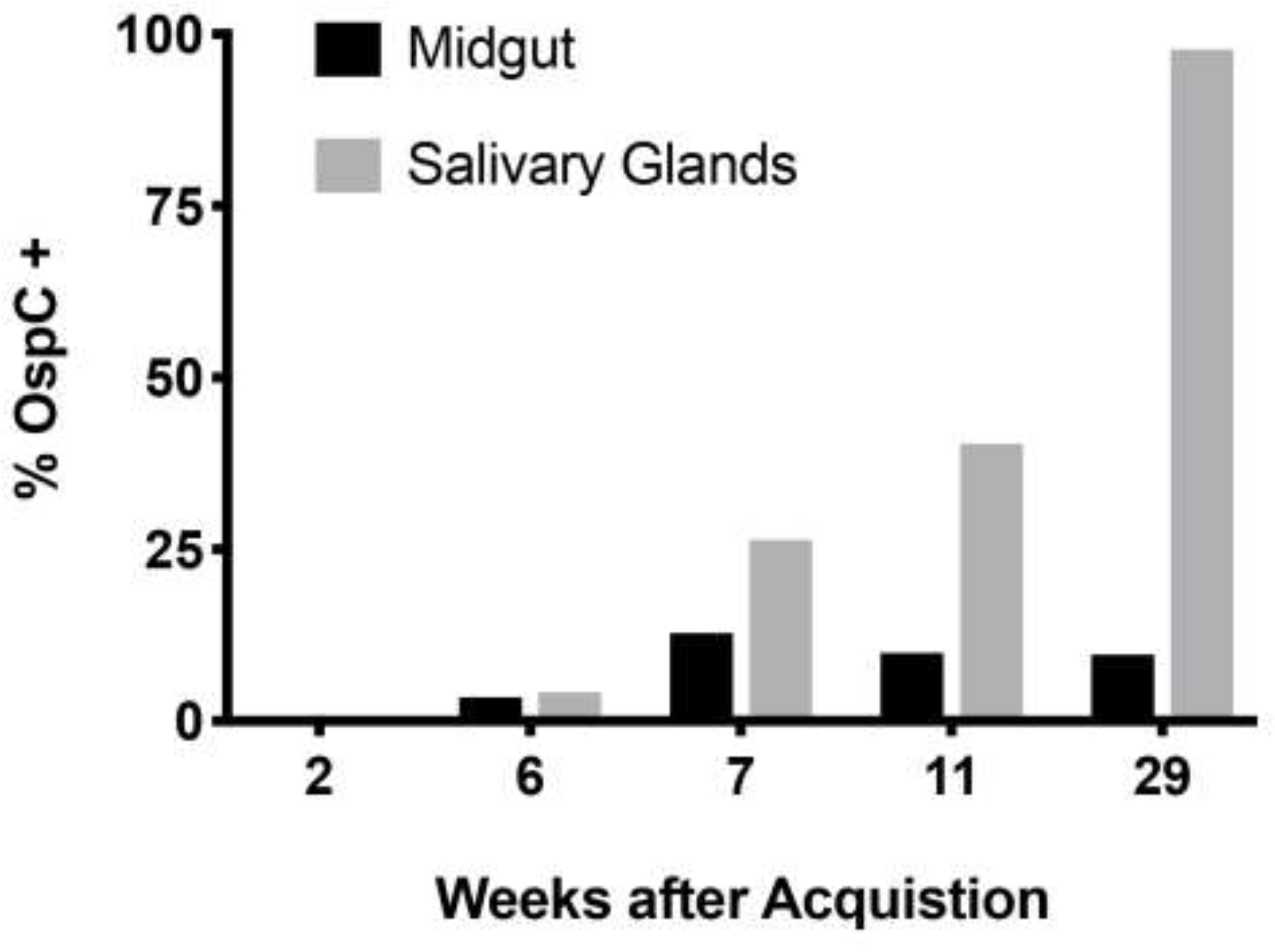
Up-regulation of OspC in Borrelia hermsii Δvtp::ospC+ transgenic spirochetes in tick midgut and salivary glands following spirochete acquisition by Ornithodoros hermsi. Percentages for midgut infections are in Table 2.
Fig. 4.
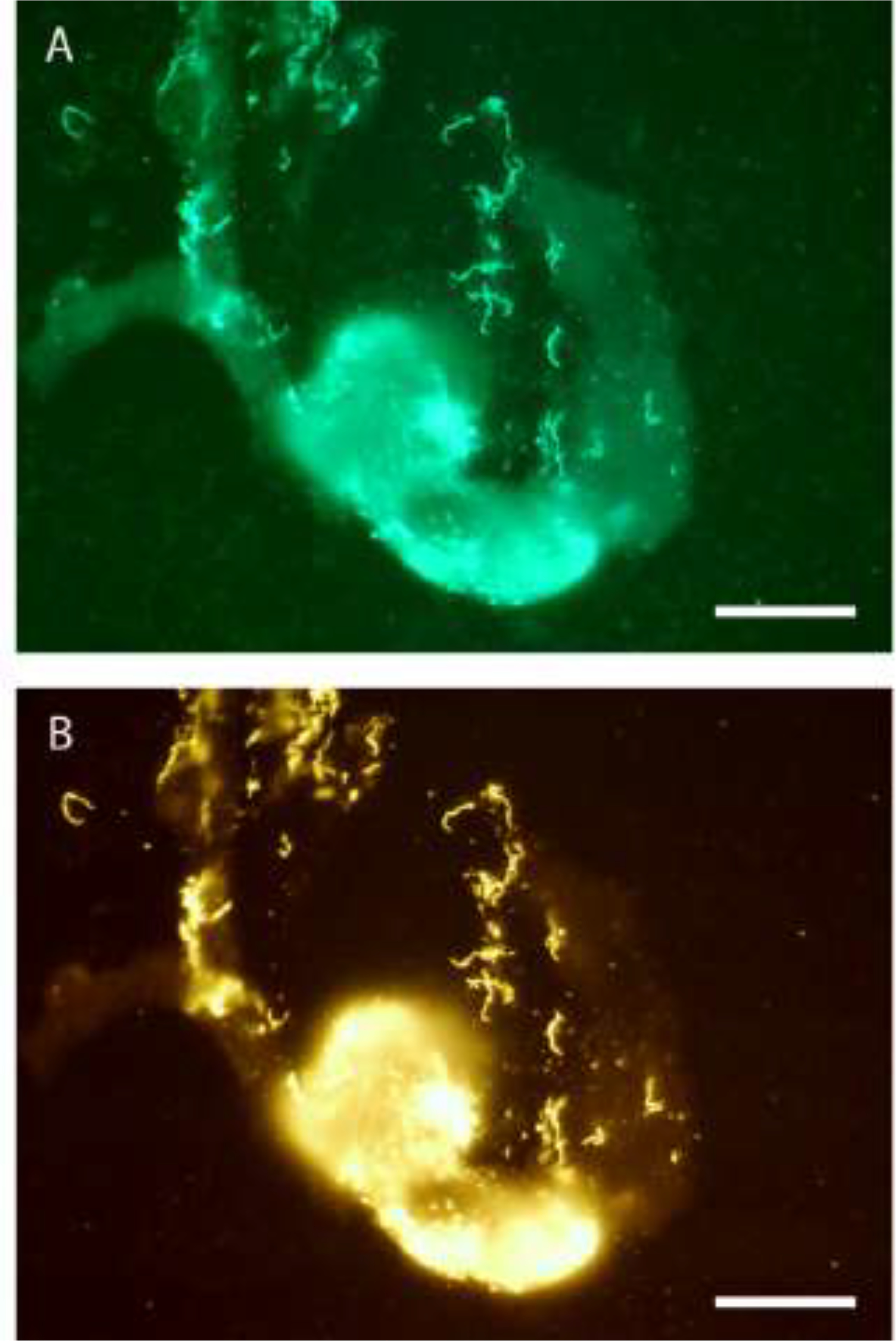
Immunofluorescence staining of Δvtp::ospC+ transgenic Borrelia hermsii in the salivary gland of Ornithodoros hermsi 29 weeks after infection. A, stained with rabbit anti-B. hermsii immune serum and FITC, and B, same field stained with mouse Mab anti-OspC antibody B5 and RITC with double-stained spirochetes yellow. Scale bars = 50 μm.
3.3. Transmission experiments with ticks infected with the transgenic B. hermsii
At 15 and 26 weeks after the previous infectious blood meal, groups of 12 to 23 nymphal and adult ticks infected with the transgenic B. hermsii were fed on 4 mice during each of the two time points (Table 3). In all, 7 of 8 mice (88%) developed microscopically detectable spirochetemias 7 to 9 days after being fed upon by these ticks. Infected blood from the three mice in the second group were inoculated into three sets of 5-ml tubes of BSK medium with either gentamycin, kanamycin, or no antibiotics. Spirochetes grew in medium with either gentamycin or no antibiotics but not with kanamycin, which confirmed the spirochetes contained the gentamycin-ospC construct. Blood smears from the 7 mice infected by tick bite were also examined by IFA during the first peak in spirochetemia (days 9 – 11) and showed the spirochetes produced neither OspC nor Vtp but reacted strongly with the mouse anti-Vlp 7 antibody (Fig. 2B). These results were anticipated based on the inverse reciprocal expression of the vmp and vtp promoters (Barbour et al., 2000). Thus, these transgenic spirochetes reverted from producing OspC in ticks (Fig. 4) to Vlp7 in mice (Fig. 2B), which was the same serotype-specific protein produced previously when the ticks acquired the infection (Fig. 2A).
Table 3.
Transmission feeding experiments with Ornithodoros hermsi ticks infected with the transgenic Borrelia hermsii Δvtp::ospC+ producing OspC.
| Weeks PIa | Single Mouse Fed Upon by ticks | Total Ticks Fed | Stageb | Mouse Infectedc |
|---|---|---|---|---|
| 15 | Mouse 1 | 12 | 1N, 7♂, 4♀ | + |
| 15 | Mouse 2 | 18 | 2 N, 5♂, 11♀ | + |
| 15 | Mouse 3 | 13 | 5 N, 1♂, 7♀ | + |
| 15 | Mouse 4 | 20 | 2 N, 7♂, 11♀ | + |
| 26 | Mouse 5 | 23 | 3 N, 5♂, 15♀ | + |
| 26 | Mouse 6 | 20 | 1 N, 8♂, 11♀ | + |
| 26 | Mouse 7 | 19 | 3 N, 2♂, 14♀ | − |
| 26 | Mouse 8 | 22 | 2 N, 14♂, 6♀ | + |
PI = post infection, time since ticks acquired spirochetes from infected mouse
N = nymph, ♂ = male, ♀ = female (numbers of each in the pool of ticks that fed)
+ = mouse became spirochetemic and seroconverted; − = mouse had no detectable spirochetemia and did not seroconvert
3.4. Serology
Convalescent serum samples collected from the mice had IFA titers of 1:2,048 to 1:8,192 for 6 infected mice (1 mouse died before sampling) and <1:16 for the single mouse that had no prior detectable spirochetemia. Thus, the serological data supported the spirochetemia results showing infectivity of the vtp-minus mutant B. hermsii was restored by the functional complementation with the ospC gene of B. burgdorferi.
4. Discussion
The transgenic B. hermsii produced OspC like wild-type spirochetes produce Vtp (Raffel et al., 2014). The prevalence of OspC+ spirochetes 1) increased with time after acquisition by ticks, 2) were more prevalent in the salivary glands than in the midgut, 3) were infectious by tick bite, and 4) switched back to serotype 7 following tick transmission to mice.
Our hypothesis prior to performing the experiments was that B. hermsii lacking vtp but transformed with ospC would behave like wild-type spirochetes, which is what we observed as summarized above. Our prediction was based on the sequence relatedness of these proteins, when these proteins are produced during the transmission cycles of relapsing fever and Lyme disease spirochetes, and their requirement for mammalian infection via tick bite (Grimm et al., 2004; Raffel et al., 2014). Vtp and OspC are orthologs belonging to a family of small outer-surface lipoproteins produced by relapsing fever and Lyme disease spirochetes (Marconi et al., 1993b; Carter et al., 1994; Margolis et al., 1994); however, OspC is the only member of this family in B. burgdorferi (Fraser et al., 1997). In contrast, a diversity of these variable small proteins (Vsps) are produced by B. hermsii as part of this spirochete’s adaptation to vary antigenically in blood (Hinnebusch et al., 1998; Dai et al., 2006; Barbour, 2016). vtp is also unique from other vsp genes by having its own promotor and only a single copy in the genome, which is inversely expressed with the promoter driving the expression of all other vmp genes (Barbour et al., 2000).
B. hermsii Vtp and B. burgdorferi OspC are also polymorphic proteins within their natural populations, in which multiple Vtp and OspC types or groups have been described (Theisen et al., 1993; Jauris-Heipke et al., 1995; Porcella et al., 2005; Barbour and Travinsky, 2010; Johnson et al., 2016). Within each type the amino acid sequences are identical or nearly so, while between types the sequences vary enough to make these proteins antigenically distinct (Gilmore et al., 1996; Probert et al.,1997; Barbour and Travinsky, 2010; Krajacich et al., 2015). To date, we have identified nine Vtp sequence types in the two genomic groups (GGI and GGII) of B. hermsii (Fig. 5) (Porcella et al., 2005; Fischer et al., 2009; Kelly et al., 2014; Christensen et al., 2015; Johnson et al., 2016) (and herein). Our previous genetic analyses showed greater diversity of sequences among the GGI isolates (Schwan et al., 2007), which is true also for the vtp locus (Fig. 5). GGI isolates (N = 42) are comprised of eight Vtp types compared to just three types in the GGII isolates (N = 37) (Fig. 5). The amino acid sequences are mostly identical within each type but include variants that have 93.4 to 99.5% identity (minus the signal peptide) to the consensus sequences. Between the types, the amino acid sequences share only 53.7 to 67.5% and 61.7 to 65.8% identities among the GGI and GGII B. hermsii, respectively. The B. burgdorferi B31 OspC is 42.2% identical to the B. hermsii DAH Vtp it replaced. Thus, the Δvtp::ospC+ transgenic spirochetes could be considered B. hermsii containing a novel vtp gene encoding a protein approximately 10 to 30% more divergent from other Vtps found in natural populations.
Fig. 5.
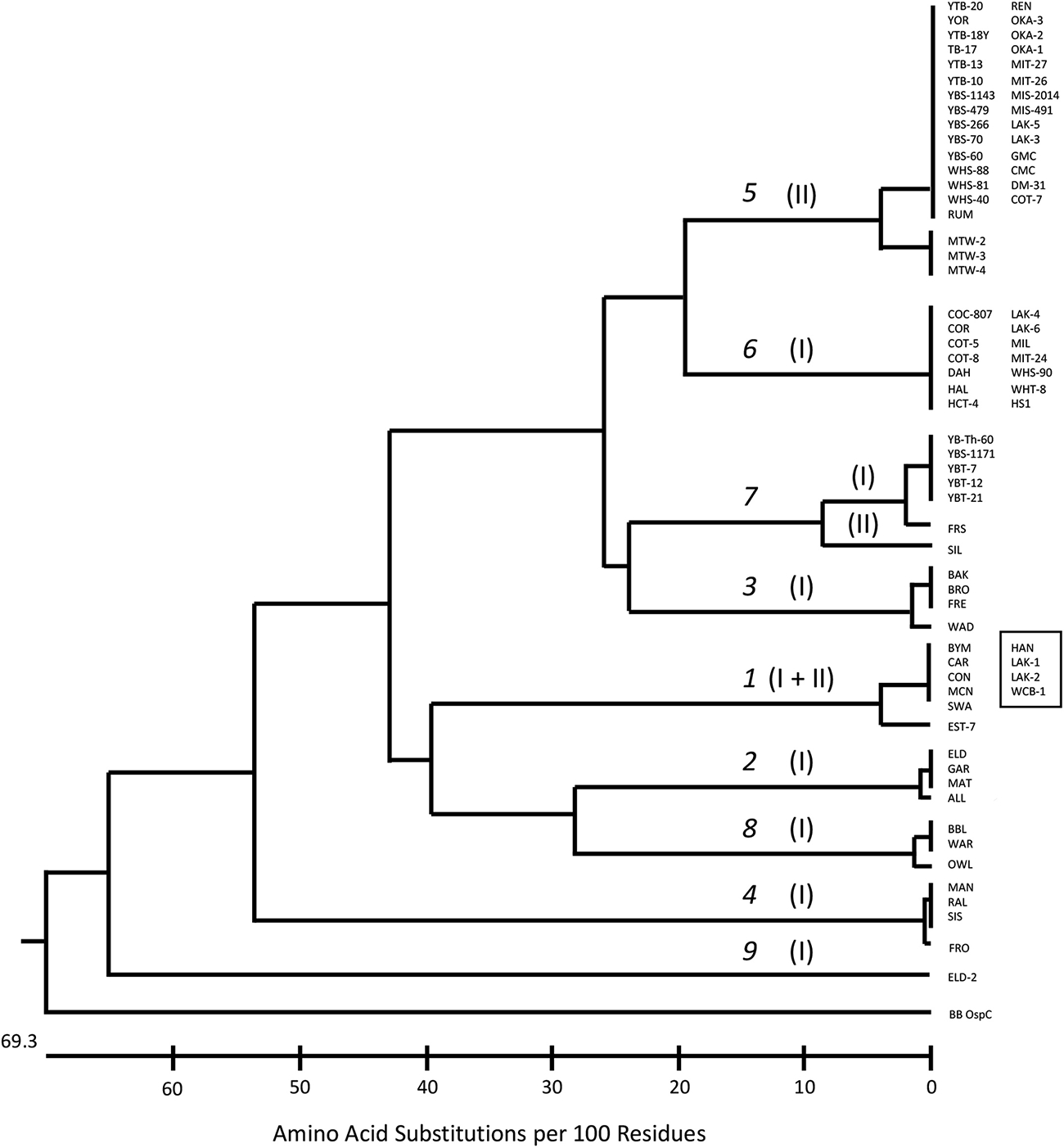
Tree showing polymorphism in Vtp amino acid sequences among 79 isolates and DNA samples of Borrelia hermsii. Borrelia burgdorferi B31 OspC, the protein used in the transformation, is included. The nine Vtp types are shown on the horizontal branches (1–9 in italics) and the genomic group of the spirochetes is shown in parentheses. Vtp Types 1 and 7 have members of both genomic groups (GG II in box). GenBank accession numbers for 8 sequences not previously deposited are MK076576 - MK076583.
Although these proteins are highly polymorphic, the signal peptide of the first 19 amino acids including the first cysteine (MKKNTLSAILMTLFLFISC) is identical in all B. hermsii Vtp we have examined (N = 79) (Porcella et al., 2005; Fischer et al., 2009; Kelly et al., 2014; Christensen et al., 2015; Johnson et al., 2016 and herein). This sequence is also identical to the OspC signal peptide of B. burgdorferi (Carter et al., 1994; Margolis et al., 1994) and in all related species for which complete OspC sequences are available in the National Center for Biotechnology Information database (www.ncbi.nlm.nih.gov) (as of February 19, 2019) (Table 4). However, one base in the nucleotide sequence of the signal peptide of the B. hermsii vtp varies in the third position of the tenth codon for leucine (TTA and TTG). Among GGI isolates, both TTA (N = 12) and TTG (N = 30) occur, while in the GGII isolates (N = 37), only TTA has been observed, again displaying less sequence variation, albeit in a small fragment, in the latter genomic group. An alignment we performed of the B. hermsii DAH vtp sequence with the B. burgdorferi B31 ospC (not shown) identified three synonymous base changes in the signal sequence at positions 7, 24 and 30, showing less conservation at the nucleotide level between the species yet still retaining the identical amino acid sequence.
Table 4.
Species identified in GenBanka containing the identical signal peptideb for the Vtp and OspC orthologous proteins.
| Species | 100% Coverage | 100% Identity |
|---|---|---|
| B. hermsii GGI | X | X |
| B. hermsii GGII | X | X |
| B. burgdorferi | X | X |
| B. garinii | X | X |
| B. afzelii | X | X |
| B. valaisiana | X | X |
| B. bissettiae | X | X |
| B. japonica | X | X |
| B. spielmanii | X | X |
| B. bavariensis | X | X |
| B. tanuki | X | X |
| B. turdi | X | X |
| B. yangtze | X | X |
| B. findlandensis | X | X |
| B. chilensis | X | X |
| B. mayonii | X | X |
| B. americana c | 89% | X |
| B. carolinensis c | 89% | X |
| B. andersonii | ?d | ? |
| B. kurtenbachii | ? | ? |
| B. lusitaniae | ? | ? |
| B. sinica | ? | ? |
| B. lanei | ? | ? |
Search performed February 19, 2019
MKKNTLSAILMTLFLFISC
Missing first two amino acids MK
? = OspC sequence not found or only partial sequence that lacked signal peptide
While all B. hermsii and B. burgdorferi sensu lato (s.l.) species appear to have Vtp and OspC containing the identical signal peptide, we have not yet found or seen reported by other investigators the same signal sequence in any of the other ~27 current valid species of relapsing fever spirochetes (Barbour and Schwan, 2018). This is somewhat puzzling, given the more distant phylogenetic relationship of B. hermsii to B. burgdorferi compared to other relapsing fever spirochetes of North America, such as Borrelia parkeri and Borrelia turicatae, and Old World species such as Borrelia crocidurae, Borrelia persica, Borrelia hispanica and Borrelia duttonii (Fukunaga et al., 1996; Ras et al., 1996; Barbour, 2014). Genomic sequences of multiple relapsing fever spirochetes completed at the RML and released to the public (GenBank accession numbers CP000048 [B. hermsii DAH], CP004146 [B. hermsii YOR], CP005680 [B. hermsii MTW], CP005706 [B. hermsii YBT], CP000049 [B. turicatae 91E135], CP005829 [B. anserina BA2], CP005745 [B. coriaceae Co53], CP004217 [B. miyamotoi FR64b], CP005851 [B. parkeri SLO], CP004267 [B. crocidurae DOU], AZIT01000001 [B. duttonii CR2A]) contain no sequences identical to the signal peptide MKKNTLSAILMTLFLFISC except for the four B. hermsii genomes. Given the plasmid location of vtp and ospC (Marconi et al., 1993a; Sadziene et al., 1993; Barbour et al., 2000; Porcella et al., 2005), the near-telomeric location of vtp on its linear plasmid (Barbour, 2016), and difficulty in sequencing the ends of linear plasmids (Casjens et al., 2000), the genome sequencing may have missed existing vtp orthologs.
Both vtp and ospC are encoded on plasmids bearing resT (Byram et al., 2004; Barbour, 2016), an essential gene encoding the enzyme telomere resolvase (Kobryn and Chaconas, 2002; Byram et al., 2004). Given the synteny between the cp26 circular plasmid of B. burgdorferi and part of the 53 kb linear plasmid of B. hermsii encoding this locus, we used resT as a potential signpost for finding possible vtp orthologs in other species. The best match was in B. anserina, which has a variable small protein (214 amino acids) with a nearly identical signal peptide (89.5% identical; 94.7% similar) (Table 5) encoded on a linear plasmid in the opposite direction and seven orfs downstream from resT (our contig sequence CP005835, and CP014520 (Elbir et al., 2017), as in B. hermsii. In B. turicatae BTE5EL on linear plasmid lp44 (CP015635) (Kingry et al., 2016), a variable small membrane protein (209 amino acids) is encoded in the opposite direction and again seven orfs downstream of resT and contains a signal peptide 73.7% identical and 89.5% similar to B. hermsii Vtp signal peptide (Table 5). In B. coriaceae Co53, its genomic sequence contains a plasmid contig (CP005749) encoding a larger variable membrane protein (353 amino acids) but with the same location and orientation to resT, with a signal peptide 80% identical and 85% similar to the B. hermsii Vtp signal peptide (Table 5). Lastly, the RML B. miyamotoi genome (CP004217) contains a contig with an orf encoding a predicted 248 amino acid membrane protein with the same orientation to resT, and a signal peptide 66.7% identical and 81% similar to the B. hermsii Vtp signal peptide (Table 5). We believe these proteins are good candidates to examine further as possible transmission-associated proteins in B. anserina, B. turicatae, B. coriaceae and B. miyamotoi, as these species alternate between their tick and vertebrate hosts.
Table 5.
Signal peptide sequences for possible transmission-associated orthologous proteins compared to B. hermsii DAH.
| Species | Amino acid sequence | % Identical | % Similar |
|---|---|---|---|
| B. anserina | MNKNALSAILMTLFLFISCa | ||
| B. hermsii | MKKNTLSAILMTLFLFISC | ||
| Identity | M-KN-LSAILMTLFLFISC | 89.5 | 94.7 |
| B. turicatae | MKRITLSALLMTLFLLMSC | ||
| B. hermsii | MKKNTLSAILMTLFLFISC | ||
| Identity | MK——TLSA-LMTLFL--SC | 73.7 | 89.5 |
| B. coriaceae | MKKNTLSAILFMTLFFLVSC | ||
| B. hermsii | MKKNTLSAIL-MTLFLFISC | ||
| Identity | MKKNTLSAIL-MTLF---SC | 80.0 | 85.0 |
| B. miyamotoi | MSKRKTLSAIIMTLFLGLVSC | ||
| B. hermsii | MKKN-TLSAILMTLFLFI-SC | ||
| Identity | M-K--TLSAI MTLFL---SC | 66.7 | 81.0 |
The lipoprotein signal peptidase cleavage site lies between serine (S) and cysteine (C) identified in bold text.
While OspC restored the wild-type infectious phenotype to B. hermsii lacking Vtp, the role these proteins play for infectivity immediately following transmission by ticks is possibly complex and remains uncertain. Significant attention has addressed possible functions of OspC and other B. burgdorferi surface proteins (Caine and Coburn, 2016). While OspC does not contribute physiologically to B. burgdorferi infection in mice (Stewart et al., 2006), its function may be, in part, to provide membrane stability that is replaceable with other outer surface proteins (Xu et al., 2008). Other possible activities and roles include its requirement for early colonization in mammals and protection from the host’s innate immune defenses (Grimm et al., 2004; Tilly et al., 2006), promote survival in blood by binding complement C4b (Caine et al., 2017), aid in spirochete dissemination and adaptation to specific host tissues (Skare et al., 2016), provide protection from phagocytosis by macrophages (Carrasco it al., 2015), bind plasminogen (Coleman et al., 1997; Hu et al., 1995; Lagal et al., 2006; Önders et al., 2012), bind fibronectin (Bierwagen et al., 2019), and bind the Ixodes scapularis salivary gland protein Salp15 (Ramamoorthi et al., 2005).
Experiments addressing the mechanistic role for Vtp and B. hermsii infectivity are lacking. The activities and possible functions described for OspC for the infectious cycle of B. burgdorferi might apply for Vtp and B. hermsii. The Vtp- mutant might have infectivity restored if complemented with other outer surface proteins rather than OspC, such as OspA, OspE or DpbA as shown for B. burgdorferi (Xu et al., 2008). The reciprocal expression of vlp7 and vtp (Barbour et al., 2000) breaks down in the salivary glands of O. hermsi, as Vlp7 was not present in either wild-type or Vtp-minus mutant spirochetes (Raffel et al., 2014). Thus, these mutant cells may lack any major outer surface protein, which reduces their fitness when transmitted to mammals.
The switch from a bloodstream Vmp to Vtp in ticks (Schwan and Hinnebusch, 1998; Raffel et al., 2014), recapitulated here with the transgenic B. hermsii, is intriguing given that low dose, cloned spirochetes producing one of any 24 Vmps (serotypes 1 to 24) are infectious by intraperitoneal inoculation in mice (Stoenner et al., 1982). Previously, we hypothesized that such a switch from a Vmp to Vtp in ticks might allow B. hermsii to achieve higher cell densities in the blood during the first spirochetemia following their next transmission by ticks, by diverting the host’s antibody response to Vtp before switching to a bloodstream serotype (Raffel et al., 2014). A similar type of parasite diversion of host immunity was described recently as a “feint attack”, coined after a military tactic to divert enemy responses, when describing antigenic variation involving different sized variant surface glycoproteins of the African trypanosome Trypanosoma brucei (Liu et al., 2018). This phrase aptly describes our notion for one reason as to why B. hermsii switches to Vtp in ticks for first entry back into mammals. Our previous study showed that B. hermsii lacking an intact vmp expression site, which was unable to switch from Vtp to a bloodstream Vmp, achieved lower cell densities in blood compared to spirochetes that could switch (Raffel et al., 2014). However, the Vtp- mutant spirochete was also less fit, producing lower cell densities in the blood of SCID mice incapable of mounting an acquired immune response (Raffel et al., 2014). In general, this first antigenic switch or any other mechanism that contributes to B. hermsii achieving higher cell densities in the host’s peripheral blood would increase the potential for spirochete acquisition by small, fast-feeding ticks (McCoy et al., 2010; Lopez et al., 2011).
Separate from why Vtp and OspC are essential to make individual spirochetes infectious by tick bite or disseminate in a vertebrate host, is what role(s) these proteins play at the population level in natural endemic foci. While not the objective of our present work and beyond the scope of this discussion, Vtp and OspC are unique among all other proteins produced by B. burgdorferi s.l. and B. hermsii, respectively, because of their temporal presence in the spirochetes’ life cycles (Schwan and Piesman, 2002), their immunogenicity (Bockenstedt et al., 1997; Gilmore et al., 1996; Izac and Marconi, 2019; Krajacich et al., 2015; Marcsisin et al., 2016; Preac-Mursic et al., 1992), their polymorphism into multiple antigenically distinct groups (Barbour and Travinsky, 2010; Brisson and Dykhuizen, 2004; Jauris-Heipke et al., 1995; Johnson et al., 2016; Wang et al., 1999), and their unique and shared signal peptide discussed herein. The immunogenicity, in part, of these proteins has resulted in a balanced polymorphism of many antigenically distinct groups that likely underlies the ability of these spirochetes to remain established among vertebrate populations that are temporarily immune to some antigenic types but not others (Bhatia et al., 2018).
In our attempt to identify orthologous transmission-associated proteins in other species of relapsing fever spirochetes, we focused on the unique Vtp-OspC signal peptide identical in all isolates of B. hermsii and B. burgdorferi s.l. species. Three species with the greatest identity were B. anserina, B. coriaceae and B. turicatae (Table 5). These species contain ospC-related sequences based on northern and Southern blot analyses (Marconi et al., 1993b). Other earlier studies identified homologous amino acid sequences in a family of small proteins (~20 kD) shared by Lyme disease and relapsing fever spirochetes (Carter et al., 1994; Margolis et al., 1994). Additionally, partial amino acid sequences of OspC in 18 isolates of B. burgdorferi s.l. and Vtp (= Vmp33) from one isolate of B. hermsii lead the authors to conclude that these spirochetes evolved from a common ancestor (Theisen et al., 1995).
We believe that the differential regulation and switching to Vtp and OspC when spirochetes are transmitted by tick bite may be a common theme shared by most if not all of these bacteria, however few species have been studied to support this notion. Borreliae may have evolved first as symbionts of ticks (Hoogstraal, 1979), which comprise a monophyletic group of arthropods with an origin dating back to the middle Permian Period approximately 260 million years ago (MYA) (Mans et al., 2011). The physiology of blood feeding in ancestral ticks then evolved separately in argasid and ixodid ticks after these families diverged during the Upper Triassic Period 223 to 234 MYA (Mans et al., 2019). The orthologous relationship of Vtp and OspC is not unique among the genomes of these spirochetes, but may represent a shared adaptation for transmission that evolved in a common ancestor before the split of these spirochetes into separate species (Fitch, 1970).
5. Conclusions
Adaptations for spirochete transmission and acquisition by ticks may be widely shared among diverse groups of these bacteria and blood-feeding arthropods (Schwan and Piesman, 2002). Vtp and OspC represent orthologous proteins shared by B. hermsii and B. burgdorferi s.l. that belong to a family of small variable major proteins involved with antigenic variation in the former species (Carter et al., 1994; Margolis et al., 1994; Hinnebusch et al., 1998). Both proteins are essential for spirochete infection when transmitted by both fast and slow feeding ticks (Grimm et al., 2004; Raffel et al., 2014). Although OspC from B. burgdorferi restored a wild-type phenotype to B. hermsii lacking Vtp, the biological roles for these proteins during tick transmission and early colonization of mammals may be unique to, or multifunctional in, each species of spirochete.
Acknowledgement
We thank Bob Gilmore for the OspC monoclonal antibody B5, Anita Mora for help with the figures, and Joe Hinnebusch and Philip Stewart for their thoughtful reviews of the manuscript. This work was supported by the Division of Intramural Research, National Institute of Allergy and Infectious Diseases, National Institute of Health.
Footnotes
Declaration of interest
None
Publisher's Disclaimer: This is a PDF file of an unedited manuscript that has been accepted for publication. As a service to our customers we are providing this early version of the manuscript. The manuscript will undergo copyediting, typesetting, and review of the resulting proof before it is published in its final form. Please note that during the production process errors may be discovered which could affect the content, and all legal disclaimers that apply to the journal pertain.
References
- Barbour AG, 1984. Isolation and cultivation of Lyme disease spirochetes. Yale J. Biol. Med. 57, 521–525. [PMC free article] [PubMed] [Google Scholar]
- Barbour AG, 1987. Immunobiology of relapsing fever, pp. 125–137. In: Cruse JM and Lewis RE Jr. (eds.), Contributions to Microbiology and Immunology, vol. 8. Karger, Basel. [PubMed] [Google Scholar]
- Barbour AG, 2014. Phylogeny of a relapsing fever Borrelia species transmitted by the hard tick Ixodes scapularis. Infect. Genet. Evol. 27, 551–558. [DOI] [PMC free article] [PubMed] [Google Scholar]
- Barbour AG, 2016. Chromosome and plasmids of the tick-borne relapsing fever agent Borrelia hermsii. Genome Announc. 4, e00528–00516. [DOI] [PMC free article] [PubMed] [Google Scholar]
- Barbour AG, Carter CJ, Sohaskey CD, 2000. Surface protein variation by expression site switching in the relapsing fever agent Borrelia hermsii. Infect. Immun. 68, 7114–7121. [DOI] [PMC free article] [PubMed] [Google Scholar]
- Barbour AG, Hayes SF, Heiland RA, Schrumpf ME, Tessier SL, 1986. A Borrelia-specific monoclonal antibody binds to a flagellar epitope. Infect. Immun. 52, 549–554. [DOI] [PMC free article] [PubMed] [Google Scholar]
- Barbour AG, Schwan TG, 2018. Borrelia. Swellengrebel 1907, 582AL, pp. DOI: 10.1002/9781118960608, Bergey’s Manual of Systematic Archaea and Bacteria. John Wiley & Sons, Inc. [DOI] [Google Scholar]
- Barbour AG, Travinsky B, 2010. Evolution and distribution of the ospC gene, a transferable serotype determinant of Borrelia burgdorferi. mBio 1, e00153–00110. [DOI] [PMC free article] [PubMed] [Google Scholar]
- Battisti JM, Raffel SJ, Schwan TG, 2008. A system for site-specific genetic manipulation of the relapsing fever spirochete Borrelia hermsii, pp. 69–84. In: DeLeo FR, Otto M (eds.), Methods in Molecular Biology 431: Bacterial Pathogenesis Methods and Protocols, vol. 431. Humana Press, Totowa. [DOI] [PubMed] [Google Scholar]
- Benach JL, Coleman JL, Skinner RA, Bosler EM, 1987. Adult Ixodes dammini on rabbits: a hypothesis for the development and transmission of Borrelia burgdorferi. J. Infect. Dis. 155, 1300–1306. [DOI] [PubMed] [Google Scholar]
- Bhatia B, Hillman C, Carracoi V, Cheff BN, Tilly K, Rosa PA, 2018. Infection history of the blood-meal host dictates pathogenic potential of the Lyme disease spirochete within the feeding tick vector. PLoS Pathog. 14: e1006959. [DOI] [PMC free article] [PubMed] [Google Scholar]
- Bierwagan P, Szpotkowski K, Jaskolski M, Urbanowicz A, 2019. Borrelia outer surface protein C is capable of human fibrinogen binding. FEBS J. 286, 2415–2428. [DOI] [PubMed] [Google Scholar]
- Bockenstedt LK, Hodzic E, Feng S, Bourrel KW, de Silva A, Montgomery RR, Fikrig E, Radolf JD, Barthold SW, 1997. Borrelia burgdorferi strain-specific Osp C-mediated immunity in mice. Infect. Immun. 65, 4661–4667. [DOI] [PMC free article] [PubMed] [Google Scholar]
- Brisson D, Dykhuizen DE, 2004. ospC diversity in Borrelia burgdorferi: different hosts are different niches. Genetics 168, 713–722. [DOI] [PMC free article] [PubMed] [Google Scholar]
- Burgdorfer W, Hayes SF, Benach JL, 1988. Development of Borrelia burgdorferi in ixodid tick vectors, pp. 172–179. In: Benach JL Bosler EM (eds.), Lyme Disease and Related Disorders. New York Academy of Sciences, New York. [DOI] [PubMed] [Google Scholar]
- Burgdorfer W, Barbour AG, Hayes SF, Benach JL, Grunwaldt E, Davis JP, 1982. Lyme disease-a tick-borne spirochetosis? Science 216, 1317–1319. [DOI] [PubMed] [Google Scholar]
- Byram R, Stewart PE, Rosa P, 2004. The essential nature of the ubiquitous 26-kilobase circular replicon of Borrelia burgdorferi. J. Bacteriol. 186, 3561–3569. [DOI] [PMC free article] [PubMed] [Google Scholar]
- Caine JA, Coburn J, 2016. Multifunctional and redundant roles of Borrelia burgdorferi outer surface proteins in tissue adhesion, colonization, and complement evasion. Front. Immunol. 7, 442. [DOI] [PMC free article] [PubMed] [Google Scholar]
- Caine JA, Lin Y-P, Kessler JR, Sato H, Leong JM, Coburn J, 2017. Borrelia burgdorferi outer surface protein C (OspC) binds complement C4b and confers bloodstream survival. Cell. Microbiol. 19:e12786. [DOI] [PMC free article] [PubMed] [Google Scholar]
- Carrasco SE, Troxell B, Yang Y, Brandt SL, Li H, Sandusky GE, Condon KW, Serezani CH, Yang XF, 2015. Outer surface protein OspC is an antiphagocytic factor that protects Borrelia burgdorferi from phagocytosis by macrophages. Infect. Immun. 83, 4848–4860. [DOI] [PMC free article] [PubMed] [Google Scholar]
- Carter CJ, Bergström S, Norris SJ, Barbour AG, 1994. A family of surface-exposed proteins of 20 kilodaltons in the genus Borrelia. Infect. Immun. 62, 2792–2799. [DOI] [PMC free article] [PubMed] [Google Scholar]
- Casjens S, Palmer N, van Vugt R, Huang WM, Stevenson B, Rosa P, Lathigra R, Sutton G, Peterson J, Dodson RJ, Haft D, Hickey E, Gwinn M, White O, Fraser CM, 2000. A bacterial genome in flux: the twelve linear and nine circular extrachromosomal DNAs in an infectious isolate of the Lyme disease spirochete Borrelia burgdorferi. Mol. Microbiol. 35, 490–516. [DOI] [PubMed] [Google Scholar]
- Christensen J, Fischer RJ, McCoy BN, Raffel SJ, Schwan TG, 2015. Tickborne relapsing fever, Bitterroot Valley, Montana, USA. Emerg. Infect. Dis. 21, 217–223. [DOI] [PMC free article] [PubMed] [Google Scholar]
- Coleman JL, Gebbia JA, Piesman J, Degen JL, Bugge TH, Benach JL, 1997. Plasminogen is required for efficient dissemination of B. burgdorferi in ticks and for enhancement of spirochetemia in mice. Cell 89, 1111–1119. [DOI] [PubMed] [Google Scholar]
- Dai Q, Restrepo BI, Porcella SF, Raffel SJ, Schwan TG, Barbour AG, 2006. Antigenic variation by Borrelia hermsii occurs through recombination between extragenic repetitive elements on linear plasmids. Mol. Microbiol. 60, 1329–1343. [DOI] [PMC free article] [PubMed] [Google Scholar]
- Dworkin MS, Schwan TG, Anderson DE, Borchardt SM, 2008. Tick-borne relapsing fever. Infect. Dis. Clin. N. Am. 22, 449–468. [DOI] [PMC free article] [PubMed] [Google Scholar]
- Elbir H, Sitlani P, Bergström S, Barbour AG, 2017. Chromosome and megaplasmid sequences of Borrelia anserina (Sakharoff 1891), the agent of avian spirochetosis and type species of the genus. Genome Announc. 5, e00018–00017. [DOI] [PMC free article] [PubMed] [Google Scholar]
- Fischer RJ, Johnson TL, Raffel SJ, Schwan TG, 2009. Identical strains of Borrelia hermsii in mammal and bird. Emerg. Infect. Dis. 15, 2064–2066. [DOI] [PMC free article] [PubMed] [Google Scholar]
- Fitch WM, 1970. Distinguishing homologous from analogous proteins. Syst. Zool. 19, 99–113. [PubMed] [Google Scholar]
- Fraser CM, Casjens S, Huang WM, Sutton GG, Clayton R, Lathigra R, White O, Ketchum KA, Dodson R, Hickey EK, Gwinn M, Dougherty B, Tomb J-F, Fleischmann RD, Richardson D, Peterson J, Kerlavage AR, Quackenbush J, Salzberg S, Hanson M, Vugt RV, Palmer N, Adams MD, Gocayne J, Weidman J, Utterback T, Watthey L, McDonald L, Artiach P, Bowman C, Garland S, Fujii C, Cotton MD, Horst K, Roberts K, Hatch B, Smith HO, Venter JC, 1997. Genomic sequence of a Lyme disease spirochaete, Borrelia burgdorferi. Nature 390, 580–586. [DOI] [PubMed] [Google Scholar]
- Fukunaga M, Okada K, Nakao M, Konishi T, Sato Y, 1996. Phylogenetic analysis of Borrelia species based on flagellin gene sequences and its application for molecular typing of Lyme disease borreliae. Int. J. Syst. Bacteriol. 46, 898–905. [DOI] [PubMed] [Google Scholar]
- Gilmore RD, Piesman J, 2000. Inhibition of Borrelia burgdorferi migration from the midgut to the salivary glands following feeding by ticks on OspC-immunized mice. Infect. Immun. 68, 411–414. [DOI] [PMC free article] [PubMed] [Google Scholar]
- Gilmore RD, Kappel KJ, Dolan MC, Burkot TR, Johnson BJB, 1996. Outer surface protein C (OspC), but not P39, is a protective immunogen against a tick-transmitted Borrelia burgdorferi challenge: evidence for a conformational protective epitope in OspC. Infect. Immun. 64, 2234–2239. [DOI] [PMC free article] [PubMed] [Google Scholar]
- Grimm D, Tilly K, Byram R, Stewart PE, Krum JG, Bueschel DM, Schwan TG, Policastro PF, Elias AF, Rosa PA, 2004. Outer-surface protein C of the Lyme disease spirochete: A protein induced in ticks for infection of mammals. Proc. Natl. Acad. Sci. USA 101, 3142–3147. [DOI] [PMC free article] [PubMed] [Google Scholar]
- Herms WB, Wheeler CM, 1936. The tick vector of the infection. Relapsing Fever Calif. State Dept. Publ. Health Special Bull. No. 61, 24–28. [Google Scholar]
- Hinnebusch BJ, Barbour AG, Restrepo BI, Schwan TG, 1998. Population structure of the relapsing fever spirochete Borrelia hermsii as indicated by polymorphism of two multigene families that encode immunogenic outer surface lipoproteins. Infect. Immun. 66, 432–440. [DOI] [PMC free article] [PubMed] [Google Scholar]
- Hu LT, Perides G, Noring R, Klempner MS, 1995. Binding of human plasminogen to Borrelia burgdorferi. Infect. Immun. 63, 3491–3496. [DOI] [PMC free article] [PubMed] [Google Scholar]
- Hoogstraal H, 1979. Ticks and spirochetes. Acta Trop. 36, 133–136. [PubMed] [Google Scholar]
- Izac JR, Marconi RT, 2019. Diversity of the Lyme disease spirochetes and its influence on immune responses to infection and vaccination. Vet. Clin. Small Anim. 49, 671–686. [DOI] [PMC free article] [PubMed] [Google Scholar]
- Jauris-Heipke S, Liegl G, Preac-Mursic V, Rössler D, Schwab E, Soutschek E, Will G, Wilske B, 1995. Molecular analysis of genes encoding outer surface protein C (OspC) of Borrelia burgdorferi sensu lato: relationship to ospA genotype and evidence of lateral gene exchange of ospC. J. Clin. Microbiol. 33, 1860–1866. [DOI] [PMC free article] [PubMed] [Google Scholar]
- Johnson TL, Fischer RJ, Raffel SJ, Schwan TG, 2016. Host associations and genomic diversity of Borrelia hermsii in an endemic focus of tick-borne relapsing fever in western North America. Parasit. Vectors 9, 575. [DOI] [PMC free article] [PubMed] [Google Scholar]
- Kelly AL, Raffel SJ, Fischer RJ, Bellinghausen M, Stevenson C, Schwan TG, 2014. First isolation of the relapsing fever spirochete, Borrelia hermsii, from a domestic dog. Ticks Tick-borne Dis. 5, 95–99. [DOI] [PMC free article] [PubMed] [Google Scholar]
- Kingry LC, Batra D, Replogle A, Sexton C, Rowe L, Stermole BM, Christensen AM, Schriefer ME, 2016. Chromosome and linear plasmid sequences of a 2015 human isolate of the tick-borne relapsing fever spirochete, Borrelia turicatae. Genome Announc. 4, e00655–00616. [DOI] [PMC free article] [PubMed] [Google Scholar]
- Kobryn K, Chaconas. G, 2002. ResT, a telemere resolvase endcoded by the Lyme disease spirochete. Mol. Cell 9, 195–201. [DOI] [PubMed] [Google Scholar]
- Krajacich BJ, Lopez JE, Raffel SJ, Schwan TG, 2015. Vaccination with the variable tick protein of the relapsing fever spirochete Borrelia hermsii protects mice from infection by tick-bite. Parasit. Vectors 8, 546. [DOI] [PMC free article] [PubMed] [Google Scholar]
- Krinsky WL,1979. Development of the tick Ixodes dammini (Acarina: Ixodidae) in the laboratory. J. Med. Entomol. 16, 354–355. [DOI] [PubMed] [Google Scholar]
- Lagal V, Portnoï D, Faure G, POstic D, Baranton G, 2006. Borrelia burgdorferi sensu stricto invasiveness is correlated with OspC-plasminogen affinity. Microbes Infect. 8, 645–652. [DOI] [PubMed] [Google Scholar]
- Lederberg J, Shope RE, Oaks SC Jr. (eds.)., 1992. Emerging Infections: Microbial Threats to Health in the United States. National Academy Press, Washington, D.C. [PubMed] [Google Scholar]
- Liu D, Albergante L, Newman TJ, Horn D, 2018. Faster growth with shorter antigens can explain a VSG hierarchy during African trypanosome infections: a feint attack by parasites. Sci. Rep. 8, 10922. [DOI] [PMC free article] [PubMed] [Google Scholar]
- Lopez JE, McCoy BN, Krajacich BJ, Schwan TG, 2011. Acquisition and subsequent transmission of Borrelia hermsii by the soft tick Ornithodoros hermsi. J. Med. Entomol. 48, 891–895. [DOI] [PubMed] [Google Scholar]
- Mans BJ, de Klerk D, Pienaar R, Latif AA 2011. Nutalliela namaqua: a living fossil and closest relative to the ancestral tick lineage: implications for the evolution of blood-feeding in ticks. PLoS ONE 6, e23675. [DOI] [PMC free article] [PubMed] [Google Scholar]
- Mans BJ, Featherston J, Kvas M, Pillay K-A, de Klerk DG, Pienaar R, de Castro MH, Schwan TG, Lopez JE, Teel P, Pérez de León AA, Sonenshine DE, Egekwu NI, Bakkes DK, Heyne H, Kanduma EG, Nyangiwe N, Bouttour A, Latif AA, 2019. Argasid and ixodid systematics: implications for soft tick evolution and systematics, with a new argasid species list. Tick Tick Borne Dis. 10, 219–240. [DOI] [PubMed] [Google Scholar]
- Marconi RT, Samuels DS, Garon CF, 1993a. Transcriptional analyses and mapping of the ospC gene in Lyme disease spirochetes. J. Bacteriol. 175, 926–932. [DOI] [PMC free article] [PubMed] [Google Scholar]
- Marconi RT, Samuels DS, Schwan TG, Garon CF, 1993b. Identification of a protein in several Borrelia species which is related to OspC of the Lyme disease spirochetes. J. Clin. Microbiol. 31:, 2577–2583. [DOI] [PMC free article] [PubMed] [Google Scholar]
- Marcsisin RA, Lewis ERG, Barbour AG, 2016. Expression of the tick-associated Vtp protein of Borrelia hermsii in a murine model of relapsing fever. PLoS ONE 11, e0149889. [DOI] [PMC free article] [PubMed] [Google Scholar]
- Margolis N, Hogan D, Cieplak W Jr., Schwan TG, Rosa PA, 1994. Homology between Borrelia burgdorferi OspC and members of the family of Borrelia hermsii variable major proteins. Gene 143, 105–110. [DOI] [PubMed] [Google Scholar]
- Mbow ML, Gilmore RD, Titus RG, 1999. An OspC-specific monoclonal antibody passively protects mice from tick-transmitted infection by Borrelia burgdorferi strain B31. Infect. Immun. 67, 5470–5472. [DOI] [PMC free article] [PubMed] [Google Scholar]
- McCoy BN, Raffel SJ, Lopez JE, Schwan TG, 2010. Bloodmeal size and spirochete acquisition of Ornithodoros hermsi (Acari: Argasidae) during feeding. J. Med. Entomol. 47, 1164–1172. [DOI] [PMC free article] [PubMed] [Google Scholar]
- Medicine, Institute of, 2008. Vector-borne diseaes: understanding the environmental, human health, and ecological connections, The National Academies Press, Washington, D.C. [PubMed] [Google Scholar]
- Önder Ö, Humphrey PT, McOmber B, Korobova F, Francella N, Greenbaum DC, Brisson D, 2012. OspC is potent plasminogen receptor on surface of Borrelia burgdorferi. J. Biol. Chem. 287, 16860–16868. [DOI] [PMC free article] [PubMed] [Google Scholar]
- Pal U, Yang X, Chen M, Bockenstedt LK, Anderson JF, Flavell RA, Norgaard MV, Fikrig E, 2004. OspC facilitates Borrelia burgdorferi invasion of Ixodes scapularis salivary glands. J. Clin. Invest. 113, 220–230. [DOI] [PMC free article] [PubMed] [Google Scholar]
- Piesman J, Schwan TG, 2010. Ecology of borreliae and their arthropod vectors, pp. 251–278. In Samuels DS, Radolf JD, (eds.), Borrelia: molecular biology, host interaction and pathogenesis. Caister Academic Press, Norfolk, UK. [Google Scholar]
- Porcella SF, Raffel SJ, Anderson DE Jr., Gilk SD, Bono JL, Schrumpf ME, Schwan TG, 2005. Variable tick protein in two genomic groups of the relapsing fever spirochete Borrelia hermsii in western North America. Infect. Immun. 73, 6647–6658. [DOI] [PMC free article] [PubMed] [Google Scholar]
- Preac-Mursic V, Wilske B, Patsouris E, Jauris S, Will G, Soutschek E, Rainhardt S, Lehnert G, Klockmann U, Mehraein P, 1992. Active immunization with pC protein of Borrelia burgdorferi protects gerbils against B. burgdorferi infection. Infection 20, 342–349. [DOI] [PubMed] [Google Scholar]
- Probert WS, Crawford M, Cadiz RB, LeFebvre RB, 1997. Immunization with outer surface protein (Osp) A, but not OspC, provides cross-protection of mice challenged with North American isolates of Borrelia burgdorferi. J. Infect. Dis. 175, 400–405. [DOI] [PubMed] [Google Scholar]
- Raffel SJ, Battisti JM, Fischer RJ, Schwan TG, 2014. Inactivation of genes for antigenic variation in the relapsing fever spirochete Borrelia hermsii reduces infectivity in mice and transmission by ticks. PLoS Pathog. 10, e1004056. [DOI] [PMC free article] [PubMed] [Google Scholar]
- Raffel SJ, Williamson BN, Schwan TG, Gherardini FC, 2018. Colony formation in solid medium by the relapsing fever spirochetes Borrelia hermsii and Borrelia turicatae. Ticks Tick-borne Dis. 9, 281–287. [DOI] [PMC free article] [PubMed] [Google Scholar]
- Ramamoorthi N, Narasimhan S, Pal U, Yang XF, Fish D, Anguita J, Norgard MV, Kantor FS, Anderson JF, Koski RA, Fikrig E, 2005. The Lyme disease agent exploits a tick protein to infect the mammalian host. Nature 436, 573–577. [DOI] [PMC free article] [PubMed] [Google Scholar]
- Ras NM, Lascola B, Postic D, Cutler SJ, Rodhain F, Baranton G, Raoult D, 1996. Phylogenesis of relapsing fever Borrelia spp. Int. J. Syst. Bacteriol. 46, 859–865. [DOI] [PubMed] [Google Scholar]
- Ribeiro JM, Mather TN, Piesman J, Spielman A, 1987. Dissemination and salivary delivery of Lyme disease spirochetes in vector ticks (Acari: Ixodidae). J. Med. Entomol. 24, 201–205. [DOI] [PubMed] [Google Scholar]
- Šadžiene A, Wilske B, Ferdows MS, Barbour AG, 1993. The cryptic ospC gene of Borrelia burgdorferi B31 is located on a circular plasmid. Infect. Immun. 61, 2192–2195. [DOI] [PMC free article] [PubMed] [Google Scholar]
- Schwan TG, Hinnebusch BJ, 1998. Bloodstream- versus tick-associated variants of a relapsing fever bacterium. Science 280, 1938–1940. [DOI] [PubMed] [Google Scholar]
- Schwan TG, Piesman J, 2000. Temporal changes in outer surface proteins A and C of the Lyme disease-associated spirochete, Borrelia burgdorferi, during the chain of infection in ticks and mice. J. Clin. Microbiol. 38, 383–388. [DOI] [PMC free article] [PubMed] [Google Scholar]
- Schwan TG, Piesman J, 2002. Vector interactions and molecular adaptations of Lyme disease and relapsing fever spirochetes associated with transmission by ticks. Emerg. Infect. Dis. 8, 115–121. [DOI] [PMC free article] [PubMed] [Google Scholar]
- Schwan TG, Piesman J, Golde WT, Dolan MC, Rosa PA, 1995. Induction of an outer surface protein on Borrelia burgdorferi during tick feeding. Proc. Natl. Acad. Sci. USA 92, 2909–2913. [DOI] [PMC free article] [PubMed] [Google Scholar]
- Schwan TG, Raffel SJ, Schrumpf ME, Porcella SF, 2007. Diversity and distribution of Borrelia hermsii. Emerg. Infect. Dis. 13, 436–442. [DOI] [PMC free article] [PubMed] [Google Scholar]
- Schwan TG, Schrumpf ME, Hinnebusch BJ, Anderson DE, Konkel ME, 1996. GlpQ: an antigen for serological discrimination between relapsing fever and Lyme borreliosis. J. Clin. Microbiol. 34, 2483–2492. [DOI] [PMC free article] [PubMed] [Google Scholar]
- Skare JT, Shaw DK, Trzeciakowski JP, Hyde JA 2016. In vivo imaging demonstrates that Borrelia burgdorferi ospC is uniquely expressed temporally and spatially throughout experimental infection. PLoS ONE 11(9): e0162501. [DOI] [PMC free article] [PubMed] [Google Scholar]
- Sonenshine DE, 1991. Biology of Ticks, vol. 1, Oxford University Press, New York. [Google Scholar]
- Steere AC, 2001. Lyme disease. N. Engl. J. Med. 345, 115–125. [DOI] [PubMed] [Google Scholar]
- Stewart PE, Wang X, Bueschel DM, Clifton DR, Grimm D, Tilly K, Carroll JA, Weiss JJ, Rosa PA 2006. Delineating the requirement for the Borrelia burgdorferi virulence factor OspC in the mammalian host. Infect. Immun. 74, 3547–3553. [DOI] [PMC free article] [PubMed] [Google Scholar]
- Stoenner HG, Dodd T, and Larsen C. 1982. Antigenic variation of Borrelia hermsii. J. Exp. Med. 156, 1297–1311. [DOI] [PMC free article] [PubMed] [Google Scholar]
- Theisen M, Borre M, Mathiesen MJ, Mikkelsen B, Lebech A-M, Hansen K, 1995. Evolution of the Borrelia burgdorferi outer surface protein OspC. J. Bacteriol. 177, 3036–3044. [DOI] [PMC free article] [PubMed] [Google Scholar]
- Theisen M, Frederiksen B, Lebech A-M, Vuust J, Hansen., K., 1993. Polymorphism in ospC gene of Borrelia burgdorferi and immunoreactivity of OspC protein: implications for taxonomy and for use of OspC protein as a diagnostic antigen. J. Clin. Microbiol. 31, 2570–2576. [DOI] [PMC free article] [PubMed] [Google Scholar]
- Tilly K, Krum JG, Bestor A, Jewett MW, Grimm D, Bueschel D, Byram R, Dorward D, Vanraden MJ, Stewart P, Rosa P, 2006. Borrelia burgdorferi OspC protein required exclusively in a crucial early stage of mammalian infection. Infect. Immun. 74, 3554–3564. [DOI] [PMC free article] [PubMed] [Google Scholar]
- Wang I-N, Dykhuizen DE, Qui W, Dunn JJ, Bosler EM, Luft BJ, 1999. Genetic diversity of ospC in a local population of Borrelia burgdorferi sensu stricto. Genetics 151, 15–30. [DOI] [PMC free article] [PubMed] [Google Scholar]
- Wheeler CM, 1943. A contribution to the biology of Ornithodoros hermsi Wheeler, Herms and Meyer. J. Parasitol. 29, 33–41. [Google Scholar]
- Xu Q, McShan K, Liang FT, 2008. Essential protective role attributed to the surface lipoproteins of Borrelia burgdorferi against innate defences. Mol. Microbiol. 69, 15–29. [DOI] [PMC free article] [PubMed] [Google Scholar]


