Abstract
Background The extent of resection for brain tumors is a critical factor in determining the oncologic outcome for a patient. However, a balance between preservation of neurological function and maximal resection is essential for true benefit.
Functional magnetic resonance imaging (fMRI) is one of the approaches that augments the neurosurgeon's ability to attain maximal safe resection by providing preoperative mapping. It may not be possible to perform awake craniotomy with intraoperative localization by direct cortical stimulation in all patients, such as children and those with neurocognitive impairment. Task-based fMRI may have limited value in these cases due to low patient cooperability.
Methods In this article we present in a case-based format, the various clinical scenarios where resting state fMRI (rs-fMRI) can be helpful in guiding neurosurgical resection. rs-fMRI of the patients has been acquired on Philips 1.5 T system. Seed voxel method has been used for processing and analysis.
Conclusion rs-fMRI does not require active patient cooperation to generate useful information and thus can be a promising tool in patients unable to cooperate for task-based studies.
Keywords: craniotomy, magnetic resonance imaging, resting state functional MRI
Introduction
What Is Functional Magnetic Resonance Imaging (fMRI)?
Functional magnetic resonance imaging (fMRI) is a technique of brain imaging that highlights functionally active areas of the brain. While magnetic resonance imaging (MRI) depicts the normal anatomy of the brain, fMRI provides both anatomical and functional information. It is a safe and noninvasive method of brain mapping not requiring administration of external gadolinium or paramagnetic contrast agents. It depicts functionally highly active areas in the brain called eloquent areas. Eloquent areas refer to specific brain areas that directly control specific function, with any damage to these areas resulting in major neurological deficits. Few of them are Broca's area, Wernicke's area, and primary motor cortex.
How Does It Work?
fMRI works on the principle of blood oxygen level dependent (BOLD) signal analysis. Because of the “neurovascular coupling,” an increase in functional activity of the brain is coupled with corresponding increase in local cerebral blood flow. This results in an increase in oxyhemoglobin levels disproportionate to the demand with a relative reduction in local deoxyhemoglobin levels. Owing to the paramagnetic nature of deoxyhemoglobin and diamagnetic properties of oxyhemoglobin, T2* hyperintensity is seen in the area of activation, also known as “BOLD signal.” It thus serves as an indirect measure to delineate the area of neural activity.
There are two ways to obtain functional information from the brain:
Task-based fMRI
Resting state fMRI (rs-fMRI; Table 1 ).
Table 1. Task-based and resting state functional magnetic resonance imaging.

|
In this article we would be discussing the rs-fMRI and its uses in clinical practice with few clinical case discussions.
What Is Resting State fMRI (rs-fMRI)?
rs-fMRI records fluctuations in the BOLD signal when the subject is not engaged in any active cognitive, motor, or linguistic task.
The brain is never completely at rest. It never stops functioning and firing neuronal signals, as well as using oxygen as long as the person in question is alive. It is seen that there are spontaneous, low-frequency (<0.15 Hz) oscillations leading to BOLD fluctuations occurring synchronously across the brain in specific areas in resting state representing resting state networks (RSN) of neural activity. RSNs are maps of areas believed to be involved in the function of the “resting” brain. The mapping of such RSNs is accomplished with the help of rs-fMRI. Using spontaneous activity, one can generate resting state correlation maps that are similar to the functional maps obtained from task activations. rs-fMRI is highly efficient: multiple RSNs (e.g., motor and speech) can be mapped simultaneously with a single imaging session lasting less than 15 minutes. The imaging can also be performed under sedation/mild anesthesia without compromise of the functional localization.
Indications
-
Mapping of eloquent cortices in a patient planned for neurological procedures such as excision/biopsy of brain tumors in the following scenarios:
(a) Failed task-based fMRI due to various factors in neurocognitively compromised patients like tumor involving eloquent cortices producing sensory, motor, or language deficits.
(b) For pediatric age group.
(c) Illiterate patients and patients with language barriers.
For research purposes, to evaluate regional interactions that occur in a resting state in a normal person and to compare them with altered regional interactions in patients with autism, attention-deficit/hyperactivity disorder, schizophrenia, bipolar disorder, and many more for better understanding of the diseases.
Recent advances include combining fMRI with transcranial magnetic stimulation and electroencephalography for better mapping of neural network.
Data Processing and Computation
Among the approaches used to analyze and process rs-fMRI, is the seed voxel method. The rs-fMRI is different from task-based fMRI as it requires the user to first select a region of interest (ROI) to start computation. The temporal course of the BOLD signal is computed for this ROI. After correlation of BOLD signal changes between the ROI and rest of the brain, functionally linked areas may be identified (indicated by BOLD coactivation).
It is the most straightforward method to analyze functional connectivity of a particular brain region. In case of suspected reorganization (further discussed below), which is usually seen in the homologous area of contralateral hemisphere, the ROI is drawn on the contralateral hemisphere also to check for any finding.
The graph method and independent component analysis are other methods of rs-fMRI processing.
Cases to Illustrate Utility of rs-fMRI
Case 1: Failed Task-Based Study
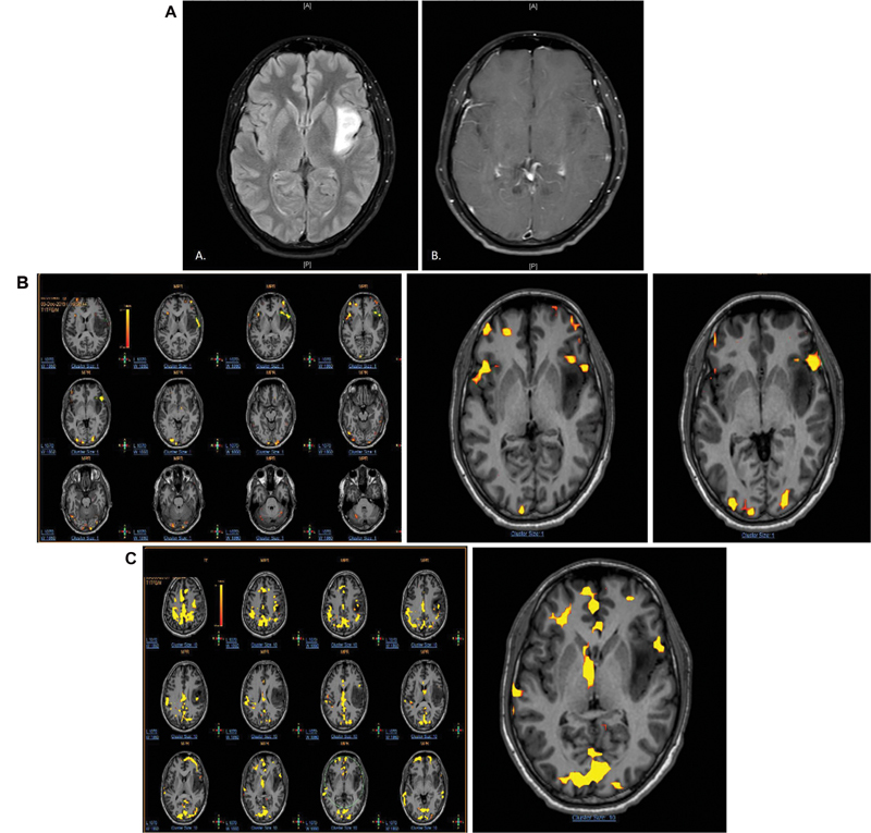
A 33-year-old male, case of left frontotemporal oligodendroglioma grade III ( Fig. 1A ): On task-based fMRI using word generation and verb generation paradigms, Broca's area was seen on both the sides in the inferior frontal gyri in pars triangularis suggestive of codominance for motor speech ( Fig. 1B ). On rs-fMRI, Broca's area was seen only on the left side ( Fig. 1C ).
Fig. 1.

( A ) Axial T2-fluid attenuated inversion recovery image shows hyperintensity infiltrating glioma in the peri-insular region in the left frontotemporal lobe. ( B ) Axial postcontrast image through same section showing no contrast enhancement. ( C ) Task-based functional magnetic resonance imaging (fMRI) using word generation and verb generation paradigms was performed. Broca's area was seen on both the sides in the pars triangularis in the inferior frontal lobes suggestive of codominance. Broca's area on the left side is abutting the tumor on its anterior aspect. Resting state fMRI showing Broca's area only on the left side, abutting the tumor.
Case 2: Language Barrier
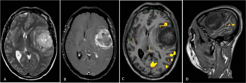
A 57-year-old right-handed male from Manipur, a place in Northeast of India, with left frontotemporal glioblastoma/World Health Organization (WHO) grade IV tumor: Patient could speak only the regional language, Manipuri. rs-fMRI was performed, which showed Broca's area in the left inferior frontal gyrus in the pars triangularis and Wernicke's area in the middle temporal gyrus on the posterosuperior aspect ( Fig. 2 ).
Fig. 2.

( A ) Axial T2 image showing hyperintense surface-based intra-axial lesion in the left frontotemporal region with T2 intermediate foci within. Perilesional edema and midline shift to the right are noted. ( B ) Axial T1 + C image showing thick peripheral shaggy rim enhancement of the lesion. ( C ) Resting state functional magnetic resonance imaging (rs-fMRI) superimposed on axial T1 images showing Broca's area placed anterior to the lesion. ( D ) rs-fMRI superimposed on sagittal T1 images showing Wernicke's area posteroinferior to the lesion.
Case 3: Limitations of Task-Based fMRI
A 52-year-old female with anaplastic astrocytoma involving left frontotemporal region with impaired comprehension: rs-fMRI was performed, which showed Broca's area in the left pars triangularis involved by the tumor ( Fig. 3A, B ).
Fig. 3.

( A ) Resting state functional magnetic resonance imaging (rs-fMRI) superimposed on axial T1-weighted images showing Broca's area in left pars triangularis, involved by the tumor. ( B ) Concordance between direct cortical stimulation (DCS) and rs-fMRI. Irregular blue region represents the Broca's area marked with rs-fMRI and green coordinates represent the area obtained by DCS. Images have been inverted for the convenience during surgery.
Case 4: Patient with Left Upper Limb Paresis
A 54-year-old female with right parietal lobe space occupying lesion, likely glioblastoma: rs-fMRI was performed due to left hemiparesis, which showed BOLD signal in the sensorimotor network ( Fig. 4 ).
Fig. 4.

( A ) Axial T2 section showing heterogeneous right parietal lobe space occupying lesion. ( B ) Resting state functional magnetic resonance imaging (rs-fMRI) overlaid on axial T1-weighted image showing signal in the sensorimotor network. ( C and D ) rs-fMRI overlaid on coronal and sagittal T1-weighted images, respectively.
Case 5: Supplementary Motor Area (SMA) Heterogeneity
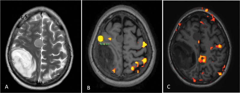
A 40-year-old female on treatment for oligometastatic carcinoma breast with adrenal metastases presented with new onset blurring of vision and focal seizures. MRI showed right parietal anaplastic astrocytoma (WHO grade III) and right parafalcine meningioma in high frontal lobe ( Fig. 5 ).
Fig. 5.

( A ) Axial T2-weighted image showing hyperintense lesion in right high parietal lobe with isointense right parafalcine meningioma. ( B ) Task-based functional magnetic resonance imaging (fMRI) superimposed on axial T1-weighted image showing 6 mm margin between tumor border and left hand representative area. However, there was no blood oxygen level dependent (BOLD) signal at expected supplementary motor area (SMA) region. ( c ) Resting state fMRI superimposed on axial T1-weighted image shows BOLD signal at SMA; the signal on contralesional side is more pronounced as compared with ipsilesional side due to plasticity.
Case 6: In Pediatric Population
A 6-year-old left-handed female operated earlier for left temporal glial/glioneuronal tumor WHO grade I, presented with worsening of symptoms and maximal safe resection was planned. rs-fMRI was performed, which showed Broca's area ∼1.4 cm anterolateral to the tumor in left inferior frontal gyrus ( Fig. 6 ).
Fig. 6.

( A ) Axial T2-weighted image showing iso- to hyperintense multilobulated lesion in left temporal lobe in a previously operated case showing subdural collection. ( B ) Axial T1-weighted postcontrast image showing patchy enhancement within the lesion. ( C and D ) Resting state functional magnetic resonance imaging superimposed, respectively, on axial and sagittal T1-weighted images showing Broca's area in the pars opercularis of the left inferior frontal lobe approximately at 1.4 cm anterior to the tumor.
Discussion
The brain attempts to overcome injury and to reacquire capabilities that are lost due to the anatomical damage by having other parts of the brain take over the lost functions; this can be termed as cortical reorganization. 1 Successful cortical reorganization in response to various insults has been described in several clinical scenarios like strokes, slow-growing tumors, and Alzheimer's disease.
Assessment of cortical reorganization of brain function in response to the growth of a tumor is crucial when considering surgical options for the excision of brain tumors. The shift of neurological functional area away from the tumor opens up the possibility of a more complete resection of the tumor without the risk of iatrogenic damage. Such changes are appreciated by preoperative fMRI. Sometimes the results of preoperative fMRI that demonstrates the relocalization of neurological function away from the expected area can convert a case, wherein a neurosurgeon earlier declared inoperable, to the successful resection of a tumor. Therefore, cortical reorganization is an important phenomenon to be considered in treatment planning of neurosurgical candidates. It is well known that oligodendroglioma are slow-growing tumors. Evidence suggests that not only can a tumor lead to neuronal hyperactivity in the immediate vicinity, but also may elicit transfer of functional control to the contralateral hemisphere. 2 There is high possibility of the cortical reorganization to the homologous area in the opposite cerebral hemisphere. In the first case of task-based fMRI, task-based functional imaging showed BOLD signal in both Broca's areas. Patient was right-handed and hemispheric dominance was calculated by laterality index (LI). According to the calculations of LI, the dominant side for motor speech was left hemisphere. rs-fMRI showed Broca's area in the left hemisphere close to the tumor-involved cortex, thus giving crucial information before the surgical procedure. Patient underwent neurosurgery under conscious sedation for the excision and the excision was limited due to speech arrest that occurred when the area was stimulated by deep nerve stimulating electrodes. Thus, task-based fMRI showed incomplete reorganization/plasticity of Broca's area. 3
In the second case patient belonged to Manipur, a state in northeast part of India. He could only speak and understand regional language Manipuri. Paradigms available with us at our institute to perform language-based tasks were in English, Hindi, Marathi, and Gujarati. Due to linguistic diversity of country, task-based fMRI could not be performed in this patient. rs-fMRI showed Broca's area in the left inferior frontal gyrus in the pars triangularis and very close to the tumor-involved cortex, that is, less than 1 cm. Chances of deficits increase if the distance between the functionally active area and the tumor is <1 cm. Studies have shown that the distance between tumor and eloquent cortex can serve as a good predictor of postoperative outcomes in patients. 4 Wernicke's area was seen in the middle temporal gyrus on the posterior aspect more than 2 cm away from the tumor. Direct cortical stimulation (DCS) during the neurosurgical procedure confirmed the locations.
DCS is considered as the gold standard procedure for functional mapping and language lateralization. The procedure involves transient disruption of function of specific region of brain by a direct electrical stimulus, mimicking neurological deficits that might be expected after resection. Functionally impaired patients or those with an altered level of consciousness or very small children may find it difficult to perform the required tasks. DCS of the gray matter within the depth of the sulci may not be readily accessible and is dependent on skill and experience of performing neurosurgeon. Not only does rs-fMRI help plan the surgery better but also it can provide useful information in this subgroup of patients where DCS is challenging, although rs-fMRI cannot be a complete replacement of DCS.
In the third case patient was having difficulties in understanding the task-based fMRI procedure and she was unable to perform due to impaired comprehension and slurred speech secondary to the anaplastic astrocytoma in left frontotemporal region. rs-fMRI showed Broca's area in left pars triangularis, which very well correlated with the area obtained by DCS during the neurosurgery as shown in Fig. 3B .
Next, in the fourth case patient had left hemiparesis due to space occupying lesion, glioblastoma in right parietal lobe. The lesion can be seen in sensory cortex and closely abutting the motor cortex due to which the power in left upper limb and lower limb was one out of five (⅕) and two out of five (⅖), respectively. The sensorimotor network includes somatosensory and motor regions and extends to the SMAs. 5 The sensorimotor network shows tight anatomico-functional coupling; 6 this network is activated during motor tasks such as finger tapping indicating that these regions may involve a premediated state that ready the brain when performing and coordinating a motor task. 5 rs-fMRI was performed because of inability to do required tasks, which showed BOLD signal along sensorimotor network and it was limited by the tumor on the right side as seen in Fig. 4 , corresponding to left hemiparesis.
Studies have shown that anatomical reviews of high-resolution T1-weighted three-dimensional sequence images by expert neuroradiologists are highly effective in locating the ipsilesional central sulcus and thus sensory and motor cortex with accuracy up to 98.6%. 6 Motor task-based fMRI also succeeded in 100%, while conventional rs-fMRI failed to localize a sensorimotor network in up to 14.1% of patients. The temporal accelerated rs-fMRI is reported with results same as that of task-based fMRI. 7 Thus, rs-fMRI is more beneficial in patients with significant neurological deficits who are unable to perform required tasks. Moreover, multiple tasks are necessary for mapping of different subregions along motor homunculus in task-based evaluation. rs-fMRI is an alternative to identify the sensorimotor network without active patient participation with almost equal potential to localize primary motor and sensory regions as in task-based fMRI.
In the fifth case , a 40-year-old female on treatment for oligometastatic carcinoma breast with adrenal metastases presented with new onset blurring of vision and focal seizures. MRI showed right parietal anaplastic astrocytoma (WHO grade III) and right parafalcine meningioma in high frontal lobe ( Fig. 5 ). Presurgery task-based fMRI failed to show signal from expected SMA, while rs-fMRI demonstrated it. SMA is involved in mediating few motor actions and speech generation through direct networks projecting into spinal cord. 8 9 It is located just anterior to primary motor cortex leg representation in the midline and is also involved in postural stabilization of the body, and bimanual action coordination. 10 Surgical resection of the SMA leads to characteristic postoperative deficits, called the SMA syndrome. The SMA syndrome is characterized by variable motor and speech impairments, severity of which ranges from complete suppression of motor and speech production to mild reduction in spontaneous motor and speech output. 11 However, the SMA syndrome shows complete or almost complete recovery within a period of around 6 months due to cortical plasticity, which are able to compensate for deficient motor and speech functions. 12 In the present case, tumor growth in the right parietal lobe has triggered preoperative reorganization of SMA to the contralateral healthy hemisphere, as seen in Fig. 5C ; more area of the left cortex showed BOLD signal as compared with right. The distance between tumor and SMA was ∼6 mm. Studies have shown 100% risk of postoperative speech deficit when the distance between fMRI centroid of activation and tumor border is less than 5 mm. 13 However, preoperative fMRI to know any plasticity of the SMA, with margin from tumor border and counseling of patient for temporary deficits, can lead to better resectability and better outcome.
In the next, sixth case , a 6-year-old left-handed female who had been operated earlier for left temporal glial/glioneuronal tumor presented with worsening of symptoms. MRI showed increase in the size of lesion; thus, progression was considered and plan made for maximal safe resection. Language lateralization mapping is often requested in right-handed patients with lesions in left cerebral hemisphere, right-handed patients with lesion in right hemisphere who presented with aphasia or any language function related deficits, and in all left-handed patients. Studies have shown that language is the purview of the left cerebral hemisphere in ∼95% of right-handed people and 70% of left-handed people. 14 15 Pediatric patients are often unable to adhere to task performance for language mapping due to age, development, and other neurocognitive factors. Moreover, they require sedation to tolerate MRI without excessive movement, thereby obviating any potential for cooperation with task-based methods. Thus, noninvasive and task-free method of rs-fMRI was performed under monitored mild sedation for language mapping, which showed Broca's area ∼1.4 cm from tumor in left inferior frontal gyrus. Sedation might impact on the magnitude of measured BOLD signal, but it has been reported that the temporal and spatial correlation of structure persists in many RSNs making rs-fMRI feasible in these patient groups. 16 Sedation, apart from alleviating anxiety, also reduces motion, thus alleviating the need of strict motion correction in processing. Pediatric patients also show smaller hemodynamic response for task-based functional studies with chances of missing out on BOLD signal. 17 Due to lack of cooperability children are often operated under general anesthesia, thus making mapping of eloquent cortices by rs-fMRI a crucial presurgical examination. In the case discussed, rs-fMRI did not show any BOLD signal in the right language representative area. Task-based fMRI for language mapping in pediatric age group may give ambiguous results due to weaker performance too. After preoperative localization of Broca's area, patient underwent cortisectomy in the region of superior temporal gyrus, inferior to identified Broca's area under general anesthesia, and residual tumor was identified with the help of neuronavigation system.
Limitation of BOLD fMRI as a test is due to use of T2* gradient echo sequences that lack the refocusing pulse of routine spin echo sequences. The susceptibility artifacts and local field inhomogeneities are accentuated as well, which cause signal distortions that mask fMRI signals from truly active sites. In patients with prior surgery, artifacts localize to areas of abrupt magnetic susceptibility changes such as air–tissue interfaces and near cavities. Titanium plates, metallic staples, hemorrhage, and blood products and residual disease add to the difficulty. As the basis for the BOLD signal is neurovascular coupling, it must also be noted that the presence of abnormal neovasculature due to tumor and the resultant neurovascular uncoupling can also lead to a muting of the hemodynamic effect and false-negative findings. 18 In glioblastoma, vessels are disorganized, highly permeable, and have abnormalities in their endothelial walls. These abnormal vessels have a decreased or absent ability to increase blood flow in response to neuronal activity, which leads to a muting of the BOLD response. 19
Conclusion
rs-fMRI is a novel technique for preoperative evaluation of eloquent cortices in patients with brain tumors. rs-fMRI is very useful with results comparable to task-based fMRI in patients with neurological deficits who cannot perform required tasks and also in pediatric patients. The information provided by rs-fMRI can be incorporated into clinical picture archiving and communication system, and thus intraoperative neuronavigation system for better guidance during resection of lesions with maximum attempt to preserve the function. rs-fMRI may also give information about cortical reorganization that, if present, can lead to better resection of tumors. However, the uses are constrained by neurovascular uncoupling, neovascularity, hemorrhage, and calcifications due to tumor.
Conflicts of Interest There are no conflicts of interest.
Declaration of Patient Consent
The authors certify that they have obtained all appropriate patient consent forms. In the form the patient(s) has/have given his/her/their consent for his/her/their images and other clinical information to be reported in the journal. The patients understand that their names and initials will not be published and due efforts will be made to conceal their identity, but anonymity cannot be guaranteed.
Financial Support and Sponsorship
Nil.
References
- 1.Fisicaro R A, Jost E, Shaw K, Brennan N P, Peck K K, Holodny A I. Cortical plasticity in the setting of brain tumors. Top Magn Reson Imaging. 2016;25(01):25–30. doi: 10.1097/RMR.0000000000000077. [DOI] [PMC free article] [PubMed] [Google Scholar]
- 2.Bogomolny D L, Petrovich N M, Hou B L, Peck K K, Kim M J, Holodny A I. Functional MRI in the brain tumor patient. Top Magn Reson Imaging. 2004;15(05):325–335. doi: 10.1097/00002142-200410000-00005. [DOI] [PubMed] [Google Scholar]
- 3.Jost E, Christiansen M H, Brennan N, Holodny A.Behavioral advantage in confrontation naming performance in brain tumor patients with left-frontal tumors In: Conference Proceedings of the Society for the Neurobiology of Language 2014;6:86
- 4.Wood J M, Kundu B, Utter A et al. Impact of brain tumor location on morbidity and mortality: a retrospective functional MR imaging study. AJNR Am J Neuroradiol. 2011;32(08):1420–1425. doi: 10.3174/ajnr.A2679. [DOI] [PMC free article] [PubMed] [Google Scholar]
- 5.Biswal B, Yetkin F Z, Haughton V M, Hyde J S. Functional connectivity in the motor cortex of resting human brain using echo-planar MRI. Magn Reson Med. 1995;34(04):537–541. doi: 10.1002/mrm.1910340409. [DOI] [PubMed] [Google Scholar]
- 6.Voets N L, Plaha P, Parker Jones O, Pretorius P, Bartsch A. Presurgical localization of the primary sensorimotor cortex in gliomas: when is resting state fMRI beneficial and sufficient? Clin Neuroradiol. 2021;31(01):245–256. doi: 10.1007/s00062-020-00879-1. [DOI] [PMC free article] [PubMed] [Google Scholar]
- 7.Posse S, Ackley E, Mutihac R et al. High-speed real-time resting-state FMRI using multi-slab echo-volumar imaging. Front Hum Neurosci. 2013;7:479. doi: 10.3389/fnhum.2013.00479. [DOI] [PMC free article] [PubMed] [Google Scholar]
- 8.Mitz A R, Wise S P. The somatotopic organization of the supplementary motor area: intracortical microstimulation mapping. J Neurosci. 1987;7(04):1010–1021. doi: 10.1523/JNEUROSCI.07-04-01010.1987. [DOI] [PMC free article] [PubMed] [Google Scholar]
- 9.Chung G H, Han Y M, Jeong S H, Jack C R., Jr Functional heterogeneity of the supplementary motor area. AJNR Am J Neuroradiol. 2005;26(07):1819–1823. [PMC free article] [PubMed] [Google Scholar]
- 10.Serrien D J, Strens L H, Oliviero A, Brown P. Repetitive transcranial magnetic stimulation of the supplementary motor area (SMA) degrades bimanual movement control in humans. Neurosci Lett. 2002;328(02):89–92. doi: 10.1016/s0304-3940(02)00499-8. [DOI] [PubMed] [Google Scholar]
- 11.Krainik A, Lehéricy S, Duffau H et al. Role of the supplementary motor area in motor deficit following medial frontal lobe surgery. Neurology. 2001;57(05):871–878. doi: 10.1212/wnl.57.5.871. [DOI] [PubMed] [Google Scholar]
- 12.Krainik A, Duffau H, Capelle L et al. Role of the healthy hemisphere in recovery after resection of the supplementary motor area. Neurology. 2004;62(08):1323–1332. doi: 10.1212/01.wnl.0000120547.83482.b1. [DOI] [PubMed] [Google Scholar]
- 13.Nelson L, Lapsiwala S, Haughton V M et al. Preoperative mapping of the supplementary motor area in patients harboring tumors in the medial frontal lobe. J Neurosurg. 2002;97(05):1108–1114. doi: 10.3171/jns.2002.97.5.1108. [DOI] [PubMed] [Google Scholar]
- 14.Corballis M C.From mouth to hand: gesture, speech, and the evolution of right-handedness Behav Brain Sci 20032602199–208., discussion 208–260 [DOI] [PubMed] [Google Scholar]
- 15.Brennan N P, Peck K K, Holodny A. Language mapping using fMRI and direct cortical stimulation for brain tumor surgery: the good, the bad, and the questionable. Top Magn Reson Imaging. 2016;25(01):1–10. doi: 10.1097/RMR.0000000000000074. [DOI] [PMC free article] [PubMed] [Google Scholar]
- 16.Palanca B JA, Mitra A, Larson-Prior L, Snyder A Z, Avidan M S, Raichle M E. Resting-state functional magnetic resonance imaging correlates of sevoflurane-induced unconsciousness. Anesthesiology. 2015;123(02):346–356. doi: 10.1097/ALN.0000000000000731. [DOI] [PMC free article] [PubMed] [Google Scholar]
- 17.Roland J L, Griffin N, Hacker C D et al. Resting-state functional magnetic resonance imaging for surgical planning in pediatric patients: a preliminary experience. J Neurosurg Pediatr. 2017;20(06):583–590. doi: 10.3171/2017.6.PEDS1711. [DOI] [PMC free article] [PubMed] [Google Scholar]
- 18.Ulmer J L, Krouwer H G, Mueller W M, Ugurel M S, Kocak M, Mark L P. Pseudo-reorganization of language cortical function at fMR imaging: a consequence of tumor-induced neurovascular uncoupling. AJNR Am J Neuroradiol. 2003;24(02):213–217. [PMC free article] [PubMed] [Google Scholar]
- 19.Jain R K, di Tomaso E, Duda D G, Loeffler J S, Sorensen A G, Batchelor T T. Angiogenesis in brain tumours. Nat Rev Neurosci. 2007;8(08):610–622. doi: 10.1038/nrn2175. [DOI] [PubMed] [Google Scholar]


