Abstract
VIP/PACAP are autocrine growth factors for lung cancer. VIP and/or PACAP mRNA is present in most lung cancer cell lines examined. Although mRNA for VPAC2-R is not common, VPAC1-R and PAC1-R mRNA is present in many lung cancer cell lines. 125I-VIP binds with high affinity to lung cancer cells and specific 125I-VIP binding is inhibited with high affinity by (Lys15, Arg16, Leu27)VIP1–7 GRF8–27, the VPAC1-R specific agonist, but not by Ro25-155318, the VPAC2-R specific agonist. VIP elevates cAMP and increases c-fos gene expression. The increase in cAMP and c-fos mRNA caused by VIP is inhibited by SN(VH). (SH)VH inhibited the proliferation of NCI-H1299 cells in the MTT assay, which is based on cytotoxicity. In a recent cell line screen, (SN)VH inhibited the growth of 51 of 56 cancer cell lines including leukemia, lung cancer, colon cancer, CNS cancer, melanoma, ovarian cancer, renal cancer, breast cancer, and prostate cancer (T. Moody, unpublished). It remains to be determined if (SN)VH will be useful for treatment of a wide variety of cancers.
INTRODUCTION
Lung cancer kills approximately 150,000 smokers in the United States annually.1 Small cell lung cancer (SCLC), which is a neuroendocrine cancer accounting for 35,000 deaths, is treated with radiation and chemotherapy. Non-SCLC (NSCLC), which is an epithelial tumor comprising adenocarcinoma, squamous cell carcinoma, and large cell carcinoma, is treated with chemotherapy and surgical resection. Because the median survival rates for SCLC and NSCLC are one and two years, respectively, better modes of treatment and detection are needed.
Numerous peptides and growth factors that stimulate lung cancer proliferation have been identified. Bombesin/gastrin releasing peptide (BB/GRP) and insulin-like growth factor (IGF)-1 are growth factors for small cell lung cancer (SCLC).2,3 The GRP receptor, a G-protein coupled receptor that causes phosphatidylinositol (PI) turnover, is blocked by peptide antagonists such as BW2258U89.4,5 In contrast, the IGF-1 receptor is a tyrosine kinase and it can be blocked by monoclonal antibody (mAb) αIR-3.6 Although SCLC is a neuroendocrine tumor, non-SCLC (NSCLC) is an epithelial tumor that exhibits high densities of vasoactive intestinal peptide (VIP) and epidermal growth factor (EGF) receptors.7,8 VIP is a G-protein coupled receptor that stimulates adenylyl cyclase and is blocked by peptide antagonists such as VIPhybrid (VH).9,10 The EGF receptor is a tyrosine kinase receptor that can be blocked by mAb108.11
VIP mRNA has been identified in several lung cancer cell lines.12 VIP immunore-activity is present in extracts derived from several lung cancer cell lines (Fahrenkrug, personal communication). The endogenous VIP-like peptides may bind to VPAC1 receptors that are present in the plasma membrane of lung cancer cells. The VPAC1 receptor interacts with a stimulatory guanine nucleotide binding protein (Gs) that activates adenylyl cyclase. The increased cAMP may activate protein kinase (PK)A. PKA may phosphorylate CREB and phosphorylated CREB may enter the nucleus and activate nuclear oncogenes such as c-fos and c-jun.13 These oncogenes may heterodimerize and stimulate growth factor genes that have an AP-1 site in the 5′ upstream regulatory region.
Numerous VIP receptor antagonists have been described including VIP10–28, (4Cl-d-Phe6, Leu17)VIP, (Ac-Tyr1,d-Phe2)GRF and neurotensin(6–11)VIP(7–28) (VH).14 VH inhibited specific 125I-VIP binding with an IC50 value of 0.5 μM using NSCLC cell line NCI-H1299. Ten μM VH inhibited the ability of 30 nM VIP to elevate cAMP using NCI-H1299 cells. Furthermore, 10 μM VH inhibited the ability of 30 nM VIP to increase c-fos mRNA using NCI-H1299 cells. Surprisingly, 10 μM VH inhibited the basal as well as 10 nM VIP stimulated proliferation of NCI-H1299 cells using a clonogenic assay. These results suggest that VIP may be an autocrine growth factor for NSCLC. Here, the effects of a new VIP receptor antagonist (N-stearyl, Nle17)VIPhybrid (SN)VH are described.
VIP/PACAP
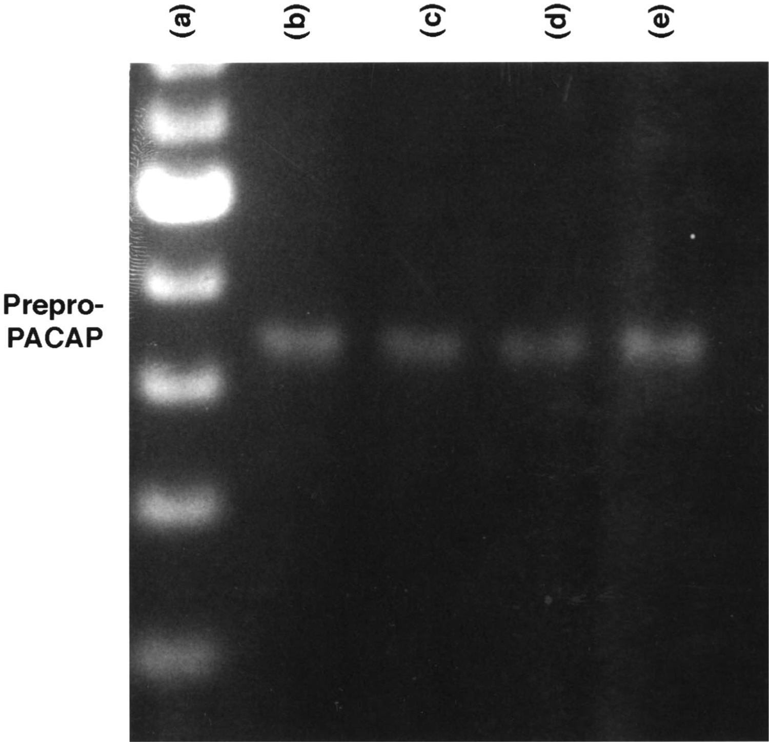
Two peptides bind with high affinity to VPAC1 receptors, VIP and pituitary adenylate cyclase-activating polypeptide (PACAP). The peptides show 69% amino acid homology and both VIP and PACAP bind with high affinity to VPAC1 and VPAC2 receptors. The mRNA for VIP and the structurally related pituitary adenylate cyclase-activating polypeptide (PACAP) was investigated in lung cancer cell lines. Using RT-PCR, a major band was detected at 443 bp for PACAP in SCLC cell lines NCI-H209, H345, N417, and H510 (see Figure 1); and NSCLC cell lines NCI-H838, H1264, and H1299, but not NSCLC cell lines NCI-H157 and H727 (see Table 1). In contrast, we found previously, using Northern blot, that VIP mRNA was present in many lung cancer cell lines including NCI-H727 and H838. Therefore, almost all lung cancer cell lines exhibit mRNA for either VIP and/or PACAP. After translation of mRNA into protein, preproVIP and preproPACAP are present in lung cancer cells. After processing by enzymes, the 28-amino acid VIP and 27-amino acid PACAP are present in vesicles ready for secretion.
FIGURE 1.

PACAP mRNA. The DNA ladder (a) and PCR products for NCI-H209 (b), H345 (c), N417 (d), and H510 (e) cells are shown. Total RNA was isolated from lung cancer cells using guanidinium isothiocyanate (GIT). The cDNA was made from reverse transcriptase. Specific primers for preproPACAP were 5′TGAGTGGCCAGGGAATCTAATA3′ (sense) and 5′AATAAGTGCCTGCATCAAACAAAA3′ (antisense). PCR was performed on a Perkin-Elmer/Cetus thermal cycler under the following conditions: 94°C for 45 sec, 55°C for 20 sec, and 72°C for 60 sec. After 35 amplification cycles using Taq polymerase, the PCR products were separated using a 2% agarose gel. Ethidium bromide fluorescence was used to assess the PCR products.
Table 1.
Messenger RNA
| Cell Line | VPAC1-R | VPAC2-R | PAC1-R | VIP | PACAP |
|---|---|---|---|---|---|
| SCLC | |||||
| H209 | + | − | + | + | + |
| H345 | − | + | + | + | + |
| N417 | + | − | + | + | + |
| H510 | + | − | + | − | + |
| NSCLC | |||||
| H157 | + | − | − | + | − |
| H727 | + | − | + | + | − |
| H838 | + | − | + | + | + |
| H1264 | − | + | + | − | + |
Note: Complementary DNA made from the mRNA by reverse transcriptase. The cDNA was added to TAQ polymerase and the following primers were used. VPAC1-R primers were 5′-ATGTGCAGATGATCGAGGTG-3′ (sense) and 5′-TGTAGCCGGTCTTCACAGAA-3′ (antisense). The VPAC2-R primers were 5′-CTTCAGGAAGCTGCACTGC-3′ (sense) and 5′-CAAACACCATGTAGTGGACG-3′ (antisense). The PAC1-R primers were 5′CATCCTTGTGCAGAAACTTC-3′ (sense) and 5′GGTGCTTGAAGTCCACAGCG-3′ (antisense). PCR was performed at 94°C for 45 sec, 62°C for 30 sec, and 72°C for 45 sec, using 40 cycles.
VIP/PACAP RECEPTORS
Previously we found that high densities of VPAC1 mRNA were present in NSCLC cells. By using Northern blot, major bands at 5, 2.5, and 1.5 kb were present in cell lines NCI-H838 and H1299, derived, respectively, from adenocarcinoma and large cell carcinoma biopsy specimens.15 Similarly, by using RT-PCR, a major band at 324 bp was detected, indicative of VPAC1 receptors. In contrast, RT-PCR products for VPAC2 (570 bp) receptors were observed in a few of the lung cancer cell lines, whereas most of the cell lines resulted in RT-PCR products for PAC1 receptors (304 bp).
125I-VIP receptor binding studies were conducted. Table 2 shows that 125I-VIP bound with high affinity to NCI-H1299 cells. (SN)VH, VIP, and PACAP-27 inhibited specific 125I-VIP binding with high affinity (IC50 values of 30, 10, and 5 nM, respectively). In contrast, VIP10–28, VIPG, and VH inhibited specific 125I-VIP binding with moderate affinity (IC50 values of 3000, 2000, and 500 nM. respectively). Reubi similarly found that malignant cells in lung cancer biopsy specimens bind 125I-VIP with high affinity using in vitro autoradiography techniques.16 Because specific 125I-VIP binding was inhibited with high affinity by the VPAC1 selective agonist,17 (Lys15, Arg16, Leu27)VIP1–7GRF8–27, but not the VPAC2 receptor selective agonist, Ro25-1553,18 the 125I-VIP was binding to VPAC1 receptors. In contrast, 125I-VIP bound with high affinity to VPAC2 receptors on lymphocytes.18 Specific binding to lymphocytes was not inhibited with high affinity by the VPAC1 selective agonist, but was inhibited with high affinity by the VPAC2 receptor selective agonist Ro25-1553. Thus, malignant cells primarily have VPAC1 receptors, whereas lymphocytes have VPAC2 receptors. Some lung cancer cell lines such as NCI-H345 and H1264 have mRNA for VPAC2-R but not for VPAC1-R. Previously, it was reported that helodermin bound with high affinity to NCI-H345 cells.19
Table 2.
Specificity of 125I-VIP binding
| Peptide | IC50, nM |
|---|---|
| PACAP-27 | 5 |
| (SN)VH | 30 |
| VIP | 10 |
| (Lys15, Arg16, Leu27)VIP1−7GRF8−27 | 20 |
| VIP10−28 | 3,000 |
| VIPG | 2,000 |
| Ro25-1553 | 1,500 |
Note: The mean IC50 value of four determinations, each repeated in quadruplicate, is indicated using cell line NCI-H1299.
Because VPAC1 receptors are present in high densities in lung cancer cells (100,000/cell) they may be used for early detection of cancer. Previously, 123I-VIP was used to localize tumors in colon cancer patients.20 Also, (18F-Arg15, Arg21)VIP was used to localize breast cancer xenografts in nude mice.21 Recently, 99mTc-VIP-Aba-Gly-Gly-(d)Ala-Gly was used to localize tumors in breast cancer patients.22 It remains to be determined whether it will be possible to detect cancer in the early stages using a radiolabeled VIP analogue.
ADENYLYL CYCLASE
VIP elevated cAMP in a dose dependent manner. VIP had little effect at 0.1 nM, but strongly elevated cAMP at 1, 10, 100, or 1000 nM. The ED50 for VIP was 3 nM. In contrast, (SN)VH had little effect on basal cAMP, but 1000 nM (SN)VH increased the ED50 for VIP to 100 nM. Furthermore, VH had little effect on basal cAMP but 10000 nM VH increased the ED50 for VIP to 30 nM. These results indicate that (SN)VH is a more potent VPAC1-R antagonist than is VH. The increase in cAMP caused by VIP causes exocytosis of vesicles using SCLC cells. As a result, 10 nM VIP increased the secretion of bombesin/GRP from lung cancer cells.23 Thus, VPAC1 receptors in lung cancer cells can be used to release growth factors.
NUCLEAR ONCOGENES
The effects of VIP on c-fos mRNA were investigated. VIP stimulated c-fos mRNA in a concentration dependent manner. Similarly, VIP stimulated c-jun in a time dependent manner, with the maximum increase occurring after one hour. The increase in c-fos or c-jun mRNA caused by VIP was inhibited by 1 μM (SN)VH. Because c-fos and c-jun proteins form heterodimers, they may reenter the nucleus and activate AP-1 sites in the 5′ regulatory region of growth factor genes. Preliminary data (T. Moody, unpublished) indicate that VIP stimulates VEGF expression in lung cancer cells.
PROLIFERATION
VH inhibited the proliferation of NCI-H1299 cells in a clonogenic growth assay that measures both cytostatic and cytotoxic effects. The effects of (SH)VH were investigated using a MTT assay. Table 3 shows that (SN)VH inhibited the proliferation of NCI-1299 cells in a concentration dependent manner, with little inhibition occurring at 0.1 μM and significant growth reduction occurring at 1 or 10 μM. Because this assay is routinely used for cytotoxic agents, (SH)VH may cause necrosis and/or apoptosis of NCI-H1299 cells. Preliminary data (T. Moody, unpublished) indicate that (SN)VH potentiates the cytotoxicty of chemotherapeutic drugs.
Table 3.
MTT assay
| Addition | Absorbance |
|---|---|
| None | 0.340 ± 0.094 |
| (SN)VH, 0.1 μM | 0.345 ± 0.051 |
| (SN)VH, 1 μM | 0.226 ± 0.009 |
| (SN)VH, 10 μM | 0.177 ± 0.022 |
Note: The mean value ± S.D. of eight determinations is indicated using NCI-H1299 cells; p < 0.05 using the Student’s t-test.
ACKNOWLEDGMENT
The authors thank Dr. J. Leyton for helpful discussions.
REFERENCES
- 1.Minna JD, Sekido Y, Fong KM & Gazdar AF. 1997. Molecular biology of lung cancer. In Cancer: Principles & Practice of Oncology. DeVita VT Jr., Hellman S & Rosenberg S, Eds.: 849–857. Lippincott-Raven, Philadelphia, New York. [Google Scholar]
- 2.Nakanishi Y, Mulshine J, Kasprzyk P, Natale R, Maneckjee R, Avis I, Treston AM, Gazdar AG, Minna JD & Cuttitta F. 1989. Insulin-like growth factor I can mediate autocrine proliferation of human small cell lung cancer cell lines in vitro. J. Clin. Invest 82: 354–359. [DOI] [PMC free article] [PubMed] [Google Scholar]
- 3.Moody TW, Pert CB, Gazdar AF, Carney DN & Minna JD. 1981. High levels of intracellular bombesin characterize human small cell lung carcinoma. Science 214: 1246–1248. [DOI] [PubMed] [Google Scholar]
- 4.Mahmoud S, Staley J, Taylor J, Bogden A, Moreau JP, Coy D, Avis I, Cuttitta F, Mulshine J & Moody TW. 1991. (Psi13,14)Bombesin analogues inhibit the growth of small cell lung cancer in vitro and in vivo. Cancer Res. 51: 1798–1802. [PubMed] [Google Scholar]
- 5.Moody TW, Zia F, Venugopal R, Patierno S, Leban J & McDermod J. 1995. BW2258: A GRP receptor antagonist which inhibits small cell lung cancer growth. Life Sci. 56: 523–529. [DOI] [PubMed] [Google Scholar]
- 6.Zia F, Jacobs S, Kull F JR., Cuttitta F, Mulshine J & Moody TW. 1996. Monoclonal antibody αIR-3 inhibits non-small cell lung cancer growth in vitro and in vivo. J. Cell Biochem 24s: 269–275. [DOI] [PubMed] [Google Scholar]
- 7.Shaffer MM, Carney DN, Korman LY, Lebovic GS & Moody TW. 1987. High affinity binding of VIP to human lung cancer cell lines. Peptides 8: 1101–1106. [DOI] [PubMed] [Google Scholar]
- 8.Siegfried JM & M Owens S. 1988. Response of primary human lung carcinomas to autocrine growth factors produced by a lung carcinoma cell line. Cancer Res. 48: 4976–4981. [PubMed] [Google Scholar]
- 9.Gozes I, McCune SK, Jacobson L, Warren D, Moody TW, Fridkin M & Brenneman D. 1991. An antagonist to vasoactive intestinal peptide affects cellular functions in the central nervous system. J. Pharm. Exp. Ther 257: 959–966. [PubMed] [Google Scholar]
- 10.Moody TW, Zia F, Brenneman D, Fridkin M, Davidson A & Gozes I. 1993. A VIP antagonist inhibits the growth of non-small cell lung cancer. Proc. Natl. Acad. Sci. USA 90: 4345–4349. [DOI] [PMC free article] [PubMed] [Google Scholar]
- 11.Lee M, Draoui M, Zia F, Gazdar A, Oie H, Tarr C, Bellot F, Kris R & Moody TW. 1992. Epidermal growth factor receptor monoclonal antibodies inhibit the growth of lung cancer cell lines. JNCI Monographs 13: 117–123. [PubMed] [Google Scholar]
- 12.Davidson A, Moody TW & Gozes I. 1996. Regulation of VIP gene expression. Mol. Cell. Neurosci 7: 99–110. [DOI] [PubMed] [Google Scholar]
- 13.Whitmarsh AJ & Davies RJ. 1996. Transcription factor AP-1 regulation by mitogen-activated protein kinase signal transduction pathways. J. Mol. Med 74: 589–607. [DOI] [PubMed] [Google Scholar]
- 14.Moody TW & Jensen RT. Bombesin/GRP and vasoactive intestinal peptide/PACAP as growth factors. 1996. In Growth Factors and Cytokines in Health and Disease. LeRoith D & Bondy C, Eds.: 491–535. JAI Press, Greenwich, CT. [Google Scholar]
- 15.Moody TW, Leyton J, Coelho T, Knight M, Takahashi K, Koh M, Fridkin M & Gozes I. 1997. (Stearyl, Norleucine17)VIPhybrid is a potent non-small cell lung cancer VIP receptor antagonist. Life Sci. 17: 1657–1666. [DOI] [PubMed] [Google Scholar]
- 16.Gourlet P, Vandermeers A, Vertongen P, Rathe J, Deneef P, Cnudde J, Waelbroeck M & Robberecht P. 1997. Development of high affinity selective VIP1 receptor agonists. Peptides 18: 1539–1545. [DOI] [PubMed] [Google Scholar]
- 17.Gourlet P, Vertongen P, Vandermeers A, Vandermeers-Piret MC, Rathe J, Deneef P, Waelbroeck M & Robberecht P. 1997. The long-acting vasoactive intestinal polypeptide agonist Ro25-1553 is highly selective of the VIP2 receptor subclass. Peptides 18: 403–408. [DOI] [PubMed] [Google Scholar]
- 18.Reubi JC 1995. In vitro identification of vasoactive intestinal peptide receptors in human tumors: implications for tumor imaging. J. Nuc. Med 36: 1846–1853. [PubMed] [Google Scholar]
- 19.Luis J & Said S. 1990. Characterization of VIP- and helodermin-preferring receptors on human small cell lung carcinoma cell lines. Peptides 11: 1239–1244. [DOI] [PubMed] [Google Scholar]
- 20.Angelberer P, Banyai B, Banyai S, Durtaran A, LI S, Pidlich J, Raderer M, Virgolini I, Yang Q, Niederle B, Scheithauer W & Balent P. 1994. Vasoactive intestinal peptide receptor imaging for the localization of intestinal adenocarcinomas and endocrine tumors. New Engl. J. Med 331: 1116–1121. [DOI] [PubMed] [Google Scholar]
- 21.Moody TW, Leyton J, Unsworth E, John C, Lang L & Eckelman W. 1998. (Arg15, Arg21)VIP: A VIP agonist for localizing breast cancer tumors. Peptides 19: 585–592. [DOI] [PubMed] [Google Scholar]
- 22.Pallela V, Tahkur ML & Chakder S. 1999. 99mTc labeled VIP receptor agonist: functional studies. J. Nucl. Med 450: 352–360. [PubMed] [Google Scholar]
- 23.Korman LY, Carney DN, Citron ML & Moody TW. 1986. Secretin/VIP stimulated secretion of bombesin-like peptides from human small cell lung cancer. Cancer Res. 46: 1214–1218. [PubMed] [Google Scholar]


