



Abstract
Head and neck paragangliomas (HNPGLs) are rare neuroendocrine tumors typically arising from nonsecretory head and neck parasympathetic ganglia. Historically thought of as aggressive tumors that warranted equally aggressive surgical intervention, evidence has emerged demonstrating that the vast majority of HNPGLs are slow growing and indolent. It is also now recognized that a large proportion of HNPGLs are hereditary with succinate dehydrogenase gene mutations typically implicated. These recent advances have led to significant changes in the way in which clinicians investigate and treat HNPGLs with most now opting for more conservative treatment strategies. However, a proportion of patients present with more aggressive disease and still require nonconservative treatment strategies. Recent studies have sought to determine in which groups of patients the morbidity associated with treatment is justified. We summarize the recent advances in the understanding and management of these tumors and we provide our recommendations regarding the management of HNPGLs.
Keywords: carotid‐body tumor, glomus tumor, neuroendocrine tumors, paraganglioma, succinate dehydrogenase (SDH)
Head and neck paragangliomas (HNPGLs) are rare neuroendocrine tumors typically arising from nonsecretory head and neck parasympathetic ganglia. Our recent improved understanding of the natural history and genetic associations has fundamentally changed our management of these tumors. We summarize the present understanding of HNPGL and provide our recommendations for the management of these tumors from the Irish national tertiary referral center.

1. INTRODUCTION
Head and neck paragangliomas (HNPGLs) are rare tumors arising from nonsecretory head and neck parasympathetic ganglia. They account for 0.6% of head and neck tumors. 1 Owing to their origin, HNPGLs are typically nonsecretory tumors. This is in contrast to paragangliomas (PGLs) in other areas of the body which usually arise from secretory sympathetic ganglia and produce catecholamines. Historically, HNPGLs have been known by various terms such as glomus tumors or chemodectomas. The World Health Organization (WHO), however, has now deemed that these alternate terms should no longer be used when referring to HNPGLs. 2 HNPGLs have been found in up to 20 anatomical locations within the head and neck. They arise predominately from the parasympathetic ganglia of the glossopharyngeal or vagus nerve. The most common HNPGLs are carotid body tumors (CBTs) followed by Jugulo‐tympanic PGLs (JTPGLs) and vagal PGLs (VPGLs). Other regions where HNPGLs have been reported include the larynx, orbit, trachea, thyroid, and nasal cavity. Approximately 35–40% of HNPGLs are associated with familial disease with mutations in the succinate dehydrogenase (SDHx) gene family typically implicated. 3 , 4
Historically, HNPGLs were considered to be aggressive tumors that warranted equally aggressive surgical intervention. This led to considerable neurovascular morbidity for patients including cranial nerve deficits and stroke. More recently, numerous series have demonstrated that the majority of HNPGLs are slow growing and indolent. 5 , 6 , 7 This has led to a shift in the management of HNPGLs with many clinicians now advocating active surveillance following diagnosis. 8 , 9 HNPGLs were recently classified by Valero et al. as CBTs and non‐CBTs. 9 This is primarily due to differing optimal management strategies but also due to differing methods of presentation. Here, we discuss what is currently known about HNPGLs and recent updates in the literature surrounding their management.
2. PATHOLOGY
Grossly HNPGLs are typically ovoid in shape with a firm or rubbery consistency. Histologically, they classically display a nested alveolar or zellballen growth pattern, comprised of chief cells, although this pattern is not universal. 10 Variants of this pattern have been observed and can make histological diagnosis challenging. 11 , 12 These variants include a sclerosing variant with rare clusters of chief cells, highly vascular PGLs and variants displaying chief cell vacuolisation. 10 Cytologically PGL chief cells range from amphophilic to pink and they are predominantly epithelioid. The PGL chief cell nucleus is round, hyperchromatic with characteristic “salt and pepper” chromatin clustering. They typically have a low mitotic rate with a Ki‐67 proliferation index <3%. 13
Both sporadic and familial HNPGLs have been described with mutations primarily affecting the SDHx gene family implicated in familial disease. Familial disease can also be associated with syndromes such as Von Hippel–Lindau (VHL) syndrome, neurofibromatosis type 1, multiple endocrine neoplasia type 2A and type 2B. Genetic alterations are discussed in detail separately. A third etiology exists for CBTs. This involves hyperplasia of the carotid body chemoreceptor induced by chronic hypoxia due to living at high altitude, cyanotic heart disease or chronic lung disease. This phenomenon is well recognized with increased incidence of CBTs observed in these populations. 14 , 15
Immunohistochemical staining of these tumors can assist in the pathological evaluation. HNPGLs stain positive for neuroendocrine markers such as chromogranin and synaptophysin on immunohistochemistry. Other positive markers include NSE and CD56. S100 will stain the sustentacular cells surrounding the zellballen. This is not specific for PGLs as tissue‐associated macrophages and Langerhans cells may infiltrate other tumors and stain positive with S100. HNPGLs stain negative for cytokeratin which is very useful in differentiating HNPGL from epithelial derived tumors mimicking HNPGLs. 10 Immunohistochemical staining for SDH is also useful in screening for SDH‐deficiency in these tumors. As the SDH complex requires all components to function a mutation in any of these components will lead to a lack of SDH expression. 16
3. GENETICS
In recent years, the recognition that a significant number of HNPGL have an underlying genetic etiology has changed our management of these patients. PGLs (encompassing both HNPGL and extra‐adrenal sites) are now recognized as having the highest risk of heritability of any tumor. 17 Up to 40% of HNPGL are associated with an identifiable germline mutation. 18 , 19 , 20 , 21 The presence of multifocal disease at presentation should raise suspicions for an underlying genetic mutation. Over 70% of multifocal PGL are associated with a germline mutation, most commonly mutations in the SDHD and SDHB genes. 22 , 23 A family history of PPGL is also highly suggestive of a germline mutation; almost all patients with HNPGL and a known family history of PPGL will carry a pathogenic germline mutation. 22 , 23 However, a significant proportion of patients carrying germline mutations will not have a known family history. Rana et al. reported in their series that only 4 of 16 patients with a germline mutation had a positive family history at initial assessment. 20 A positive family history may be obscured for several reasons including incomplete penetrance, parent of origin effect 24 and failure to recognize combinations of tumors in various family members as linked. 20
There are several reasons why identifying a genetic mutation is important when managing a patient with HNPGL. 20 The likelihood of synchronous, metachronous, and metastatic PPGL, and risk of non‐PPGL tumors will depend on the underlying mutation. Therefore, knowledge of an individual's mutation status will inform treatment and ongoing surveillance. There are also implications for family members of affected patients, including the early identification of asymptomatic disease and ongoing surveillance of affected relatives. 20 , 25 The Endocrine Society and European Society of Endocrinology both therefore recommend that genetic testing should be considered and discussed with all patients diagnosed with PPGL and that patients presenting with HNPGL should all be screened for SDHD, SDHB, and SDHC mutations. 25 , 26 Additional, targeted genetic screening may be appropriate in patients with a syndromic presentation (e.g., vHL). 25
Genetic mutations which result in a predisposition to the development of pheochromocytomas or PGLs (PPGLs) can be grouped into two main clusters. Cluster 1 gene mutations lead to an alteration in the cellular hypoxic response, and cluster 2 mutations alter activation of cellular kinase signaling. 26 Germline mutations associated with the development of HNPGL are typically associated with cluster 1 genes. These include mutations to the genes encoding the four subunits of SDH and its accessory proteins (SDHD, SDHB, SDHC, SDHA, SDHAF2) 17 , 18 , 19 , 22 , 23 and VHL syndrome. 27 SDH is a heterotetrameric protein which forms the mitochondrial complex II of the electron transport chain and converts succinate to fumarate. 28 , 29 SDHx genes are considered to be tumor suppressor genes. 29 SDH mutations are also associated with non‐PPGL tumors such as renal cell carcinoma (RCC), pituitary adenomas, and gastrointestinal stromal tumors (GIST). 17 , 30
Rarely, HNPGL can arise in association with cluster 2 mutations (RET, NF1, TMEM127), 27 , 31 , 32 these mutations are more commonly associated with non‐head and neck sympathetic PGL or pheochromocytoma. 29 The genotype:phenotype correlation of some of the more common germline mutations reported in association with HNPGL is discussed below.
3.1. SDHD mutation
The most common mutation associated with HNPGL occurs in the SDHD gene. It accounts for over 80% of familial HNPGL. In one series, SDHD mutation was detected in 24 of 95 patients with suspected sporadic HNPGL. 18 , 33 The SDHD gene is located on chromosome 11q23 and disease is generally expressed only after paternal inheritance. 34 HNPGLs are the most common manifestation of SDHD germline mutations, and SDHD‐associated disease can be multifocal. 22 SDHD affected carriers are also at risk of pheochromocytomas, and less commonly PGL of the thorax, abdomen or pelvis, RCC, GIST, and rarely pituitary adenomas. 35 SDHD mutations are associated with a higher disease penetrance overall compared to SDHB mutation carriers. 36 , 37
3.2. SDHB mutation
The SHDB gene is encoded on chromosome 1p36. SDHB mutations account for up to 20% familial HNPGL and 8–10% of familial PPGL disease overall 17 , 19 , 21 SDHB mutations are associated with a higher risk of malignancy and metastatic disease than other SDH mutations. 37 The risk of associated RCC is greatest in SDHB mutation carriers. 30
3.3. SDHC mutation
SDHC mutations account for a small proportion of familial HNPGL. 21 Age‐related risk of HNPGL is similar to paternally inherited SDHD mutation. 30 Carriers can also develop PPGL beyond the head and neck region. 21 , 35 , 38 SDHC‐associated HNPGLs are also less likely to be multifocal than SDHD‐associated tumors and have a lower malignant potential than SDHB‐related HNPGL. 21
3.4. SDHA mutation
SDHA mutations are reported in up to 3% of apparently sporadic PPGL disease, occurring primarily in HNPGL. 29 , 39 SDHA mutations are strongly associated with wild‐type SDH deficient GIST (wtGIST) with germline mutations to SDHA gene present in up to 47% wtGIST. 31 , 40
3.5. SDHAF2 mutation
SDHAF2 is a rare cause of HNPGL. Similarly to SDHD mutations, disease is only penetrant following paternal inheritance with up to 75% of carriers developing multifocal HNPGL at a young age. 38 , 41
4. DIAGNOSIS
4.1. Presentation of HNPGL
The presentation of HNPGLs can be highly variable and they have been described in as many as 20 different anatomical locations in the head and neck. 4 HNPGL can present as an enlarging neck mass, pulsatile tinnitus, with cranial nerve deficits, as part of familial screening or incidentally. As discussed approximately 5% of HNPGLs secrete noradrenalin. These tumors cause symptoms of sympathetic overactivity such as tachycardia, hypertension, and sweating. 42 , 43 HNPGLs typically present in the 5th decade of life although familial disease often presents earlier. 44 , 45 HNPGL may present with multifocal disease and the incidence of this varies within the literature (9–37%). 42 , 46 , 47 , 48 As discussed, multifocal disease is more common in familial disease, particularly SDHD mutations. 8 , 47 Approximately one‐in‐ten of HNPGLs present incidentally, a figure which is increasing due to patients undergoing more frequent cross‐sectional imaging. 42 Our recommended proforma for the full multidisciplinary investigation of HNPGL is demonstrated in Figure 1.
FIGURE 1.
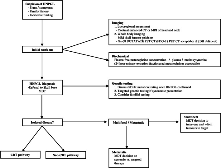
Investigation of a suspected HNPGL. CBT, carotid body tumor; CT, computed tomography; FDG‐18, fluorodeoxyglucose‐18; Ga‐68, Gallium‐68; HNPGL, head and neck paragangliomas; MDT, multidisciplinary team meeting; MRI, magnetic resonance imaging; non‐CBT, non‐carotid body tumor; PET, Positron emission tomography; SDH, succinate dehydrogenase
4.1.1. Presentation of CBTs
CBTs are the most common type of HNPGL accounting for over half of HNPGLs. 42 CBTs arise from paraganglia at the carotid body. They occur more commonly in females. 48 CBTs are classified according to the Shamblin classification which describes the tumor relationship to the carotid vessel's (Appendix 1). 49 The majority present as a painless slowly enlarging lateral neck mass. A minority of cases present with cranial nerve dysfunction. 48 Large CBTs may induce vagal dysfunction causing dysphagia and dysphonia. Less frequently, they have also been reported to affect other cranial nerves (VII, IX, XI, and XII) and in rare instances, they can cause a Horner's syndrome. 45 , 48 , 50 On clinical examination, the mass may be pulsatile and a carotid bruit may be detected. Classically on clinical examination, CBTs are more mobile in the horizontal plane compared with the vertical plane, a finding known as Fontaine's sign. 50 , 51
4.1.2. Presentation of non‐CBTs
Non‐CBTs account for all other types of HNPGLs. JTPGLs are the next most frequent after CBTs accounting for 20–30% of HNPGLs while VPGLs account for 5–10%. 42 Other locations such as the larynx, orbit, trachea, thyroid, and nasal cavity are extremely rare. Non‐CBTs are more likely to present with cranial nerve dysfunction when compared with CBTs. 9 , 52 They again occur more frequently in females. 52
4.2. Jugulo‐tympanic PGLs
JTPGLs arise from either the jugular bulb or within the ear along Arnold's or Jacobsen's nerve. They are classified according to either the Fisch or the Glassock–Jackson classification, with the Fisch classification more widely cited in the literature (Appendix 2). 53 , 54 The majority of patients present with pulsatile tinnitus and the many of patients have some degree of hearing loss (usually conductive) at presentation. 55 , 56 Cranial nerve dysfunction is common in JTPGLs at presentation. Neskey et al. observed 9 out of 21 patients operated on with JTPGLs had cranial nerve deficits at presentation. 52 Cranial nerves IX and X are most commonly affected but dysfunction of cranial nerves VII, VIII, XI, and XII have all been observed. 56 , 57 On otoscopy, JTPGLs may be visualized as a purple lesion behind the eardrum and they classically blanch on pneumatic otoscopy, a finding known as Brown's sign. 58
4.3. Vagal PGLs
The clinical presentation of VPGLs is highly variable as they may arise anywhere along the course of the vagus nerve. 59 The majority of these tumors arise from the ganglion nodosum of the vagus nerve which is found between the jugular vein and internal carotid artery, in close relation to the jugular foramen. They classically present with a painless slow growing mass in the superior aspect of the neck, posterior to the angle of the mandible and they are often associated with pulsatile tinnitus. Rarely when extremely large they can cause bulging of the lateral pharyngeal wall with medialization of the tonsil seen on examination. Between 25 and 36% of these tumors present with cranial nerve deficits, primarily affecting cranial nerves IX, X, XI, and XII. 46 , 59 Owing to their anatomical location near the jugular foramen intracranial extension of VPGLs has been observed. In a series of 46 patients with VPGLs Netterville et al. observed that 10 cases had associated intracranial extension. 46
4.4. Biochemical
HNPGL typically arise from the nonsecretory parasympathetic ganglia of the head and neck region, and rarely secrete catecholamines. 25 The conversion of noradrenalin to adrenalin by phenylethanolamine‐N‐methyltransferase is confined to the adrenal medulla, and therefore unlike pheochromocytomas, HNPGL do not secrete adrenaline. 60 Noradrenalin secretion is rare, only 5% HNPGL will produce noradrenalin, in contrast to over one‐third of PGLs in the thorax or abdomen. 17 , 61 The presence of markedly elevated noradrenalin/adrenalin concentration in a HNPGL should therefore prompt the clinician to consider the presence of a synchronous pheochromocytoma or secretory paraganglioma elsewhere, which would require treatment prior to the management of HNPGL. 62 , 63
Patients with noradrenalin secreting HNPGL will typically display symptoms of catecholamine excess and as such the term “secretory” HNPGL is reserved for these tumors. Although noradrenalin excess is rare in HNPGL, it is important that it is identified preoperatively if present as these patients will require preoperative alpha adrenergic blockade. 25 These patients should be managed with centers with a multidisciplinary team with experience in the perioperative management of catecholamine‐secreting lesions.
Excess dopamine production in HNPGL is seen in approximately one third of all HNPGL tumors, both sporadic and familial. 63 , 64 , 65 Dopamine producing tumors are not typically associated with signs or symptoms of catecholamine excess. 28
Catecholamines and their metabolites can be measured in both plasma and 24‐h urinary collection. Measurement of O‐methylated metabolites of catecholamines and dopamine (metanephrine, normetanephrine, and 3‐methoxytyramine [3MT]) has a higher sensitivity than direct catecholamine (adrenalin, noradrenalin, and dopamine) measurement, as the secretion of O‐methylated metabolites occurs independently of episodic tumoral catecholamine release and is therefore the preferred method. 25 , 28 Urinary VMA concentration is no longer recommended due to poor sensitivity. 66
In cases where the elevation in plasma metanephrines is modest, both the degree of clinical suspicion and pre‐analytical factors should be considered. 25 Where the pretest probability is low, it may be appropriate to repeat a plasma metanephrine screen after optimizing any possible interfering factors. These include food or caffeine intake, nicotine use, stress and exposure to medications including antidepressants, beta blockers, paracetamol, and dopamine containing medication. 66 Supine sampling, where the patient is recumbent for at least 30 min prior to phlebotomy is associated with a greater diagnostic accuracy than seated sampling of plasma metanephrines. 25 , 67
Measurement of urinary fractionated metanephrines can be performed as an alternative to plasma sampling, using a 24‐h urinary collection with a similar sensitivity and specificity. 25 However, performing the collection may be inconvenient for patients, and incomplete 24‐h urinary collections can yield false negative results.
It is important to remember that as the majority of HNPGL are not associated with catecholamine excess, a negative plasma or urinary metanephrine screen does not exclude the diagnosis of HNPGL. 28
4.5. Radiology
Imaging forms an integral part of the diagnosis and surveillance of HNPGL, particularly in biochemically inactive disease. In new presentations of HNPGL imaging can both confirm the diagnosis of HNPGL in biochemically silent disease and excluding multifocal synchronous tumors and metastases, which will inform treatment decisions. Radiological investigations are also a key component in monitoring HNPGL following treatment (particularly biochemically silent disease), in the screening and ongoing surveillance of germline mutation carriers.
Imaging techniques used in the diagnosis and surveillance of HNPGL include cross‐sectional anatomical imaging such as CT and MRI, and functional imaging techniques. Both CT and MRI have similar sensitivity (80–90%) and specificity (90%) for the diagnosis of HNPGL and are useful for locoregional staging. 63 MRI is recommended for both the initial diagnosis and surveillance of PPGL/HNPGL. 68 , 69 CT is recommended by the Endocrine Society for the initial investigation of catecholamine excess 25 ; however, the associated ionizing radiation makes it a less desirable option for ongoing tumor surveillance. 70
Several indications exist for the use of functional or nuclear imaging in patients with HNPGL. In biochemically silent disease, functional imaging may help to confirm the diagnosis of PPGL. Functional imaging is useful at initial disease staging and at restaging after treatment. 25 , 71 More recently, imaging has been used to provide theranostic information when considering treatments such as somatostatin or MIBG (131I‐metaiodobenzylguanidine) therapy. 72 The choice of imaging modality will therefore depend on the individual clinical scenario.
4.6. Somatostatin receptor imaging
There are three main 68Gallium labeled somatostatin receptor (SSTR) ligands used with PET CT; DOTATATE, DOTATOC, and DOTANOC, each with a different combination of SSTR subtype affinity. SSTR imaging modalities have a high lesion‐based sensitivity for HNPGL. 71 , 73 , 74 They can detect up to 96% lesions including sub‐centimeter tumors, which may influence treatment decisions. 72 , 75
68Ga‐DOTATATE has the highest affinity for SSTR2, the most commonly expressed SSTR in PPGL and is particularly sensitive in the detection of HNPGL. 71 , 76 Increased expression of SSTR 2 and 3 has been reported in SDH deficient tumors, which has led to the suggestion that Ga‐DOTATATE PET CT be considered as part of the initial assessment of asymptomatic carriers of SDH germline mutations in conjunction with MRI. 72 , 75 Ga‐DOTATATE PET CT is also useful in metastatic disease; it has a superior reported performance to FDG or F‐DOPA PET‐CT, CT or MRI for both sporadic and SDH mutated metastatic disease. 75 , 77 , 78 , 79 The exception to this is in the liver, where high background uptake of Ga‐DOTATATE may obscure hepatic PPGL metastases. 75 Ga‐DOTANOC binds SSTR 2‐5 and has been reported to offer superior detection rates of synchronous and metastatic tumors in HNPGL over MIBG scintigraphy, CT, or MRI. 80 Ga‐DOTATOC binds SSTR 2 and 5 and also has excellent sensitivity for the detection of metastatic or multifocal extra‐adrenal PGL. 71 , 81
SSTR expression by HNPGL can also be exploited to deliver targeted therapy in the setting of inoperable or metastatic disease using 177Lu‐DOTATATE.
4.7. 18F‐FDG PET CT
FDG PET‐CT is a useful imaging modality in SDH deficient HNPGL/PPGL. 82 The radiotracer is taken up via cell membrane glucose transporters and phosphorylated by hexokinase to FDG‐6‐phosphate, which accumulates intracellularly. 72 SDH deficient tumors show an increase in glycolysis activity, as SDH deficiency induces the pseudo‐hypoxic response and shifts cellular metabolism from oxidative phosphorylation to aerobic glycolysis. 83 Aerobic glycolysis is less energy efficient, and therefore glucose demand increases within the tumor cells, thereby increasing FDG tracer uptake. This may explain why SDH deficient PPGL tumors have a higher SUV mean and SUV max than their sporadic counterparts, and why FDG PET‐CT is a particularly useful imaging modality for SDH‐associated PPGL and metastatic disease, albeit with low specificity. 26 , 71 , 72 , 82
4.8. 18F‐FDOPA PET CT
18F‐dihydroxyphenylanine is taken up by the L‐type amino acid transporter and converted by L‐aromatic amino acid decarboxylase to 18F‐fluorodopa in catecholamine secreting tissues and can be used to image HNPGL. 25 , 63 , 72 F‐DOPA has excellent per patient sensitivity and specificity (91 and 95%, respectively) and per lesion specificity (95%), but a lower per lesion sensitivity for PPGL than other functional modalities. 78 , 82 In functional PPGL tumors, F‐DOPA uptake has been reported to correlate with tumor metabolic activity, as the tracer is taken up the catecholamine synthesis pathway and therefore may play a role in localization of PPGL with catecholamine excess. 84 Biochemically silent HNPGL, however, also demonstrate high avidity for F‐DOPA despite absent catecholamine production, irrespective of the presence or absence of germline mutation. 85
4.9. MIBG scintigraphy
MIBG has structural similarities with noradrenalin and is taken up via a common cell membrane transporter (noradrenaline transporter) into catecholamine secreting tissues, where it accumulates in neurosecretory granules. 71 MIBG scintigraphy has excellent specificity for PPGL, but its sensitivity varies significantly depending on tumor location. 42 Approximately 50% of metastatic PPGL are MIBG avid. However, among PPGL, its sensitivity is lowest for HNPGL; therefore, in HNPGL, its role is limited to where MIBG therapy is under consideration. 24 , 71 , 86
4.10. Current radiological recommendations
The British Skull Base Society and the European Society of Hypertension working group on Endocrine Hypertension have both released recent guidelines suggesting that contrast enhanced MRI head and neck should be performed for locoregional assessment of the primary HNPGL tumor, and in the case of temporal bone HNPGL, a CT of the skull base should also be performed. 86 , 87 In addition, all new HNPGL should have imaging of thorax/abdomen/pelvis to exclude metastatic or synchronous PPGL, using either MRI skull base to pelvis, or alternatively 68Ga DOTATATE PET CT where available. 63 , 86 , 87 68Ga‐labeled somatostatin analogue imaging is recommended as first line nuclear imaging for sporadic HNPGL but FDG‐PET CT can also be considered in SDH deficient HNPGL. 26 , 71
5. NATURAL HISTORY
Historically, HNPGLs were considered aggressive tumors that warranted equally aggressive management. However, several studies have shed light into the fact that these tumors are slow growing and indolent and that they usually remain asymptomatic for a prolonged period. 5 , 6 , 7 , 44 , 88 , 89 Death secondary to nonmetastatic HNPGLs is almost unheard of. 8 Jansen et al. were the first to describe an observational series of HNPGLs. Within this series, 29 of the 48 observed HNPGLs grew over the follow‐up period with a mean growth rate of 0.8 mm/year in maximal tumor dimensions. 6 Further studies over the years have added to this work. These series have demonstrated smaller proportions of observed HNPGLs have grown during active surveillance with one such study showing that 10 out of 15 HNPGLs either remained stable in size or regressed during follow‐up. 5 The mean growth rate of HNPGLs in these studies remains small and is estimated between 0.8 and 2 mm/year in maximal tumor dimensions. 5 , 6 , 7 , 44 , 45 , 88 , 89 When comparing growth rates by tumor location, Jansen et al. recently demonstrated for enlarging HNPGLs the median growth rate of JTPGLs is 0.4 mm/year and the median growth rate is 1.6 mm/year for cervical HNPGLs (including both CBTs and VPGLs). 7 Younger age at presentation appears to be the main predictor of tumor growth with patients younger than 50 at presentation demonstrating significantly greater tumor growth. 7 , 90 Retrospective studies have shown that approximately 10–30% of patients with HNPGLs will develop new or worsening cranial nerve deficits when undergoing active surveillance and that tumor growth does not always predict worsening symptoms. 5 , 7 , 88 , 91
One worrisome feature that has been observed in HNPGL is the risk of malignant change. While certain cytological and histological changes such as increased mitotic index, central necrosis or perineural invasion may suggest malignant change, malignancy can only be confirmed by the presence of metastatic disease within non‐neuroendocrine tissue. 92 Metastatic disease has been observed developing years after the initial presentation of HNPGLs. Better outcomes are observed in metastatic disease presenting later following initial presentation. 93 The risk of metastatic disease in HNPGLs is estimated to be in the order of 5–10% overall. 1 , 13 , 93 , 94 However, this risk is highly variable depending on the individual case and clinicians should be aware of the factors associated with an increased risk of metastatic disease. These include: tumor location (16% for VPGLs and 2–6% for CBTs and JTPGLs), rapidly increasing tumor size, secretory tumors, younger age at diagnosis and most importantly the presence of SDHB mutations. 13 , 93 , 95 , 96 When observing metastatic HNPGLs there appears to be two distinct patterns of progression. Those which have spread solely to cervical lymph nodes follow a more indolent pattern with long‐term survival observed in multiple series. 1 , 13 , 97 A series of 59 patients with malignant HNPGL by Lee et al. demonstrated much lower 5‐year survival among HNPGL with distant metastatic disease. 1
6. TREATMENT
Historically, the management of HNPGLs was upfront surgical resection which poses a risk to surrounding neurovascular structures. Complications associated with surgical resection of HNPGL include: neurological complications such as dysphagia, dysphonia, hearing loss, and facial nerve palsy; vascular complications such as stroke; cerebrospinal fluid leak and in some cases even death. 48 , 52 Due to increased recognition regarding the largely indolent nature of this disease, this strategy has changed over the years with increasing numbers of HNPGLs being managed conservatively in an attempt to avoid the potential morbidity associated with surgery. 8 , 9
The present management strategies for all nonmetastatic HNPGLs can largely be divided into: surgery, RT (RT), and active surveillance. 86 The aim of each treatment modality is different. Surgery typically aims for total tumor extraction, RT aims to arrest tumor growth and active surveillance aims to avoid the potential morbidity of either surgery or RT while determining an appropriate time for intervention if necessary. Individual patient management plans should always be determined at an appropriate multidisciplinary meeting. 86 Tumor location is of critical importance when making decisions regarding management of HNPGLs. Valero et al. recently classified HNPGLs as CBTs and non‐CBTs. 9 This is mainly due to the higher rates of adverse outcomes and the increased morbidity when operating on non‐CBT HNPGLs. As such, when planning management of HNPGL we would advocate using this distinction.
6.1. Isolated CBTs
Many CBTs are slow growing and are not associated with significant symptoms. 9 , 48 As such, active surveillance is a suitable strategy for the many of these tumors and should always be considered prior to surgery or RT. However, active surveillance not risk free. Patients undergoing active surveillance may develop new onset cranial nerve deficits and CBTs may undergo malignant change in 2–6% of cases. 95 , 96
Surgery remains the most common strategy for managing CBTs. 8 , 9 , 48 , 98 , 99 It is associated with long‐term control greater than 95%. 48 , 98 The Shamblin classification has long been used to predict operative morbidity and mortality in CBTs. 49 A recent meta‐analysis supported this use and demonstrated cranial nerve complications in 2.8% (grade 1), 18.0% (grade 2), and 32 % (grade 3) of surgically resected cases, respectively. 98 The Shamblin classification also appears to predict a higher risk of stoke within 30 days postoperatively which is reported in 1.9% (grade 1), 2.7% (grade 2), and 4.0% (grade 3) of cases, respectively. 48 The risk of complications is lower when surgery is performed for smaller tumors in young healthy patients by a suitably experienced either vascular or head and neck surgeon. 86 , 100 These figures would support primary surgery in this patient cohort. In the case of secretory CBTs, surgery is indicated to alleviate symptoms of sympathetic overactivity. 48 , 86 Other instances when surgery is appropriate include: where a concern regarding malignancy exists, patients with established cranial nerve deficits and in tumors demonstrating rapid growth on surveillance. 86 Should an associated SDHB mutation be detected surgery may also be considered due to the increased risk of malignancy.
RT has demonstrated a similar efficacy (~95%) when compared with surgery for long‐term control of CBTs. 86 , 101 It has conventionally been reserved for highly complex Shamblin III lesions or for high risk surgical patients. 102 , 103 It is associated with minimal risk of worsening an existing or causing a new cranial nerve palsy. 104 Concerns exist regarding the long‐term risks such as radiation induced malignancy, bone and brain necrosis and stroke following radiation to the neck and skull‐base region, particularly in young patients. 104 , 105 Other symptoms such as xerostomia, mucositis, and nausea may also occur with RT. Then, 45 Gy at 1.8 Gy per once‐daily fraction appears to be the optimal dosing for fractionated RT when treating CBTs at present. Higher doses confer no benefit in terms of local control while significantly increasing complications. 101 Less data surround the use of stereotactic radiosurgery (SRS) in CBTs. It may be preferable to fractionated therapy when managing large CBTs approaching the skull base where the risk of damaging surrounding structures is high but further studies are required to support the efficacy and safety of SRS in this population. 106 Due to concerns regarding the long‐term risks of RT, it is difficult to recommend its widespread use in younger patients. On the other hand, the proven efficacy and low rates of cranial nerve deficits associated with RT make it the optimal strategy in older patients with large CBTs at high risk of operative morbidity. RT should also be considered when tumor recurrence occurs following surgery.
Our recommended treatment algorithm for CBTs is demonstrated in Figure 2.
FIGURE 2.
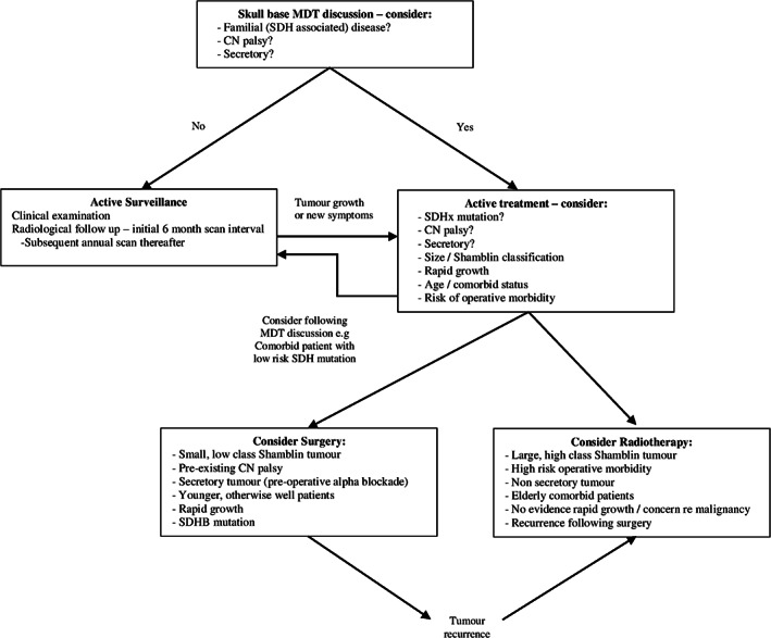
Management of isolated carotid body tumor. CN, cranial nerve; MDT, multidisciplinary team; SDH, succinate dehydrogenase;
6.2. Isolated non‐CBTs
The main non‐CBT HNPGLs include VPGLs and JTPGLs. As with CBTs, Non‐CBTs are frequently an indolent disease process displaying slow or no growth. For example, Jansen et al. demonstrated that 28 of 66 observed Fisch class C/D JTPGL displayed no growth during follow‐up. 107 This would support active surveillance as the primary strategy in the management of many non‐CBTs. However, clinicians should again be aware that this strategy is not risk free as these patients may develop worsening cranial nerve deficits and malignant change has been observed. 5 , 95 , 96
Surgical resection of non‐CBTs can be challenging due to their proximity to and frequent involvement of critical neurovascular structures in the skull‐base. Deficits of cranial nerves VII, IX, X, XI, and XII have all been reported. These cranial nerve deficits can be associated with symptoms such as dysphagia, dysphonia, and facial paralysis. CSF leaks are another feared complication of non‐CBT resection seen in some 1–3% of cases 9 , 104 , 108
Resection of VPGLs usually requires resection of the associated vagus nerve. This results in speech, swallow, and pharyngeal sensory deficits. 86 Cranial nerves VII, IX, X, and XII are also at risk during resection of VPGLs. A systematic review demonstrated 155 new non‐vagal cranial nerve deficits among 226 patients as a result of VPGL resection. 109
When considering JTPGLs a clear distinction should be made between Fisch class A/B tumors and Fisch class C/D tumors. Class A/B tumors limited to the tympanic region can be safely extracted with minimal risk of postoperative morbidity. 110 Early surgery is therefore indicated for these tumors in patients who are good surgical candidates. On the other hand, Fisch Class C/D JTPGLs involving either the jugular bulb or extending intracranially, pose an operative challenge. Total tumor extraction usually requires facial nerve rerouting, a practice which is known to significantly increase postoperative morbidity. 107 , 108 , 111 Therefore, most authors advocate subtotal resection of JTPGLs to reduce surgical morbidity. 86 , 107 With this approach, one series still demonstrated cranial nerve deficits in 20 out of 36 patients following resection of Fisch class C/D tumors. 107
However, despite the reported morbidity, studies still demonstrate good long‐term control of disease following surgery 107 , 108 , 111 , 112 Surgery is therefore an appropriate strategy in cases with prior cranial nerve deficits and those with secretory disease. For both VPGL and JTPGL, it is important to identify an associated SDHB mutation prior to making treatment decisions. In these cases, surgery may be considered due to an increased risk of malignancy. In experienced centers, when tumor location and relationship to surrounding structures allows for resection with low risk of morbidity it may also be considered.
RT provides excellent long‐term tumor control for non‐CBTs. Rates of local control are similar to or in some cases better than with surgery, particularly for Fisch class C/D JTPGLs. 101 , 110 , 113 Morbidity includes symptoms such as xerostomia, mucositis, and nausea. Serious complications such as bone or brain necrosis, dysphagia or cranial nerve deficits are rarely observed. 113 There is again concern regarding long‐term side effects of RT in this region such as radiation induced malignancy and stroke, particularly in younger patients. 104 , 105 Most studies have treated larger tumors with fractionated RT at doses between 45 and 50 Gy. The literature supports the same dosing regimen of 45 Gy delivered at 1.8 Gy daily fractions proposed for CBTs when treating these tumors with fractionated RT. 101
The use of various forms of SRS has been assessed in the management of non‐CBT most notably for treating tumors involving the jugular bulb. Current evidence does not favor one form of SRS over another. 114 SRS has obvious benefits when compared with conventional fractionated RT with less damage to critical local neurovascular structures and treatment at a single visit. SRS has been seen to be an effective treatment with rates of local control comparable to surgery or fractionated RT observed in small series. 114 Marginal doses of 12–15 Gy to the tumor appear to be the most appropriate dose. 101 , 114 Considering the current evidence SRS should be considered for cases where use of conventional fractionated RT is undesirable such as tumors involving the skullbase. 101
Figure 3 demonstrates our recommended treatment algorithm for non‐CBTs.
FIGURE 3.
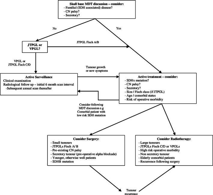
Management of isolated non‐carotid body tumor. CN, cranial nerve; HNPGL, head and neck paraganglioma; JTPGL, Jugulo‐tympanic paraganglioma; MDT, multidisciplinary team; non‐CBT, non‐carotid body tumor; SDH, succinate dehydrogenase; VPGL, vagal paraganglioma
6.3. Surgical considerations
6.3.1. Secretory tumors
Secretory HNPGLs should always be surgically resected if possible to alleviate the symptoms of sympathetic overactivity. 86 Patients with noradrenalin‐secreting HNPGLs should be managed in the same manner as secretory PPGL at other sites. It is recommended that preoperative α‐adrenergic blockade should be commenced as the first choice of medication to minimize perioperative complications in secretory PPGL. These complications include: a hypertensive crisis, cardiac arrhythmias, or myocardial infarction at initiation of anesthesia or during tumor manipulation, or hypotension following tumor removal. 115
Close collaboration with endocrinology is required to prepare patients with catecholamine excess for surgery. Both nonselective α‐blockers (e.g., phenoxybenzamine) and selective α blockers (e.g., doxazocin) are effective, and should be introduced at least 7–14 days preoperatively, to allow sufficient time to titrate medication to reach target blood pressure. 25 Exposure to excess catecholamine over a prolonged period can lead to significant volume contraction, and patients should be encouraged to increase dietary salt and water intake preoperatively. Intravenous saline infusion (1–2 L) is often required and in our practice, this reduces the risk of postoperative hypotension significantly. Caution is required in patients with underlying heart failure or catecholamine‐induced cardiomyopathy. 25 The role of β adrenergic blockade is limited to the management of persistent tachycardia unresponsive to adequate α‐blockade and volume expansion. Β‐blockade should never be initiated without α‐blockade, as to do so risks provoking a catecholamine crisis due to unopposed α adrenergic stimulation. 25
6.3.2. Preoperative embolization
There is significant debate in the literature surrounding the practice of preoperative embolization of HNPGL. Some authors recommend preoperative embolization within 48 h of surgery for patients undergoing surgical resection of HNPGLs due to their vascularity. These authors point to reduced intraoperative blood loss and reduced rates of sacrifice of other vascular structures. 116 , 117 This view is not universally endorsed and other studies suggest that preoperative embolization of these tumors does not attenuate the risks of surgery. 48 , 118 , 119 These authors state that preoperative embolization does not adequately embolize the tumor feeding vessels in HNPGLs and exposes the patient to an unnecessary procedure. They also point to increased risk of stroke in patients who have undergone preoperative embolization. Issues also surround inter‐clinician efficacy when performing tumor embolization with wildly varied results between different institutions. 48 , 118 , 119 As such it is difficult to endorse this practice at present.
6.3.3. Multifocal disease
As mentioned previously, presentation with multifocal HNPGLs is not infrequent. 42 , 46 , 47 , 48 Management of these cases poses a challenge for clinicians. Most series advocate they are treated surgically. 63 , 120 Every effort should be made to preserve cranial nerve function on one side, particularly in patients with non‐CBTs at higher risk of posttreatment cranial nerve deficits. Bilateral cranial nerve deficits can lead to devastating consequences such as patients requiring a lifelong tracheostomy in the case of bilateral vagus nerve dysfunction. Therefore, it is important to distinguish between unilateral and bilateral multifocal disease. 120 Provided patients have no pre‐existing contralateral cranial nerve deficit unilateral multifocal HNPGLs can be treated in a single stage with minimal excess morbidity. 121
Bilateral HNPGLs provide more of a challenge. As mentioned the goal of treatment should be preservation of cranial nerve function on one side. 109 , 122 This does not always require active treatment and indeed in many cases patients are best served with active surveillance, particularly elderly comorbid patients. 63 When patients are to undergo active treatment of bilateral HNPGLs, it should be undertaken in a staged manner with unilateral resection initially to avoid iatrogenic bilateral cranial nerve palsies as well as the risk of baroreflex failure syndrome. 123 Patients should be observed following initial management and in the absence of postoperative cranial nerve deficits treatment of the contralateral side may be undertaken. There is conflicting evidence advocating which tumors should be treated first. We would propose that secretory tumors (to alleviate symptoms), tumors causing pre‐existing cranial nerve dysfunction (reduced risk of further morbidity) and small CBTs (small risk of surgical morbidity) should be considered first for resection. 120 , 124 , 125 All patients with multifocal disease should be discussed at an appropriate MDT to decide if surgery is indicated and if so which tumors are to be treated first.
6.4. Management of malignant HNPGL
HNPGL can only be considered malignant once spread to non‐neuroendocrine tissue is demonstrated. 92 Data from malignant HNPGL series are scarce and treatment regimens are often based on series of non‐HNPGLs and pheochromocytomas. 63 , 126 Treatment options include: surgery, RT, chemotherapy, peptide therapy, and immunotherapy. With metastatic disease confined to the neck, surgery followed by postoperative RT is associated with good outcomes, particularly in younger patients. 13
Management of distant metastases is more challenging. Surgical resection of the primary tumor is indicated when symptomatic due to either catecholamine excess or mass effect. RT can be considered for tumor shrinkage in those who are not surgical candidates. Conventional chemotherapy is historically the most frequently used therapy for distant disease with moderate outcomes. 127 , 128 Cyclophosphamide, vincristine, and dacarbazine is the regime most often cited in the literature.
Recently, peptide therapy and immunotherapy have shown hope in the treatment of distant disease. 129 , 130 , 131 At present, 131I‐MIBG (Azdera) is the most studied treatment in metastatic PPGL and has recently received FDA approval for treatment of metastatic PPGL. Prospective data have shown stable disease or tumor response in 59 of 64 patients with metastatic PPGL treated with 131I‐MIBG. 132 177Lu‐DOTATATE (Luthera) is another peptide therapy that has shown promise for treatment of metastatic PPGL with one series showing either tumor response or stable disease in 12 of 14 patients with biochemically active metastatic PPGL evaluated. 133 Higher baseline SUV max on baseline imaging appears to predict early response to treatment. 131 Data are currently awaited from a phase II trial undertaken by the National Institute of Health evaluating the safety, tolerability, and survival among patients with inoperable PPGL treated with 177Lu‐DOTATATE (NCT03206060). Tyrosine kinase inhibitors (Sunitinib) and programmed death‐ligand 1 inhibitors are currently under investigation for management of metastatic rare tumors including metastatic PPGL (FIRSTMAPPP NCT01371201; NCT02834013; NCT02721732).
All malignant cases should be discussed and treatment decisions made at appropriate multidisciplinary meetings involving: surgeons, endocrinologists, radiation oncologists, and medical oncologists.
7. SURVEILLANCE
7.1. Active surveillance
All patients with HNPGLs undergoing active surveillance should be followed for a prolonged period of time by an appropriately experienced expert. Follow‐up should include at minimum: repeat clinical examination and repeat imaging. 86 Clinical examination should focus on full cranial nerve examination particularly cranial nerves VII–XII and formal audiology testing may also be considered. Individuals with germline mutations should undergo regular biochemical screening measuring plasma or urinary metanephrines and plasma 3MT to screen for additional PPGLs.
MRI head and neck is an appropriate imaging modality for surveillance of sporadic HNPGLs. Imaging from skull base to pelvis should be considered for individuals with germline mutations at risk of PPGLs outside of the head and neck. The British Skull Base Society suggests an initial 6‐month interval, with annual scans thereafter. 86 As the majority of cases are slow‐growing, a yearly follow‐up interval appears to be appropriate. 7 The follow‐up interval may be extended or reduced as appropriate depending on patient specific risk factors and tumor growth during surveillance. Clinicians should consider active management when: rapid growth is observed or new onset cranial nerve dysfunction is noted. Further evidence is required to determine the optimal follow‐up intervals and the necessary follow‐up duration.
7.2. Posttreatment surveillance
7.2.1. Biochemical surveillance of HNPGL patients
Patients with elevated preoperative metanephrines/3MT levels should undergo repeat testing 2–6 weeks postoperatively to confirm complete resection. Persistent catecholamine excess implies either an incomplete resection, or an occult synchronous tumor. 26
Patients at high risk of recurrent or metastatic disease (including germline mutation carriers), should be offered lifelong annual biochemical surveillance measuring plasma or urinary metanephrines and plasma 3MT to screen for recurrent, metastatic, or metachronous tumors, irrespective of the secretory status of the primary tumor, in conjunction with imaging surveillance of the entire sympathetic chain. 25 , 26
7.2.2. Radiological surveillance
For patients who undergo resection of HNPGL, it is recommended that baseline postoperative imaging should be performed 3 months after surgery to assess for residual disease. 86 The extent, modality, and frequency of surveillance imaging of HNPGL depends on presence or absence of a germline mutation predisposing to PPGL (e.g., SDH mutations). It may be reasonable to monitor a patient with an isolated, sporadic HNPGL using MRI head and neck only. Germline mutation carriers are at greater risk of both metastatic disease and metachronous tumors, and therefore will require ongoing imaging surveillance encompassing the skull base to pelvis. 86 Biannual rapid sequence, non‐contrast MRI skull base to pelvis has been reported to be an effective surveillance tool in SDH‐associated PPGL or for postoperative surveillance of biochemically inactive disease. 26 , 70 The addition of a DWI sequence to MRI may increase sensitivity for surveillance of SDH patients. 134 Imaging surveillance should also be offered to asymptomatic carriers of germline mutations identified during cascade genetic testing. 62 , 86 The age of commencement of image‐based surveillance of asymptomatic germline mutation carriers depends on the individual mutation but is recommended from the age of 10–15 years in SDHB and D germline mutations. 35
7.2.3. Duration of surveillance
The risk of recurrence following treatment overall in HNPGLs is <10%, but is higher in those with familial disease. 26 , 48 , 110 , 112 Recurrence does not necessarily occur early and Jackson et al. determined a median time to recurrence of 5.1 years (mean 8.2 years) in HNPGL. 58 As such, long‐term follow‐up with serial imaging and thorough clinical examination is warranted. 26 , 86 There is no ideal follow‐up pro forma widely agreed on in the literature. We would agree with the follow‐up proposed by the British Skull Base Society Clinical Consensus on the Management of Head and Neck Paragangliomas. This recommends yearly imaging for the first 3 years with reduced follow‐up intervals thereafter if appropriate. 86 Little guidance exists within the literature to suggest an ideal follow‐up duration. The European Society of Endocrinology recommends a minimum of 10 years follow‐up for these patients following treatment and that lifelong follow‐up should be considered for high‐risk patients, and those with a genetic predisposition to PPGL. 26
8. POTENTIAL AREAS FOR FUTURE RESEARCH
Many recent advances have been made in the understanding of the pathophysiology, genetic associations, radiological investigations and natural history of HNPGLs. Despite this limitations, in our understanding of these tumors remain. The area of most interest at present remains immunotherapy and targeted peptide therapy for metastatic disease. The results of numerous prospective trials including the NCT03206060 and the FIRSTMAPPP trials are eagerly awaited to guide future management of distant metastatic disease.
Second, further efforts are necessary to investigate the utility of SRS in the management of HNPGL, particularly in cases with tumor involving the skull base where surgical management is challenging. Successful application of SRS for management of HNPGL, particularly these challenging cases, offers the potential to reduce morbidity for patients while providing comparable oncologic outcomes.
Finally, access to genetic testing and our improved understanding of the mutations and associated phenotypes associated with HNPGL has fundamentally altered our understanding of these tumors. Further data are still required to allow for improved substantification and risk assessment of tumors at risk of rapid progression and distant spread in order to adequately council and treat our patients.
9. CONCLUSIONS
HNPGLs are rare, typically benign neuroendocrine tumors that should be managed in appropriate centers with an experienced multidisciplinary team incorporating both head and neck or vascular surgeons and endocrinologists. It is now recognized that a significant proportion of HNPGL are hereditary, predominantly associated with germline mutations in the SDHx gene family. If detected, germline mutations have important implications for both the investigation and surveillance of the affected individual and their family members. Thus, genetic screening should be discussed with all patients diagnosed with HNPGL. Historically considered aggressive tumors necessitating surgical intervention, recent evidence has demonstrated that the vast majority can be safely observed. Classification of HNPGL into CBTs and non‐CBTs is useful for clinicians when planning treatment strategies, due to differing morbidity associated with these entities. We would advocate that surgical treatment strategies are more frequently indicated in CBTs while nonsurgical strategies should be utilized in non‐CBT where possible.
CONFLICT OF INTEREST
The authors declare no conflict of interest.
Supporting information
Appendix S1. Shamblin Classification of Carotid Body Tumors
Appendix S2. Fisch Classification of Jugulo‐tympanic paragangliomas
Cleere EF, Martin‐Grace J, Gendre A, Sherlock M, O'Neill JP. Contemporary management of paragangliomas of the head and neck. Laryngoscope Investigative Otolaryngology. 2022;7(1):93‐107. doi: 10.1002/lio2.706
REFERENCES
- 1. Lee JH, Barich F, Karnell LH, et al. National Cancer Data Base report on malignant paragangliomas of the head and neck. Cancer. 2002;94(3):730‐737. [DOI] [PubMed] [Google Scholar]
- 2. El‐Naggar AK, JKC C, Grandis JR, Takata T, Slootweg PJ. WHO Classification of Head and Neck Tumours. 4th ed. WHO; 2017. [Google Scholar]
- 3. Fishbein L, Merrill S, Fraker DL, Cohen DL, Nathanson KL. Inherited mutations in pheochromocytoma and paraganglioma: why all patients should be offered genetic testing. Ann Surg Oncol. 2013;20(5):1444‐1450. [DOI] [PMC free article] [PubMed] [Google Scholar]
- 4. Galan SR, Kann PH. Genetics and molecular pathogenesis of pheochromocytoma and paraganglioma. Clin Endocrinol (Oxf). 2013;78(2):165‐175. [DOI] [PubMed] [Google Scholar]
- 5. Harrison L, Corbridge R. Active surveillance management of head and neck paragangliomas: case series and review of the literature. J Laryngol Otol. 2017;131(7):580‐584. [DOI] [PubMed] [Google Scholar]
- 6. Jansen JC, van den Berg R, Kuiper A, van der Mey AG, Zwinderman AH, Cornelisse CJ. Estimation of growth rate in patients with head and neck paragangliomas influences the treatment proposal. Cancer. 2000;88(12):2811‐2816. [PubMed] [Google Scholar]
- 7. Jansen TTG, Timmers H, Marres HAM, Kunst HPM. Feasibility of a wait‐and‐scan period as initial management strategy for head and neck paraganglioma. Head Neck. 2017;39(10):2088‐2094. [DOI] [PubMed] [Google Scholar]
- 8. Rijken JA, de Vos B, van Hest LP, et al. Evolving management strategies in head and neck paragangliomas: a single‐centre experience with 147 patients over a 60‐year period. Clin Otolaryngol. 2019;44(5):836‐841. [DOI] [PMC free article] [PubMed] [Google Scholar]
- 9. Valero C, Ganly I, Shah JP. Head and neck paragangliomas: 30‐year experience. Head Neck. 2020;42(9):2486‐2495. [DOI] [PMC free article] [PubMed] [Google Scholar]
- 10. Williams MD. Paragangliomas of the head and neck: an overview from diagnosis to genetics. Head Neck Pathol. 2017;11(3):278‐287. [DOI] [PMC free article] [PubMed] [Google Scholar]
- 11. Papathomas TG, de Krijger RR, Tischler AS. Paragangliomas: update on differential diagnostic considerations, composite tumors, and recent genetic developments. Semin Diagn Pathol. 2013;30(3):207‐223. [DOI] [PubMed] [Google Scholar]
- 12. Tischler AS. Pheochromocytoma and extra‐adrenal paraganglioma: updates. Arch Pathol Lab Med. 2008;132(8):1272‐1284. [DOI] [PubMed] [Google Scholar]
- 13. Javidiparsijani S, Brickman A, Lin DM, et al. Is regional lymph node metastasis of head and neck paraganglioma a sign of aggressive clinical behavior: a clinical/pathologic review. Ear Nose Throat J. 2019;145561319863373. 100(6):447–453. [DOI] [PubMed] [Google Scholar]
- 14. Kay JM, Laidler P. Hypoxia and the carotid body. J Clin Pathol Suppl (R Coll Pathol). 1977;11:30‐44. [DOI] [PMC free article] [PubMed] [Google Scholar]
- 15. Rodriguez‐Cuevas S, Lopez‐Garza J, Labastida‐Almendaro S. Carotid body tumors in inhabitants of altitudes higher than 2000 meters above sea level. Head Neck. 1998;20(5):374‐378. [DOI] [PubMed] [Google Scholar]
- 16. Gill AJ, Benn DE, Chou A, et al. Immunohistochemistry for SDHB triages genetic testing of SDHB, SDHC, and SDHD in paraganglioma‐pheochromocytoma syndromes. Hum Pathol. 2010;41(6):805‐814. [DOI] [PubMed] [Google Scholar]
- 17. Williams MD, Tischler AS. Update from the 4th edition of the World Health Organization classification of head and neck tumours: paragangliomas. Head Neck Pathol. 2017;11(1):88‐95. [DOI] [PMC free article] [PubMed] [Google Scholar]
- 18. Bayley JP, van Minderhout I, Weiss MM, et al. Mutation analysis of SDHB and SDHC: novel germline mutations in sporadic head and neck paraganglioma and familial paraganglioma and/or pheochromocytoma. BMC Med Genet. 2006;7:1. [DOI] [PMC free article] [PubMed] [Google Scholar]
- 19. Group NGSiPS , Toledo RA, Burnichon N, et al. Consensus statement on next‐generation‐sequencing‐based diagnostic testing of hereditary phaeochromocytomas and paragangliomas. Nat Rev Endocrinol. 2017;13(4):233‐247. [DOI] [PubMed] [Google Scholar]
- 20. Rana MU, Osthus AA, Heimdal K, Jebsen P, Revheim MR, Osnes TA. Head and neck paragangliomas in Norway, importance of genetics, updated diagnostic workup and treatment. Acta Otolaryngol. 2021;141(3):303‐308. [DOI] [PubMed] [Google Scholar]
- 21. Schiavi F, Boedeker CC, Bausch B, et al. Predictors and prevalence of paraganglioma syndrome associated with mutations of the SDHC gene. JAMA. 2005;294(16):2057‐2063. [DOI] [PubMed] [Google Scholar]
- 22. Hensen EF, Siemers MD, Jansen JC, et al. Mutations in SDHD are the major determinants of the clinical characteristics of Dutch head and neck paraganglioma patients. Clin Endocrinol (Oxf). 2011;75(5):650‐655. [DOI] [PubMed] [Google Scholar]
- 23. Piccini V, Rapizzi E, Bacca A, et al. Head and neck paragangliomas: genetic spectrum and clinical variability in 79 consecutive patients. Endocr Relat Cancer. 2012;19(2):149‐155. [DOI] [PubMed] [Google Scholar]
- 24. Hensen EF, Jordanova ES, van Minderhout IJ, et al. Somatic loss of maternal chromosome 11 causes parent‐of‐origin‐dependent inheritance in SDHD‐linked paraganglioma and phaeochromocytoma families. Oncogene. 2004;23(23):4076‐4083. [DOI] [PubMed] [Google Scholar]
- 25. Lenders JW, Duh QY, Eisenhofer G, et al. Pheochromocytoma and paraganglioma: an endocrine society clinical practice guideline. J Clin Endocrinol Metab. 2014;99(6):1915‐1942. [DOI] [PubMed] [Google Scholar]
- 26. Plouin PF, Amar L, Dekkers OM, et al. European Society of Endocrinology Clinical Practice Guideline for long‐term follow‐up of patients operated on for a phaeochromocytoma or a paraganglioma. Eur J Endocrinol. 2016;174(5):G1‐G10. [DOI] [PubMed] [Google Scholar]
- 27. Boedeker CC, Erlic Z, Richard S, et al. Head and neck paragangliomas in von Hippel‐Lindau disease and multiple endocrine neoplasia type 2. J Clin Endocrinol Metab. 2009;94(6):1938‐1944. [DOI] [PMC free article] [PubMed] [Google Scholar]
- 28. Eisenhofer G, Klink B, Richter S, Lenders JW, Robledo M. Metabologenomics of phaeochromocytoma and paraganglioma: an integrated approach for personalised biochemical and genetic testing. Clin Biochem Rev. 2017;38(2):69‐100. [PMC free article] [PubMed] [Google Scholar]
- 29. Melmed SAR, Goldfine A, Koenig R, Rosen C. Williams Textbook of Endocrinology. 14th ed. Elsevier; 2019. [Google Scholar]
- 30. Andrews KA, Ascher DB, Pires DEV, et al. Tumour risks and genotype‐phenotype correlations associated with germline variants in succinate dehydrogenase subunit genes SDHB, SDHC and SDHD. J Med Genet. 2018;55(6):384‐394. [DOI] [PMC free article] [PubMed] [Google Scholar]
- 31. DeAngelis LM, Kelleher MB, Post KD, Fetell MR. Multiple paragangliomas in neurofibromatosis: a new neuroendocrine neoplasia. Neurology. 1987;37(1):129‐133. [DOI] [PubMed] [Google Scholar]
- 32. Neumann HP, Sullivan M, Winter A, et al. Germline mutations of the TMEM127 gene in patients with paraganglioma of head and neck and extraadrenal abdominal sites. J Clin Endocrinol Metab. 2011;96(8):E1279‐E1282. [DOI] [PubMed] [Google Scholar]
- 33. Taschner PE, Jansen JC, Baysal BE, et al. Nearly all hereditary paragangliomas in the Netherlands are caused by two founder mutations in the SDHD gene. Genes Chromosomes Cancer. 2001;31(3):274‐281. [DOI] [PubMed] [Google Scholar]
- 34. Baysal BE, Ferrell RE, Willett‐Brozick JE, et al. Mutations in SDHD, a mitochondrial complex II gene, in hereditary paraganglioma. Science. 2000;287(5454):848‐851. [DOI] [PubMed] [Google Scholar]
- 35. MacFarlane J, Seong KC, Bisambar C, et al. A review of the tumour spectrum of germline succinate dehydrogenase gene mutations: beyond phaeochromocytoma and paraganglioma. Clin Endocrinol (Oxf). 2020;93(5):528‐538. [DOI] [PubMed] [Google Scholar]
- 36. Benn DE, Gimenez‐Roqueplo AP, Reilly JR, et al. Clinical presentation and penetrance of pheochromocytoma/paraganglioma syndromes. J Clin Endocrinol Metab. 2006;91(3):827‐836. [DOI] [PubMed] [Google Scholar]
- 37. Neumann HP, Pawlu C, Peczkowska M, et al. Distinct clinical features of paraganglioma syndromes associated with SDHB and SDHD gene mutations. JAMA. 2004;292(8):943‐951. [DOI] [PubMed] [Google Scholar]
- 38. Benn DE, Robinson BG, Clifton‐Bligh RJ. 15 Years of paraganglioma: clinical manifestations of paraganglioma syndromes types 1‐5. Endocr Relat Cancer. 2015;22(4):T91‐T103. [DOI] [PMC free article] [PubMed] [Google Scholar]
- 39. Korpershoek E, Favier J, Gaal J, et al. SDHA immunohistochemistry detects germline SDHA gene mutations in apparently sporadic paragangliomas and pheochromocytomas. J Clin Endocrinol Metab. 2011;96(9):E1472‐E1476. [DOI] [PubMed] [Google Scholar]
- 40. Evenepoel L, Papathomas TG, Krol N, et al. Toward an improved definition of the genetic and tumor spectrum associated with SDH germ‐line mutations. Genet Med. 2015;17(8):610‐620. [DOI] [PubMed] [Google Scholar]
- 41. Kunst HP, Rutten MH, de Monnink JP, et al. SDHAF2 (PGL2‐SDH5) and hereditary head and neck paraganglioma. Clin Cancer Res. 2011;17(2):247‐254. [DOI] [PubMed] [Google Scholar]
- 42. Erickson D, Kudva YC, Ebersold MJ, et al. Benign paragangliomas: clinical presentation and treatment outcomes in 236 patients. J Clin Endocrinol Metab. 2001;86(11):5210‐5216. [DOI] [PubMed] [Google Scholar]
- 43. Smith JD, Harvey RN, Darr OA, et al. Head and neck paragangliomas: a two‐decade institutional experience and algorithm for management. Laryngoscope Investig Otolaryngol. 2017;2(6):380‐389. [DOI] [PMC free article] [PubMed] [Google Scholar]
- 44. Kunzel J, de Tristan J, Mantsopoulos K, et al. Experiences in the treatment of patients with multiple head and neck paragangliomas. Am J Otolaryngol. 2014;35(3):294‐299. [DOI] [PubMed] [Google Scholar]
- 45. Langerman A, Athavale SM, Rangarajan SV, Sinard RJ, Netterville JL. Natural history of cervical paragangliomas: outcomes of observation of 43 patients. Arch Otolaryngol Head Neck Surg. 2012;138(4):341‐345. [DOI] [PubMed] [Google Scholar]
- 46. Netterville JL, Jackson CG, Miller FR, Wanamaker JR, Glasscock ME. Vagal paraganglioma: a review of 46 patients treated during a 20‐year period. Arch Otolaryngol Head Neck Surg. 1998;124(10):1133‐1140. [DOI] [PubMed] [Google Scholar]
- 47. Papaspyrou K, Mewes T, Rossmann H, et al. Head and neck paragangliomas: report of 175 patients (1989‐2010). Head Neck. 2012;34(5):632‐637. [DOI] [PubMed] [Google Scholar]
- 48. Robertson V, Poli F, Hobson B, Saratzis A, Ross NA. A systematic review and meta‐analysis of the presentation and surgical management of patients with carotid body tumours. Eur J Vasc Endovasc Surg. 2019;57(4):477‐486. [DOI] [PubMed] [Google Scholar]
- 49. Shamblin WR, ReMine WH, Sheps SG, Harrison EG Jr. Carotid body tumor (chemodectoma). Clinicopathologic analysis of ninety cases. Am J Surg. 1971;122(6):732‐739. [DOI] [PubMed] [Google Scholar]
- 50. Boedeker CC, Ridder GJ, Schipper J. Paragangliomas of the head and neck: diagnosis and treatment. Fam Cancer. 2005;4(1):55‐59. [DOI] [PubMed] [Google Scholar]
- 51. Patetsios P, Gable DR, Garrett WV, et al. Management of carotid body paragangliomas and review of a 30‐year experience. Ann Vasc Surg. 2002;16(3):331‐338. [DOI] [PubMed] [Google Scholar]
- 52. Neskey DM, Hatoum G, Modh R, et al. Outcomes after surgical resection of head and neck paragangliomas: a review of 61 patients. Skull Base. 2011;21(3):171‐176. [DOI] [PMC free article] [PubMed] [Google Scholar]
- 53. Fisch U. Infratemporal fossa approach for glomus tumors of the temporal bone. Ann Otol Rhinol Laryngol. 1982;91(5 Pt 1):474‐479. [DOI] [PubMed] [Google Scholar]
- 54. Jackson CG, Glasscock ME 3rd, Harris PF. Glomus tumors. Diagnosis, classification, and management of large lesions. Arch Otolaryngol. 1982;108(7):401‐410. [DOI] [PubMed] [Google Scholar]
- 55. Fayad JN, Keles B, Brackmann DE. Jugular foramen tumors: clinical characteristics and treatment outcomes. Otol Neurotol. 2010;31(2):299‐305. [DOI] [PubMed] [Google Scholar]
- 56. Moe KS, Li D, Linder TE, Schmid S, Fisch U. An update on the surgical treatment of temporal bone paraganglioma. Skull Base Surg. 1999;9(3):185‐194. [DOI] [PMC free article] [PubMed] [Google Scholar]
- 57. Sanna M, Jain Y, De Donato G, Rohit LL, Taibah A. Management of jugular paragangliomas: the Gruppo Otologico experience. Otol Neurotol. 2004;25(5):797‐804. [DOI] [PubMed] [Google Scholar]
- 58. Jackson CG. Glomus tympanicum and glomus jugulare tumors. Otolaryngol Clin North Am. 2001;34(5):941‐970. [DOI] [PubMed] [Google Scholar]
- 59. Miller RB, Boon MS, Atkins JP, Lowry LD. Vagal paraganglioma: the Jefferson experience. Otolaryngol Head Neck Surg. 2000;122(4):482‐487. [DOI] [PubMed] [Google Scholar]
- 60. Eisenhofer G, Huynh TT, Hiroi M, Pacak K. Understanding catecholamine metabolism as a guide to the biochemical diagnosis of pheochromocytoma. Rev Endocr Metab Disord. 2001;2(3):297‐311. [DOI] [PubMed] [Google Scholar]
- 61. Osinga TE, Korpershoek E, de Krijger RR, et al. Catecholamine‐synthesizing enzymes are expressed in parasympathetic head and neck paraganglioma tissue. Neuroendocrinology. 2015;101(4):289‐295. [DOI] [PubMed] [Google Scholar]
- 62. Corssmit EP, Romijn JA. Clinical management of paragangliomas. Eur J Endocrinol. 2014;171(6):R231‐R243. [DOI] [PubMed] [Google Scholar]
- 63. Taieb D, Kaliski A, Boedeker CC, et al. Current approaches and recent developments in the management of head and neck paragangliomas. Endocr Rev. 2014;35(5):795‐819. [DOI] [PMC free article] [PubMed] [Google Scholar]
- 64. Rao D, Peitzsch M, Prejbisz A, et al. Plasma methoxytyramine: clinical utility with metanephrines for diagnosis of pheochromocytoma and paraganglioma. Eur J Endocrinol. 2017;177(2):103‐113. [DOI] [PMC free article] [PubMed] [Google Scholar]
- 65. van Duinen N, Corssmit EP, de Jong WH, Brookman D, Kema IP, Romijn JA. Plasma levels of free metanephrines and 3‐methoxytyramine indicate a higher number of biochemically active HNPGL than 24‐h urinary excretion rates of catecholamines and metabolites. Eur J Endocrinol. 2013;169(3):377‐382. [DOI] [PubMed] [Google Scholar]
- 66. van Berkel A, Lenders JW, Timmers HJ. Diagnosis of endocrine disease: biochemical diagnosis of phaeochromocytoma and paraganglioma. Eur J Endocrinol. 2014;170(3):R109‐R119. [DOI] [PubMed] [Google Scholar]
- 67. Darr R, Kuhn M, Bode C, et al. Accuracy of recommended sampling and assay methods for the determination of plasma‐free and urinary fractionated metanephrines in the diagnosis of pheochromocytoma and paraganglioma: a systematic review. Endocrine. 2017;56(3):495‐503. [DOI] [PMC free article] [PubMed] [Google Scholar]
- 68. Brink I, Hoegerle S, Klisch J, Bley TA. Imaging of pheochromocytoma and paraganglioma. Fam Cancer. 2005;4(1):61‐68. [DOI] [PubMed] [Google Scholar]
- 69. Koopmans KP, Jager PL, Kema IP, Kerstens MN, Albers F, Dullaart RP. 111In‐octreotide is superior to 123I‐metaiodobenzylguanidine for scintigraphic detection of head and neck paragangliomas. J Nucl Med. 2008;49(8):1232‐1237. [DOI] [PubMed] [Google Scholar]
- 70. Daniel E, Jones R, Bull M, Newell‐Price J. Rapid‐sequence MRI for long‐term surveillance for paraganglioma and phaeochromocytoma in patients with succinate dehydrogenase mutations. Eur J Endocrinol. 2016;175(6):561‐570. [DOI] [PubMed] [Google Scholar]
- 71. Taieb D, Hicks RJ, Hindie E, et al. European Association of Nuclear Medicine Practice Guideline/Society of Nuclear Medicine and Molecular Imaging Procedure Standard 2019 for radionuclide imaging of phaeochromocytoma and paraganglioma. Eur J Nucl Med Mol Imaging. 2019;46(10):2112‐2137. [DOI] [PMC free article] [PubMed] [Google Scholar]
- 72. Ryder SJ, Love AJ, Duncan EL, Pattison DA. PET detectives: molecular imaging for phaeochromocytomas and paragangliomas in the genomics era. Clin Endocrinol (Oxf). 2020;95(1):13–28. [DOI] [PubMed] [Google Scholar]
- 73. Gimenez‐Roqueplo AP, Caumont‐Prim A, Houzard C, et al. Imaging work‐up for screening of paraganglioma and pheochromocytoma in SDHx mutation carriers: a multicenter prospective study from the PGL.EVA investigators. J Clin Endocrinol Metab. 2013;98(1):E162‐E173. [DOI] [PubMed] [Google Scholar]
- 74. Michalowska I, Cwikla JB, Peczkowska M, et al. Usefulness of somatostatin receptor scintigraphy (Tc‐[HYNIC, Tyr3]‐octreotide) and 123I‐metaiodobenzylguanidine scintigraphy in patients with SDHx gene‐related pheochromocytomas and paragangliomas detected by computed tomography. Neuroendocrinology. 2015;101(4):321‐330. [DOI] [PubMed] [Google Scholar]
- 75. Chang CA, Pattison DA, Tothill RW, et al. (68)Ga‐DOTATATE and (18)F‐FDG PET/CT in paraganglioma and pheochromocytoma: utility, patterns and heterogeneity. Cancer Imaging. 2016;16(1):22. [DOI] [PMC free article] [PubMed] [Google Scholar]
- 76. Reubi JC, Waser B, Schaer JC, Laissue JA. Somatostatin receptor sst1‐sst5 expression in normal and neoplastic human tissues using receptor autoradiography with subtype‐selective ligands. Eur J Nucl Med. 2001;28(7):836‐846. [DOI] [PubMed] [Google Scholar]
- 77. Janssen I, Blanchet EM, Adams K, et al. Superiority of [68Ga]‐DOTATATE PET/CT to other functional imaging modalities in the localization of SDHB‐associated metastatic pheochromocytoma and paraganglioma. Clin Cancer Res. 2015;21(17):3888‐3895. [DOI] [PMC free article] [PubMed] [Google Scholar]
- 78. Janssen I, Chen CC, Millo CM, et al. PET/CT comparing (68)Ga‐DOTATATE and other radiopharmaceuticals and in comparison with CT/MRI for the localization of sporadic metastatic pheochromocytoma and paraganglioma. Eur J Nucl Med Mol Imaging. 2016;43(10):1784‐1791. [DOI] [PMC free article] [PubMed] [Google Scholar]
- 79. Kan Y, Zhang S, Wang W, Liu J, Yang J, Wang Z. (68)Ga‐somatostatin receptor analogs and (18)F‐FDG PET/CT in the localization of metastatic pheochromocytomas and paragangliomas with germline mutations: a meta‐analysis. Acta Radiol. 2018;59(12):1466‐1474. [DOI] [PubMed] [Google Scholar]
- 80. Sharma P, Thakar A, Suman KCS, et al. 68Ga‐DOTANOC PET/CT for baseline evaluation of patients with head and neck paraganglioma. J Nucl Med. 2013;54(6):841‐847. [DOI] [PubMed] [Google Scholar]
- 81. Kroiss A, Putzer D, Frech A, et al. A retrospective comparison between 68Ga‐DOTA‐TOC PET/CT and 18F‐DOPA PET/CT in patients with extra‐adrenal paraganglioma. Eur J Nucl Med Mol Imaging. 2013;40(12):1800‐1808. [DOI] [PubMed] [Google Scholar]
- 82. Timmers HJ, Kozupa A, Chen CC, et al. Superiority of fluorodeoxyglucose positron emission tomography to other functional imaging techniques in the evaluation of metastatic SDHB‐associated pheochromocytoma and paraganglioma. J Clin Oncol. 2007;25(16):2262‐2269. [DOI] [PubMed] [Google Scholar]
- 83. Warburg O. On the origin of cancer cells. Science. 1956;123(3191):309‐314. [DOI] [PubMed] [Google Scholar]
- 84. Fiebrich HB, Brouwers AH, Kerstens MN, et al. 6‐[F‐18]Fluoro‐L‐dihydroxyphenylalanine positron emission tomography is superior to conventional imaging with (123)I‐metaiodobenzylguanidine scintigraphy, computer tomography, and magnetic resonance imaging in localizing tumors causing catecholamine excess. J Clin Endocrinol Metab. 2009;94(10):3922‐3930. [DOI] [PubMed] [Google Scholar]
- 85. Reichert T, Fakhry N, Lavieille JP, et al. Exploring the link between tumour metabolism and succinate dehydrogenase deficiency: a (18) F‐FDOPA PET/CT study in head and neck paragangliomas. Clin Endocrinol (Oxf). 2019;91(6):879‐884. [DOI] [PMC free article] [PubMed] [Google Scholar]
- 86. Lloyd S, Obholzer R, Tysome J, Group BC. British skull base society clinical consensus document on management of head and neck paragangliomas. Otolaryngol Head Neck Surg. 2020;163(3):400‐409. [DOI] [PubMed] [Google Scholar]
- 87. Lenders JWM, Kerstens MN, Amar L, et al. Genetics, diagnosis, management and future directions of research of phaeochromocytoma and paraganglioma: a position statement and consensus of the Working Group on Endocrine Hypertension of the European Society of Hypertension. J Hypertens. 2020;38(8):1443‐1456. [DOI] [PMC free article] [PubMed] [Google Scholar]
- 88. Carlson ML, Sweeney AD, Wanna GB, Netterville JL, Haynes DS. Natural history of glomus jugulare: a review of 16 tumors managed with primary observation. Otolaryngol Head Neck Surg. 2015;152(1):98‐105. [DOI] [PubMed] [Google Scholar]
- 89. Cosetti M, Linstrom C, Alexiades G, Tessema B, Parisier S. Glomus tumors in patients of advanced age: a conservative approach. Laryngoscope. 2008;118(2):270‐274. [DOI] [PubMed] [Google Scholar]
- 90. Heesterman BL, de Pont LMH, Verbist BM, et al. Age and tumor volume predict growth of carotid and vagal body paragangliomas. J Neurol Surg B Skull Base. 2017;78(6):497‐505. [DOI] [PMC free article] [PubMed] [Google Scholar]
- 91. Prasad SC, Mimoune HA, D'Orazio F, et al. The role of wait‐and‐scan and the efficacy of radiotherapy in the treatment of temporal bone paragangliomas. Otol Neurotol. 2014;35(5):922‐931. [DOI] [PubMed] [Google Scholar]
- 92. Obholzer RJ, Hornigold R, Connor S, Gleeson MJ. Classification and management of cervical paragangliomas. Ann R Coll Surg Engl. 2011;93(8):596‐602. [DOI] [PMC free article] [PubMed] [Google Scholar]
- 93. Mediouni A, Ammari S, Wassef M, et al. Malignant head/neck paragangliomas. Comparative study. Eur Ann Otorhinolaryngol Head Neck Dis. 2014;131(3):159‐166. [DOI] [PubMed] [Google Scholar]
- 94. Manolidis S, Shohet JA, Jackson CG, Glasscock ME 3rd. Malignant glomus tumors. Laryngoscope. 1999;109(1):30‐34. [DOI] [PubMed] [Google Scholar]
- 95. Kahn LB. Vagal body tumor (nonchromaffin paraganglioma, chemodectoma, and carotid body‐like tumor) with cervical node metastasis and familial association: ultrastructural study and review. Cancer. 1976;38(6):2367‐2377. [DOI] [PubMed] [Google Scholar]
- 96. Kloppel G. Tumors of the adrenal medulla and the paraganglia. Pathologe. 2003;24(4):280‐286. [DOI] [PubMed] [Google Scholar]
- 97. Nishijima H, Asakage T, Sugasawa M. Malignant carotid body tumor with systemic metastases. Ann Otol Rhinol Laryngol. 2011;120(6):381‐385. [DOI] [PubMed] [Google Scholar]
- 98. Jansen TTG, Marres HAM, Kaanders J, Kunst HPM. A meta‐analysis on the surgical management of paraganglioma of the carotid body per Shamblin class. Clin Otolaryngol. 2018;43(4):1104–1116. [DOI] [PubMed] [Google Scholar]
- 99. Lozano FS, Munoz A, de Las Heras JA, Gonzalez‐Porras JR. Simple and complex carotid paragangliomas. Three decades of experience and literature review. Head Neck. 2020;42(12):3538‐3550. [DOI] [PubMed] [Google Scholar]
- 100. Mascia D, Esposito G, Ferrante A, Grandi A, Melissano G, Chiesa R. Carotid body tumor contemporary management in a high‐volume center. J Cardiovasc Surg (Torino). 2020;61(4):459‐466. [DOI] [PubMed] [Google Scholar]
- 101. Gilbo P, Morris CG, Amdur RJ, et al. Radiotherapy for benign head and neck paragangliomas: a 45‐year experience. Cancer. 2014;120(23):3738‐3743. [DOI] [PubMed] [Google Scholar]
- 102. Meyer FB, Sundt TM Jr, Pearson BW. Carotid body tumors: a subject review and suggested surgical approach. J Neurosurg. 1986;64(3):377‐385. [DOI] [PubMed] [Google Scholar]
- 103. Pacheco‐Ojeda LA. Carotid body tumors: surgical experience in 215 cases. J Craniomaxillofac Surg. 2017;45(9):1472‐1477. [DOI] [PubMed] [Google Scholar]
- 104. Suarez C, Rodrigo JP, Mendenhall WM, et al. Carotid body paragangliomas: a systematic study on management with surgery and radiotherapy. Eur Arch Otorhinolaryngol. 2014;271(1):23‐34. [DOI] [PubMed] [Google Scholar]
- 105. Krych AJ, Foote RL, Brown PD, Garces YI, Link MJ. Long‐term results of irradiation for paraganglioma. Int J Radiat Oncol Biol Phys. 2006;65(4):1063‐1066. [DOI] [PubMed] [Google Scholar]
- 106. Zabel A, Milker‐Zabel S, Huber P, et al. Fractionated stereotactic conformal radiotherapy in the management of large chemodectomas of the skull base. Int J Radiat Oncol Biol Phys. 2004;58(5):1445‐1450. [DOI] [PubMed] [Google Scholar]
- 107. Jansen TTG, Kaanders J, Beute GN, Timmers H, Marres HAM, Kunst HPM. Surgery, radiotherapy or a combined modality for jugulotympanic paraganglioma of Fisch class C and D. Clin Otolaryngol. 2018;43(6):1566‐1572. [DOI] [PubMed] [Google Scholar]
- 108. Bacciu A, Ait Mimoune H, D'Orazio F, Vitullo F, Russo A, Sanna M. Management of facial nerve in surgical treatment of previously untreated Fisch class C tympanojugular paragangliomas: long‐term results. J Neurol Surg B Skull Base. 2014;75(1):1‐7. [DOI] [PMC free article] [PubMed] [Google Scholar]
- 109. Suarez C, Rodrigo JP, Bodeker CC, et al. Jugular and vagal paragangliomas: systematic study of management with surgery and radiotherapy. Head Neck. 2013;35(8):1195‐1204. [DOI] [PubMed] [Google Scholar]
- 110. Jansen TTG, Timmers H, Marres HAM, Kaanders J, Kunst HPM. Results of a systematic literature review of treatment modalities for jugulotympanic paraganglioma, stratified per Fisch class. Clin Otolaryngol. 2018;43(2):652‐661. [DOI] [PubMed] [Google Scholar]
- 111. Odat H, Shin SH, Odat MA, Alzoubi F. Facial nerve management in jugular paraganglioma surgery: a literature review. J Laryngol Otol. 2016;130(3):219‐224. [DOI] [PubMed] [Google Scholar]
- 112. Moore MG, Netterville JL, Mendenhall WM, Isaacson B, Nussenbaum B. Head and neck paragangliomas: an update on evaluation and management. Otolaryngol Head Neck Surg. 2016;154(4):597‐605. [DOI] [PubMed] [Google Scholar]
- 113. Hinerman RW, Amdur RJ, Morris CG, Kirwan J, Mendenhall WM. Definitive radiotherapy in the management of paragangliomas arising in the head and neck: a 35‐year experience. Head Neck. 2008;30(11):1431‐1438. [DOI] [PubMed] [Google Scholar]
- 114. Fatima N, Pollom E, Soltys S, Chang SD, Meola A. Stereotactic radiosurgery for head and neck paragangliomas: a systematic review and meta‐analysis. Neurosurg Rev. 2020;44:741–752. [DOI] [PubMed] [Google Scholar]
- 115. Challis BG, Casey RT, Simpson HL, Gurnell M. Is there an optimal preoperative management strategy for phaeochromocytoma/paraganglioma? Clin Endocrinol (Oxf). 2017;86(2):163‐167. [DOI] [PubMed] [Google Scholar]
- 116. Li J, Wang S, Zee C, et al. Preoperative angiography and transarterial embolization in the management of carotid body tumor: a single‐center, 10‐year experience. Neurosurgery. 2010;67(4):941‐948. [DOI] [PubMed] [Google Scholar]
- 117. Texakalidis P, Charisis N, Giannopoulos S, et al. Role of preoperative embolization in carotid body tumor surgery: a systematic review and meta‐analysis. World Neurosurg. 2019;129:503‐13 e2. [DOI] [PubMed] [Google Scholar]
- 118. Abu‐Ghanem S, Yehuda M, Carmel NN, Abergel A, Fliss DM. Impact of preoperative embolization on the outcomes of carotid body tumor surgery: a meta‐analysis and review of the literature. Head Neck. 2016;38(suppl 1):E2386‐E2394. [DOI] [PubMed] [Google Scholar]
- 119. Gözen ED, Tevetoğlu F, Kara S, Kızılkılıç O, Yener HM. Is Preoperative Embolization Necessary for Carotid Paraganglioma Resection: Experience of a Tertiary Center. Ear, Nose & Throat Journal. 2020; 10.1177/0145561320957236 [DOI] [PubMed] [Google Scholar]
- 120. Szymanska A, Szymanski M, Czekajska‐Chehab E, Golabek W, Szczerbo‐Trojanowska M. Diagnosis and management of multiple paragangliomas of the head and neck. Eur Arch Otorhinolaryngol. 2015;272(8):1991‐1999. [DOI] [PMC free article] [PubMed] [Google Scholar]
- 121. Sobol SM, Dailey JC. Familial multiple cervical paragangliomas: report of a kindred and review of the literature. Otolaryngol Head Neck Surg. 1990;102(4):382‐390. [DOI] [PubMed] [Google Scholar]
- 122. van den Berg R. Imaging and management of head and neck paragangliomas. Eur Radiol. 2005;15(7):1310‐1318. [DOI] [PubMed] [Google Scholar]
- 123. Shah‐Becker S, Pennock M, Sinoway L, Goldenberg D, Goyal N. Baroreceptor reflex failure: review of the literature and the potential impact on patients with head and neck cancer. Head Neck. 2017;39(10):2135‐2141. [DOI] [PubMed] [Google Scholar]
- 124. Capatina C, Ntali G, Karavitaki N, Grossman AB. The management of head‐and‐neck paragangliomas. Endocr Relat Cancer. 2013;20(5):R291‐R305. [DOI] [PubMed] [Google Scholar]
- 125. Myssiorek D, Ferlito A, Silver CE, et al. Screening for familial paragangliomas. Oral Oncol. 2008;44(6):532‐537. [DOI] [PubMed] [Google Scholar]
- 126. Xing J, Cheng Y, Ying H, Guan M, Jia N, Bai C. Systemic treatment of a metastatic carotid body tumor: a case report and literature review. Medicine (Baltimore). 2020;99(47):e22811. [DOI] [PMC free article] [PubMed] [Google Scholar]
- 127. Huang H, Abraham J, Hung E, et al. Treatment of malignant pheochromocytoma/paraganglioma with cyclophosphamide, vincristine, and dacarbazine: recommendation from a 22‐year follow‐up of 18 patients. Cancer. 2008;113(8):2020‐2028. [DOI] [PMC free article] [PubMed] [Google Scholar]
- 128. Moskovic DJ, Smolarz JR, Stanley D, et al. Malignant head and neck paragangliomas: is there an optimal treatment strategy? Head Neck Oncol. 2010;2:23. [DOI] [PMC free article] [PubMed] [Google Scholar]
- 129. Fanciulli G, Di Molfetta S, Dotto A, et al. Emerging therapies in pheochromocytoma and paraganglioma: immune checkpoint inhibitors in the starting blocks. J Clin Med. 2020;10(1):88. [DOI] [PMC free article] [PubMed] [Google Scholar]
- 130. Jungels C, Karfis I. 131I‐metaiodobenzylguanidine and peptide receptor radionuclide therapy in pheochromocytoma and paraganglioma. Curr Opin Oncol. 2021;33(1):33‐39. [DOI] [PubMed] [Google Scholar]
- 131. Jaiswal SK, Sarathi V, Memon SS, et al. 177Lu‐DOTATATE therapy in metastatic/inoperable pheochromocytoma‐paraganglioma. Endocr Connect. 2020;9(9):864‐873. [DOI] [PMC free article] [PubMed] [Google Scholar]
- 132. Pryma DA, Chin BB, Noto RB, et al. Efficacy and safety of high‐specific‐activity (131)I‐MIBG therapy in patients with advanced pheochromocytoma or paraganglioma. J Nucl Med. 2019;60(5):623‐630. [DOI] [PMC free article] [PubMed] [Google Scholar]
- 133. Kong G, Grozinsky‐Glasberg S, Hofman MS, et al. Efficacy of peptide receptor radionuclide therapy for functional metastatic paraganglioma and pheochromocytoma. J Clin Endocrinol Metab. 2017;102(9):3278‐3287. [DOI] [PubMed] [Google Scholar]
- 134. Tufton N, White G, Drake WM, Sahdev A, Akker SA. Diffusion‐weighted imaging (DWI) highlights SDHB‐related tumours: a pilot study. Clin Endocrinol (Oxf). 2019;91(1):104‐109. [DOI] [PubMed] [Google Scholar]
Associated Data
This section collects any data citations, data availability statements, or supplementary materials included in this article.
Supplementary Materials
Appendix S1. Shamblin Classification of Carotid Body Tumors
Appendix S2. Fisch Classification of Jugulo‐tympanic paragangliomas