Abstract
Introduction Vestibular schwannomas (VS) are histologically benign tumors arising from cranial nerve VIII. Far from a homogenous proliferation of Schwann cells, mounting evidence has highlighted the complex nature of the inflammatory microenvironment in these tumors.
Methods A review of the literature pertaining to inflammation, inflammatory molecular pathways, and immune-related therapeutic targets in VS was performed. Relevant studies published up to June 2020 were identified based on a literature search in the PubMed and MEDLINE databases and the findings were synthesized into a concise narrative review of the topic.
Results The VS microenvironment is characterized by a dense infiltrate of inflammatory cells, particularly macrophages. Significantly higher levels of immune cell infiltration are observed in growing versus static tumors, and there is a demonstrable interplay between inflammation and angiogenesis in growing VS. While further mechanistic studies are required to ascertain the exact role of inflammation in angiogenesis, tumor growth, and Schwann cell control, we are beginning to understand the key molecular pathways driving this inflammatory microenvironment, and how these processes can be monitored and targeted in vivo .
Conclusion Observational research has revealed a complex and heterogeneous tumor microenvironment in VS. The functional landscape and roles of macrophages and other immune cells in the VS inflammatory infiltrate are, however, yet to be established. The antiangiogenic drug bevacizumab has shown the efficacy of targeted molecular therapies in VS and there is hope that agents targeting another major component of the VS microenvironment, inflammation, will also find a place in their future management.
Keywords: vestibular schwannoma, inflammation, tumor-associated macrophages, tumor immunology, immunotherapy, immunomodulation, angiogenesis, antiangiogenic, bevacizumab, biomarkers
Introduction
Vestibular schwannomas (VS) are slow growing, World Health Organization (WHO) grade-I tumors that form on the vestibulocochlear nerve. These tumors are rare, with an annual incidence of 15 to 20/million and are usually sporadic, but also form part of the schwannoma predisposition syndromes, Neurofibromatosis type 2 (NF2) and LZTR1 schwannomatosis. 1 2 3 These tumors display a wide variation in their natural history; the majority have an indolent growth pattern characterized by either no growth or very slow growth over prolonged follow-up, whereas a significant minority demonstrate early, persistent growth. 4 5 6 Previously, VS were considered to be a largely homogenous proliferation of Schwann's cells with bland intervening stroma, but it is now understood that there is a complex microenvironment characterized by an inflammatory cell infiltrate that may correlate with tumor progression. 7 8 9 Analyses of VS tissue have demonstrated greater levels of inflammatory cell infiltration in growing as opposed to static VS and there is evidence that inflammatory cells account for a substantial proportion of cells in growing VS. 6 10 This has led to the suggestion that tumor inflammation promotes growth, although we are yet to determine if immune infiltrates are fueling tumor cell proliferation or eliminating tumor cells and maintaining growth control in more active tumors. These developments in VS pathobiology create new avenues for translational research, both in terms of the early identification of growing tumors, and the development of targeted therapeutic agents.
In this review we describe and synthesize the key evidence in this evolving field of research and how it may impact on the care of patients with VS in the future. Through a literature search of the terms “inflammation” and “vestibular schwannoma” in the PubMed and MEDLINE databases and through review of other sources including the reference lists of published articles, relevant studies pertaining to the inflammatory microenvironment in VS were identified. Studies published up to and including June 2020 were included and the findings were synthesized into a narrative review of the topic.
The Inflammatory Microenvironment in VS: Evidence Emerges
It has been known since 1920 and the time of Nils Antoni that there are inflammatory cells in VS, especially within the loosely cellular so called Antoni B areas. 11 12 It was not until recently, however, that the clinical significance of these inflammatory cell infiltrates in VS began to be elucidated. 7 8 10 In an early publication addressing this issue, semiquantitative analysis of inflammatory cell infiltration in VS was reported, and demonstrated that higher levels of inflammation in resected VS correlated with longer symptom duration. 8 A further clinicopathological study expanded this work and demonstrated that within larger VS and tumors with an elevated growth index (tumor maximum diameter/patient age), there was a significantly greater infiltration of both CD45+ (a pan-leucocyte marker) and CD68+ (a monocyte/macrophage marker) inflammatory cells. Although tumor growth index is an imprecise measure of VS growth, this was nonetheless the first study to link macrophage influx specifically to VS growth. 7 13
In several malignant tumors, inflammation has been implicated in the initiation of tumor growth and the maintenance of an inflammatory microenvironment that can promote dysregulated cellular proliferation, angiogenesis and metastasis. 14 Table 1 provides a summary on the terminology used in this review with regard to the tumor microenvironment. Tumor-associated macrophages (TAM) have been shown to be pivotal in the generation of a protumorigenic inflammatory microenvironment, and in many cancers their presence has significant treatment and prognostic implications. In breast and pancreatic cancer, for example, higher TAM density correlates with both a worse prognosis and a higher probability of distant metastases. 15 16
Table 1. A refresher of terminology relevant to the vestibular schwannoma microenvironment.
| Terminology | Description |
|---|---|
| Macrophage | These tissue resident cells, derived from circulating monocytes, serve numerous functions but their primary role is to phagocytose infective organisms and serve as antigen presenting cells. Their importance in the immune response to tumors is increasingly understood. There is a broad spectrum of macrophage activity, outlined in Fig. 1 |
| Innate immunity | A nonspecific, direct response to antigens of rapid onset, mediated by macrophages, the hallmark of innate immunity, as well as neutrophils and natural killer cells. Both the innate and adaptive responses are involved the immunologic response to tumor development |
| Adaptive immunity | A highly specific response to antigenic stimuli, facilitated by antigen presenting cells and mediated by B- and T-lymphocytes. Less rapidly responsive than the innate response, but allows for the generation of immunological “memory” and faster responses with second exposure |
| Cytokine | Circulating protein mediators of intercellular signaling. Their secretion by leukocytes plays a central role in the regulation of the immune system and they can be broadly divided into pro and anti-inflammatory cytokines. Proinflammatory cytokines act to recruit leukocytes to sites of injury/insult and induce antigen presentation. Anti-inflammatory cytokines act to attenuate the inflammatory response |
| Chemokine | A subset of cytokines, acting specifically as chemotactic factors for leukocytes |
| Immunomodulation | An alteration of the immune response, which can be induced externally by therapeutic agents. Examples of this in clinical practice include drugs for rheumatoid disease and inflammatory bowel disease, directed against proinflammatory cytokines, as well as immune checkpoint inhibitors, now in widespread use in the treatment of melanoma |
| Merlin | An acronym for moesin–ezrin–radixin–like protein, a tumor suppressor protein centrally implicated in vestibular schwannoma development. It mediates contact mediated inhibition of growth but has also been shown to act as an inhibitor of nuclear factor-κB, a proinflammatory transcription factor |
| Angiogenesis | The formation of new blood vessels from preexisting vasculature. This process is largely mediated by vascular endothelial growth factor (VEGF). VEGF expression is higher in faster growing vestibular schwannomas (VS), and anti-VEGF therapeutic agents are established in the treatment of neurofibromatosis type-2–associated VS |
Closer to home for the neurosurgeon, TAM may play a role in the progression of central nervous system (CNS) tumors, both malignant and benign. TAM are a feature of the glioblastoma microenvironment and numerous studies have demonstrated an association between a higher proportion of tumor-promoting, M2 (see below for definition) macrophages and a worse prognosis in affected patients. 17 18 Studies of meningioma have similarly shown that TAM comprise up to a quarter of all cells in the tumor mass. 19 The presence of a higher proportion of tumor promoting, M2 macrophages may also be associated with a higher histological grade in meningioma. 20 Furthermore, recurrent meningiomas may be characterized by an increased population of M2 macrophages compared with primary tumors. 19 21
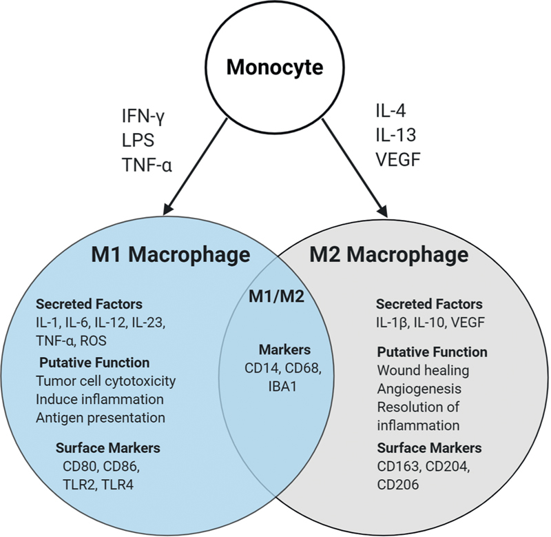
Macrophages were traditionally understood to play a central role in inflammation, acting to phagocytose pathogens, and arise in response to microbial agents and proinflammatory cytokines. However, it is now clear that macrophages are not a homogenous, monolithic cell population, and that they exhibit a differential response depending on their stimuli. 20 The concept of macrophage polarity broadly dichotomizes macrophages into M1 “classically activated” macrophages and M2 “alternatively activated” macrophages that have widely divergent functions in the setting of cancer ( Fig. 1 ). 22 M1 macrophages are observed to attack tumor cells in the manner of the traditional antigen presenting macrophage, stimulating cytotoxic T-cells, phagocytosing tumor cells, and secreting proinflammatory cytokines. 23 Conversely, M2 macrophages are considered to encourage tumor growth, attenuating the cytotoxic T-cell response, facilitating angiogenesis, and promoting break down of the extracellular matrix to facilitate tumor invasion and metastasis. 24 Macrophages are induced to an M1 phenotype by exposure to interferon-γ and lipopolysaccharide. 25 In contrast, exposure to immunomodulatory cytokines, such as interleukin (IL)-4 and IL-13, is thought to drive macrophages toward the protumorigenic M2 phenotype. 24 Although it is now recognized that this dichotomy is overly simplistic with macrophage activity better conceptualized as a continuum, it nonetheless remains of clinicopathological relevance, with many cancers demonstrating a positive prognostic association with greater M1 and lower M2 TAM abundance, respectively. 26 27
Fig. 1.

Macrophage polarity. Typically, monocytes/macrophages can be polarized to an M1 phenotype by Toll like receptor (TLR) ligands such as lipopolysaccharide (LPS), interferon-g (IFN-g), or tumor necrosis factor-a (TNF-a), and secrete proinflammatory cytokines to be antitumorigenic. Alternatively, they can be polarized to an M2 phenotype by vascular endothelial growth factor (VEGF), Interleukin-4 (IL-4) and IL-13, which secrete anti-inflammatory cytokines to be pro-tumorigenic. CD14 and CD68 act as cell surface markers for macrophages, common to M1 and M2 types. Although this dichotomous polarity is somewhat of an oversimplification, the concept of pro and anti-tumoral activity of macrophages is highly clinically relevant. Readers interested in exploring this topic in more detail are directed to an excellent review by Mantovani et al.
In VS, a higher infiltration of CD163 expressing M2 macrophages have been observed within fast-growing tumors compared with slow-growing tumors. 10 Macrophage colony-stimulating factor (M-CSF) may play a role in inducing the macrophage M2 polarization and elevated expression of this cytokine has been observed as a feature of rapidly growing VS. 10 28 An association between CD163+ macrophage infiltration and shorter progression-free survival has also been demonstrated in NF2 associated VS. 29 In a study of VS patients who underwent initial subtotal resection (STR), higher TAM density in the resected tumor was associated with tumor recurrence and poorer postoperative facial nerve function. In apparent contrast to earlier results, however, higher M2/M1 macrophage ratio corresponded to a lower rate of postoperative recurrence. 30
The precise function of macrophages following their infiltration into VS is yet to be determined and the discordant findings of the previously cited study serves to highlight the necessity for further research to aide our mechanistic understanding of the VS microenvironment. From a pragmatic perspective, however, the size of VS correlates with outcome and as it stands macrophage infiltration seems to correlate with tumor size and tumor growth rate. 1 2 3 4 5 7 The mechanisms underlying macrophage infiltration and their function within the tumor may represent promising therapeutic targets and are explored further below.
Molecular Pathways and Chemical Mediators
It has been known since the 1980s that VS tissue elicits a cell-mediated immune response in the blood of affected patients, as measured through indirect leukocyte migration assays. 31 It is only recently, however, that the mediators of this immune cell migration have started to be elucidated. A comparative analysis of resected VS tissue and normal vestibular nerve demonstrated increased expression of proinflammatory cytokines IL-1, IL-6, tumor necrosis factor-α (TNF-α), and the leukocyte adhesion molecule, intercellular adhesion molecule-1 (ICAM-1), in the resected tumors. 32 Furthermore, a recent analysis of 60 specimens from patients with sporadic and NF2 associated VS found upregulation of the C-X-C chemokine receptor type 4 (CXCR4) on VS Schwann's cells when compared with Schwann's cells derived from normal control nerves. 33 As well as expression on the tumors themselves, VS tissue has been shown to secrete proinflammatory cytokines which are ototoxic in a murine cochlear explant model, and TNF-α is a mediator of this ototoxicity. 34
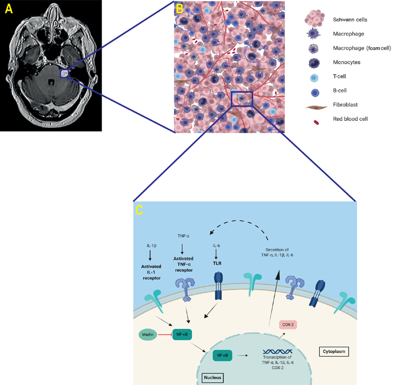
At the subcellular level, key transcription factors involved in potentiating the inflammatory processes in VS have been identified. Nuclear factor-κB (NF-κB) is a master immune regulator, that is activated by proinflammatory cytokines such as TNF-α and IL-1 and which controls expression of numerous inflammation related genes. It has been implicated in the maintenance of cancer-related inflammation in several malignancies and in VS tissue specimens NF-κB levels have been shown to be upregulated. 35 36 In human Schwann's cells, NF-κB activity is inhibited by merlin, the tumor suppressor protein whose loss is pivotal to the development of VS and in derived VS cell lines pharmacological targeting of NF-κB has decreased cellular proliferation rates. 35 37 Following its activation, NF-κB induces the secretion of multiple proinflammatory cytokines and there is, therefore, a potential for an autocrine positive feedback loop in VS, whereby IL-1 and TNF-α activate NF-κB, leading to further potentiation of these candidate proinflammatory cytokine levels ( Fig. 2 ).
Fig. 2.

Overview of biology of vestibular schwannoma (VS). ( A ) An axial T1-W Post-Gd MRI of a VS. ( B ) Magnified image depicting a schematic representation of the tumor microenvironment and the cell populations implicated in the VS microenvironment ( C ) Schematic representation of a positive feedback loop of inflammation in VS. Cytokines bind to cell surface receptors of Schwann's cells, activating the transcription factor NF-κB which demonstrably upregulates TNF-α, IL-1β, IL-6 and COX-2 expression. The cytoskeleton tumor suppressor protein merlin blocks NF-κB activity. COX-2 production is upregulated by NF-κB. Dashed line illustrates the purported positive feedback loop perpetuating the inflammation within VS. COX, cyclooxygenase; Gd, gadolinium; IL, Interleukin; MRI, magnetic resonance imaging; NF, nuclear factor; TNF, tumor necrosis factor; W, weighted.
Another molecular cascade which has been implicated in driving intratumoral inflammation in VS is the cyclooxygenase 2 (COX-2) pathway. COX-2 is an enzyme found to be upregulated at sites of inflammation and acts to metabolize arachidonic acid. 38 COX-2 has been found to be overexpressed in a wide variety of tumors, including meningiomas, and this also holds true for VS; analysis of over 1,000 resected VS found that 99% of tumors demonstrated immunopositivity for COX-2. 39 40 The most interesting observation, however, was that COX-2 positivity was found to be positively associated with tumor size and cellular proliferation index, as quantified by MIB1 expression, on multivariate analysis. 39 Inhibitors of COX-2 have been found to attenuate VS cellular proliferation in vitro. 41 Analogous to the situation with NF-κB, COX-2 is known to stimulate macrophages to produce proinflammatory cytokines such as IL-1 and IL-6, which are themselves activators of both COX-2 and NF-κB. This once again raises the possibility of a proinflammatory positive feedback loop within VS ( Fig. 2 ), and in other neurosurgical conditions, such as intracranial aneurysms formation, there is evidence that such feedback loops occur. 42
Inflammation and Angiogenesis in VS: The Chicken or the Egg?
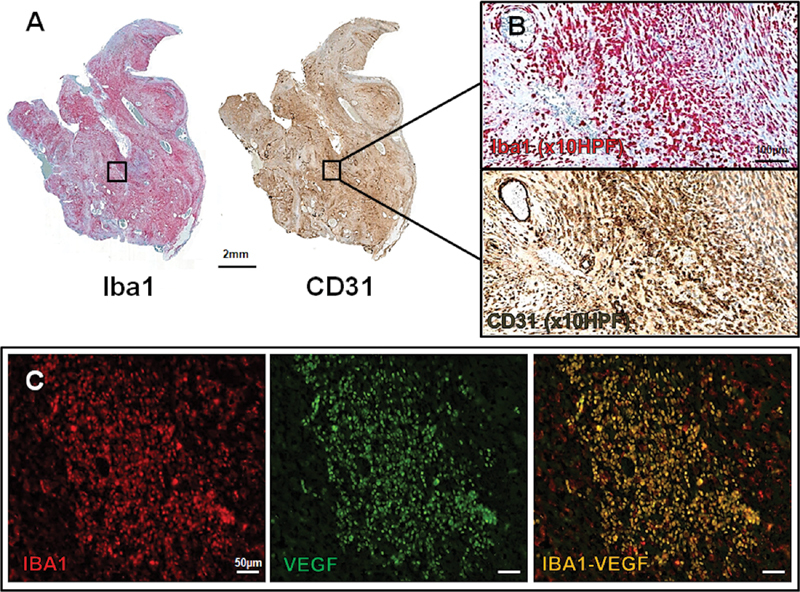
The growth of any tumor is dependent on new blood vessel formation or angiogenesis, and VS angiogenesis is now established as a key driver of tumor growth targetable with the agent bevacizumab. In many solid tumors, it is recognized that TAM abundance and vascular density are closely associated and previous tissue studies in both sporadic, and NF2-related VS have similarly demonstrated a close association between tissue microvessel density, tissue vascular permeability, and TAM density in these tumors. 7 43 44 45 Previous VS tissue studies have documented expression of both the proangiogenic cytokine, vascular endothelial growth factor (VEGF) and its receptor VEGFR-1, but until recently the cellular origin of these proteins and their relationship to inflammation in VS had not been studied. In a recent tissue study of resected sporadic and NF2-related VS, it was demonstrated that VS TAM were a source of both VEGF and VEGFR1's expression ( Fig. 3 ). 6
Fig. 3.

Relationship between hypoxia, angiogenesis and inflammation in VS. ( A ) Immunostains from a growing NF2-related VS demonstrating macroscopic spatial colocalization between Iba1 (ionized calcium-binding adapter molecule 1, marker of active macrophages) and regions of high CD31+ (expressed on endothelial cells, indicative of angiogenesis) microvessel density. Left: Iba1, red, immunoperoxidase, whole mount; CD31, brown, immunoperoxidase, whole mount. ( B ) Higher magnification (×10 HPF) of the area framed in the whole mounts) demonstrating an intratumoral region of high Iba1 + TAM density and high CD31 + microvessel density respectively. Top: Iba1, red; bottom: CD31, brown (immunoperoxidase, ×10). ( C ) Higher magnification (×20 HPF) immunofluoresence images of the area framed in the whole mounts demonstrating areas of cellular colocalization between Iba1 and VEGF (yellow). From left to right: Iba1 (red), VEGF (green) and VEGF-Iba1 (red-green) (immunofluorescence, ×20). HPF, high power field; NF2, neurofibromatosis type 2; VEGF, vascular endothelial growth factor; TAM, tumor-associated macrophages; VS, vestibular schwannoma.
VEGF is known to promote vasodilatation, increase vascular permeability, and induce angiogenesis. 46 47 There is, however, a growing body of evidence that VEGF also acts as a potent chemoattractant for circulating VEGFR1 expressing monocytes and that once these cells are in the tumor, VEGF serves to drive monocytes toward the proangiogenic and tumor-promoting M2 macrophage phenotype. 47 48 49 50 51 Tumor hypoxia and the hypoxia-sensing transcription factor HIF1α are known to be key drivers of angiogenesis and proangiogenic cytokine production, and HIF1α expression has been demonstrated in VS. In other tumors, TAMs preferentially localize within hypoxic regions, and low oxygen conditions promote HIF1α expression within these cells, driving them toward a specific proangiogenic activation profile. 45 Alongside stimulating angiogenesis, HIF1α is thought to induce the recruitment of monocytes through regulating the expression of key chemokines, such as C-X-C motif chemokine 12 (CXCL12), and its receptor CXCR4 and also directly influence the polarization of TAMs into protumorigenic M2 phenotypes.
The above evidence suggests that within growing VS, a self-perpetuating cycle may arise, whereby TAM infiltration drives VEGF production and angiogenesis, which in turn promotes further monocyte infiltration and polarization into protumorigeneic and proangiogenic M2-type TAM. 52 The evidence for this hypothesis at present, however, is observational and further in vivo mechanistic studies are required to better establish the relationship between angiogenesis and TAM infiltration in VS.
Immune Regulation and Evasion in VS
In many tumors, both malignant and benign, neoplastic tumor cells are able to evade and suppress the immune response. 53 One mechanism through which this immune suppression is achieved is through the stimulation of so called regulatory T-cells or T reg , which are characterized by the expression of the transcription factor fork-head box P3 (Foxp3). 53 Within the tumor microenvironment, T reg help to establish an immunosuppresive phenotype and facilitate tumor cell immune evasion through suppressing the proliferation and cytolytic activity of CD4 + and CD8 + T-cells and inducing an immunosuppressive TAM phenotype. In many solid tumors, such as melanoma and non–small cell lung cancer, heavy tumor infiltration of T reg cells is seen and there is a demonstrable association between high tumoral T reg density and poor patient prognosis. 53 While T reg have also been demonstrated in histologically benign tumors, such as neurofibroma, their role in VS has only recently been investigated. 54 Tamura et al demonstrated expression of Foxp3 + regulatory T-cells (T reg ) in resected VS specimens and an association between increased Foxp3 + T reg density and progressive tumor growth. 29 55
Another mechanism by which tumor cells evade immunosurveillance is through activation of immune checkpoint pathways, such as the PD-L1/PD1 (Programmed death-ligand 1 [PD-L1]/Programmed cell death protein 1 [PD1]) signaling axis. Programmed death-ligand 1 or PD-L1 (or B7 homolog 1) is a transmembrane protein upregulated in several tumors and is the target ligand for the T cell receptor PD-1. 56 Binding within the tumor microenvironment leads to several downstream effects on T-cell homeostasis including apoptosis and reduced proliferation of activated cytotoxic T-cells. As a result, there is diminished immune clearance of tumor cells, and in many tumors PD-L1 positive tumor cells have demonstrable resistance to cytolytic T-cell–mediated destruction. 57 Similar to T reg , PD-1/ PD-L1 signaling has not been extensively studied in VS. An early study demonstrated PD-L1 expression in 48 resected VS including surgically salvaged VS tumors that had failed stereotactic radiosurgery. 58 While later studies have confirmed this expression and demonstrated an association of higher PD-L1 expression with both tumor progression and unfavorable facial nerve outcomes following STR, other authors have demonstrated low rates of PD-L1 expression in VS tumor cells. 29 30 57 Larger studies are therefore required to better understand the role of immune checkpoint pathways in VS progression and growth.
Alongside the proposed relationship between TAM infiltration, hypoxia, and angiogenesis, VEGF signaling may similarly play a role in the development of an immunosuppressive microenvironment in VS. 29 In systemic tumors VEGF-A/VEGFR signaling has a key role in inhibiting dendritic cell maturation, stimulating T reg proliferation, and upregulating expression of PD-1/PD-L1 on CD8 + T cells, T reg , and TAMs within the tumor microenvironment. 29 Our understanding of the interaction between angiogenesis and immune regulation in VS is nascent but is strengthened by recent demonstrations in NF2-related VS that selective vaccination against the VEGFR1/2 receptor results in both microvascular changes and a reduction in the number of Foxp3 + T reg cells within the tumor microenvironment. 55
Detecting VS Inflammation in the Clinic
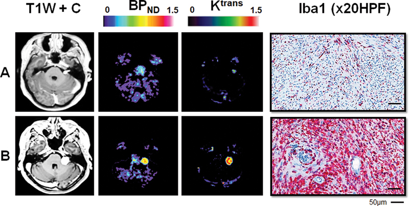
Successful translation of immune-related therapeutic targets into VS clinical practice will require both in vivo animal studies and early phase clinical trials. 59 Of equal importance, however, are diagnostic biomarkers which can be used to identify tumors with a high inflammatory burden are likely to benefit most from immunomodulatory therapy. There has been interest in the use of novel positron emission tomography tracers (PET) as markers of local intratumoral inflammation in VS. PET imaging utilizing [ 68 Ga]-pentixafor, a ligand targeted against the CXCL12 chemokine receptor CXCR4, has been performed to provide real-time, in vivo evidence of active inflammation in VS. 33 In the largest in vivo imaging study of inflammation in VS, thus far, use of an established translocator protein (TSPO) PET tracer for macrophages [ 11 C]-(R)PK11195 demonstrated that compared with static tumors, growing sporadic VS displayed higher specific binding of [ 11 C]-(R) PK11195 ( Fig. 4 ). 43 60 Perhaps more practical in the clinical environment than PET is the role of magnetic resonance imaging (MRI) markers of inflammation. K trans , a dynamic contrast-enhanced (DCE) MRI-derived biomarker of vascular permeability, might have value as an indirect measure of inflammation in VS, as it correlates strongly with both binding of [ 11 C]-(R)PK11195 and tissue macrophage density. Previous studies in NF2-related VS have shown that DCE-MRI based biomarkers can be used as early predictive biomarkers of bevacizumab treatment response, and there is hope that permeability metrics could serve as future clinically applicable predictors of response to immune-targeted therapies in these tumors. 52 61
Fig. 4.

Imaging biomarkers of intratumoral inflammation in VS. Representative imaging and histology from a patient with a static VS ( A ); and a growing VS ( B ) is shown. From left to right: axial T1-W post-Gd; parametric map of [ 11 C]-( R )-PK11195 PET specific binding (BP ND ); map of DCE-MRI derived K trans ; and immunostains demonstrating Iba1 (Iba1 red, immunoperoxidase) TAM density in each tumor. Within the growing VS there is demonstrably higher binding of the inflammation PET tracer, [ 11 C]-( R )-PK11195, and elevated K trans suggesting indicating increased vascular density/permeability compared with the static tumor. Comparative immunohistochemistry (Iba1 red, immunoperoxidase) demonstrates that within the growing VS, there is an abundance of intratumoral Iba1 + macrophages. DEC-MRI, dynamic contrast-enhanced magnetic resonance imaging; Gd, gadolinium; PET, positron emission tomography; TAM, tumor-associated macrophages; VS, vestibular schwannoma; W, weighted.
Arguably the “holy grail” of clinical biomarkers in VS would be a blood test which allows evaluation of the inflammatory profile in any given tumor and prediction of future tumor growth. In patients with extracranial malignancies, the peripheral blood neutrophil-to-lymphocyte blood ratio (NLR) has shown utility as a marker of systemic subclinical inflammation. 62 63 64 In the CNS, an increased NLR has been shown to be an independent prognostic indicator for poor outcome in patients with glioblastoma. 65 66 In VS, a study of 161 patients demonstrated that the peripheral blood NLR was independently predictive of tumor growth status. 67 The cause of the apparent systemic neutrophilia seen is incompletely understood but tumoral induction of VEGF, IL-1β, and IL-6 secretion have all been implicated. 68 As such future prospective studies of peripheral biomarkers of inflammation in VS should focus on candidate cytokines and chemokines thought to be driving immune cell chemotaxis into the tumors.
A Basis for New Therapies?
Given our nascent appreciation of the contribution of the immune microenvironment to VS pathobiology, it is unsurprising that few therapeutic interventions aimed at targeting this process in vivo have been trialed. There has been considerable interest in aspirin, an anti-inflammatory and known inhibitor of both COX-2 activity, and NF-κB activation, as a pharmacological therapy to inhibit VS growth. 41 69 To date, however, there have been conflicting results from observational studies aimed at determining if aspirin usage is associated with a decreased rate of VS growth. 69 70 This question will be better addressed by the results of a currently recruiting phase-II clinical trial, randomizing patients to 325-mg twice daily, or placebo, with tumor progression the outcome measure (NCT03079999).
Treatments aimed at “reeducating” protumorigenic M2 macrophages toward an antitumor, cytotoxic M1 phenotype have proven effective in in vitro models of other malignancies, although further work is required to validate their use in a clinical setting. 71 72 IL-1 and TNF-α antagonists are well established in the treatment of chronic inflammatory conditions, such as rheumatoid arthritis and inflammatory bowel disease, but their use in VS would require extensive preclinical validation prior to their use in this patient cohort. 73
Given the evidence, supporting the upregulation of PD-L1 within VS that have not responded to primary treatment with SRS or surgery, there may be a rationale for immune checkpoint inhibition strategies targeted against the immunosuppressive activity of the PD-L1/PD-1 axis. 30 58 Recombinant anti-PD-1 antibodies have been used to dramatic effect in tumors with high levels of cytotoxic T-cell inhibition, such as melanoma. 74 There is also emerging evidence of the efficacy of such therapies in a subset of glioma patients, and they may represent a promising therapeutic avenue for patients with VS, particularly those with tumors that recur following surgery or SRS with high levels of PD-L1 expression. 75 Remaining cognizant of the putative dual angiogenic and immunosuppressive role of VEGF in VS, consideration may be given to the coadministration of anti-VEGF therapy and immune checkpoint inhibitors. 55 The administration of bevacizumab in glioblastoma led to prolonged increases in cytotoxic T-cell activity, and this may aid in the creation of immunologically active “hot” tumors that are more responsive to immune checkpoint inhibition. 76 This therapeutic strategy has been pursued with some success in tumors with high levels of immune evasion, such as renal cell carcinoma and melanoma. 77 78
Conclusion and Clinical Impact
In this review, we have outlined the current state of the science underpinning our understanding of the VS immune microenvironment. More than aberrant Schwann's cells alone, VS have a complex microenvironment, characterized by an inflammatory and angiogenic milieu, that is likely to play a critical role in tumor growth. Our challenge now is to harness our increased understanding of the tumor microenvironment to bring tangible benefits to patients with VS.
In the near future, there is a clear rationale to justify the addition of immune checkpoint inhibitors to bevacizumab in progressive NF2-associated disease, where other treatment options are limited or undesirable. Similarly, there may be a selected group of sporadic VS patients who fail repeat radiosurgery and warrant consideration of a comparable strategy with or without the addition of subtotal tumor resection. For the majority of sporadic VS patients, combining drug therapy with radiosurgery or subtotal surgery might seem an example of “using a sledgehammer to crack a nut.” However, over the coming decade, the development of imaging biomarkers of tumor progression and early identification of radiosurgery failure, may allow us to predict patients for whom the addition of limited cycles of immune checkpoint inhibitors and/or VEGF inhibition may be of adjuvant benefit to prevent failure of primary treatment. It is also plausible that targeted anti-inflammatories may allow a degree of hearing preservation in some conservatively managed VS patients with recent hearing loss, given the purported ototoxic role of TNF-α in VS-associated hearing loss. It might also be possible to further improve surgical facial nerve outcomes in large tumors by decreasing inflammation perioperatively. As such, strategies for inhibiting immune evasion, promoting immune-mediated tumor clearance and/or targeting the inflammatory cytokine pathways represent promising candidate treatments in VS.
Funding Statement
Funding Aspects of this work were funded by Cancer Research, UK, and the Countess Dowager Eleanor Peel Trust. D.G.E. and S.K.L. are supported by the NIHR Manchester Biomedical Research Centre (IS-BRC-1215–20007).
The authors declare no personal or financial interest in the investigative or treatment modalities described in this review article.
Conflict of Interest D.G.E. reports personal fees from Astrazeneca, outside the submitted work. All the other authors report no conflict of interest.
Note
Aspects of this work were presented at the British Skull Base Society Meetings in January 2019 in Glasgow, UK, and January 2020 in London, UK. Further presentations were made at the Society of British Neurological Surgeons Meeting in March 2019 in Manchester, UK.
Contributed equally and share first authorship.
References
- 1.Evans D G, Moran A, King A, Saeed S, Gurusinghe N, Ramsden R. Incidence of vestibular schwannoma and neurofibromatosis 2 in the North West of England over a 10-year period: higher incidence than previously thought. Otol Neurotol. 2005;26(01):93–97. doi: 10.1097/00129492-200501000-00016. [DOI] [PubMed] [Google Scholar]
- 2.Halliday D, Parry A, Evans D G. Neurofibromatosis type 2 and related disorders. Curr Opin Oncol. 2019;31(06):562–567. doi: 10.1097/CCO.0000000000000579. [DOI] [PubMed] [Google Scholar]
- 3.Pathmanaban O N, Sadler K V, Kamaly-Asl I D. Association of genetic predisposition with solitary schwannoma or meningioma in children and young adults. JAMA Neurol. 2017;74(09):1123–1129. doi: 10.1001/jamaneurol.2017.1406. [DOI] [PMC free article] [PubMed] [Google Scholar]
- 4.Lees K A, Tombers N M, Link M J. Natural history of sporadic vestibular schwannoma: a volumetric study of tumor growth. Otolaryngol Head Neck Surg. 2018;159(03):535–542. doi: 10.1177/0194599818770413. [DOI] [PubMed] [Google Scholar]
- 5.Stangerup S E, Caye-Thomasen P, Tos M, Thomsen J. The natural history of vestibular schwannoma. Otol Neurotol. 2006;27(04):547–552. doi: 10.1097/01.mao.0000217356.73463.e7. [DOI] [PubMed] [Google Scholar]
- 6.Lewis D, Donofrio C A, O'Leary C. The microenvironment in sporadic and neurofibromatosis type II-related vestibular schwannoma: the same tumor or different? A comparative imaging and neuropathology study. J Neurosurg. 2020;29:1–11. doi: 10.3171/2020.3.JNS193230. [DOI] [PubMed] [Google Scholar]
- 7.de Vries M, Hogendoorn P C, Briaire-de Bruyn I, Malessy M J, van der Mey A G. Intratumoral hemorrhage, vessel density, and the inflammatory reaction contribute to volume increase of sporadic vestibular schwannomas. Virchows Arch. 2012;460(06):629–636. doi: 10.1007/s00428-012-1236-9. [DOI] [PMC free article] [PubMed] [Google Scholar]
- 8.Labit-Bouvier C, Crebassa B, Bouvier C, Andrac-Meyer L, Magnan J, Charpin C. Clinicopathologic growth factors in vestibular schwannomas: a morphological and immunohistochemical study of 69 tumours. Acta Otolaryngol. 2000;120(08):950–954. doi: 10.1080/00016480050218681. [DOI] [PubMed] [Google Scholar]
- 9.Hannan C J, Lewis D, O'Leary C. The inflammatory microenvironment in vestibular schwannoma. Neuro Oncol Adv. 2020;2(01):1–12. doi: 10.1093/noajnl/vdaa023. [DOI] [PMC free article] [PubMed] [Google Scholar]
- 10.de Vries M, Briaire-de Bruijn I, Malessy M J, de Bruïne S F, van der Mey A G, Hogendoorn P C. Tumor-associated macrophages are related to volumetric growth of vestibular schwannomas. Otol Neurotol. 2013;34(02):347–352. doi: 10.1097/MAO.0b013e31827c9fbf. [DOI] [PubMed] [Google Scholar]
- 11.Antoni N RE. Munich: JF Bergmann; 1920. Über Rückenmarkstumoren und Neurofibrome. [Google Scholar]
- 12.Escalona-Zapata J, Diez Nau M D. The nature of macrophages (foam cells) in neurinomas. Tissue culture study. Acta Neuropathol. 1978;44(01):71–75. doi: 10.1007/BF00691642. [DOI] [PubMed] [Google Scholar]
- 13.Bedavanija A, Brieger J, Lehr H-A, Maurer J, Mann W J. Association of proliferative activity and size in acoustic neuroma: implications for timing of surgery. J Neurosurg. 2003;98(04):807–811. doi: 10.3171/jns.2003.98.4.0807. [DOI] [PubMed] [Google Scholar]
- 14.Mantovani A, Allavena P, Sica A, Balkwill F.Cancer-related inflammation Nature 2008454(7203):436–444. [DOI] [PubMed] [Google Scholar]
- 15.Zhang S C, Hu Z Q, Long J H. Clinical implications of tumor-infiltrating immune cells in breast cancer. J Cancer. 2019;10(24):6175–6184. doi: 10.7150/jca.35901. [DOI] [PMC free article] [PubMed] [Google Scholar]
- 16.Di Caro G, Cortese N, Castino G F. Dual prognostic significance of tumour-associated macrophages in human pancreatic adenocarcinoma treated or untreated with chemotherapy. Gut. 2016;65(10):1710–1720. doi: 10.1136/gutjnl-2015-309193. [DOI] [PubMed] [Google Scholar]
- 17.Hambardzumyan D, Gutmann D H, Kettenmann H. The role of microglia and macrophages in glioma maintenance and progression. Nat Neurosci. 2016;19(01):20–27. doi: 10.1038/nn.4185. [DOI] [PMC free article] [PubMed] [Google Scholar]
- 18.Vidyarthi A, Agnihotri T, Khan N. Predominance of M2 macrophages in gliomas leads to the suppression of local and systemic immunity. Cancer Immunol Immunother. 2019;68(12):1995–2004. doi: 10.1007/s00262-019-02423-8. [DOI] [PMC free article] [PubMed] [Google Scholar]
- 19.Proctor D T, Huang J, Lama S, Albakr A, Van Marle G, Sutherland G R. Tumor-associated macrophage infiltration in meningioma. Neuro Oncol Adv. 2019;1(01):1–10. doi: 10.1093/noajnl/vdz018. [DOI] [PMC free article] [PubMed] [Google Scholar]
- 20.Stein M, Keshav S, Harris N, Gordon S. Interleukin 4 potently enhances murine macrophage mannose receptor activity: a marker of alternative immunologic macrophage activation. J Exp Med. 1992;176(01):287–292. doi: 10.1084/jem.176.1.287. [DOI] [PMC free article] [PubMed] [Google Scholar]
- 21.Domingues P H, Teodósio C, Ortiz J. Immunophenotypic identification and characterization of tumor cells and infiltrating cell populations in meningiomas. Am J Pathol. 2012;181(05):1749–1761. doi: 10.1016/j.ajpath.2012.07.033. [DOI] [PubMed] [Google Scholar]
- 22.Mills C D, Kincaid K, Alt J M, Heilman M J, Hill A M. M-1/M-2 macrophages and the Th1/Th2 paradigm. J Immunol. 2000;164(12):6166–6173. doi: 10.4049/jimmunol.164.12.6166. [DOI] [PubMed] [Google Scholar]
- 23.Gordon S, Taylor P R. Monocyte and macrophage heterogeneity. Nat Rev Immunol. 2005;5(12):953–964. doi: 10.1038/nri1733. [DOI] [PubMed] [Google Scholar]
- 24.Mantovani A, Sozzani S, Locati M, Allavena P, Sica A. Macrophage polarization: tumor-associated macrophages as a paradigm for polarized M2 mononuclear phagocytes. Trends Immunol. 2002;23(11):549–555. doi: 10.1016/s1471-4906(02)02302-5. [DOI] [PubMed] [Google Scholar]
- 25.Dale D C, Boxer L, Liles W C. The phagocytes: neutrophils and monocytes. Blood. 2008;112(04):935–945. doi: 10.1182/blood-2007-12-077917. [DOI] [PubMed] [Google Scholar]
- 26.Mantovani A. Reflections on immunological nomenclature: in praise of imperfection. Nat Immunol. 2016;17(03):215–216. doi: 10.1038/ni.3354. [DOI] [PubMed] [Google Scholar]
- 27.Ohri C M, Shikotra A, Green R H, Waller D A, Bradding P. Macrophages within NSCLC tumour islets are predominantly of a cytotoxic M1 phenotype associated with extended survival. Eur Respir J. 2009;33(01):118–126. doi: 10.1183/09031936.00065708. [DOI] [PubMed] [Google Scholar]
- 28.Kawamura K, Komohara Y, Takaishi K, Katabuchi H, Takeya M. Detection of M2 macrophages and colony-stimulating factor 1 expression in serous and mucinous ovarian epithelial tumors. Pathol Int. 2009;59(05):300–305. doi: 10.1111/j.1440-1827.2009.02369.x. [DOI] [PubMed] [Google Scholar]
- 29.Tamura R, Morimoto Y, Sato M. Difference in the hypoxic immunosuppressive microenvironment of patients with neurofibromatosis type 2 schwannomas and sporadic schwannomas. J Neurooncol. 2020;146(02):265–273. doi: 10.1007/s11060-019-03388-5. [DOI] [PubMed] [Google Scholar]
- 30.Perry A, Graffeo C S, Carlstrom L P. Predominance of M1 subtype among tumor-associated macrophages in phenotypically aggressive sporadic vestibular schwannoma. J Neurosurg. 2019:1–9. doi: 10.3171/2019.7.JNS19879. [DOI] [PubMed] [Google Scholar]
- 31.Rasmussen N, Bendtzen K, Thomsen J, Tos M. Specific cellular immunity in acoustic neuroma patients. Otolaryngol Head Neck Surg. 1983;91(05):532–536. doi: 10.1177/019459988309100511. [DOI] [PubMed] [Google Scholar]
- 32.Taurone S, Bianchi E, Attanasio G. Immunohistochemical profile of cytokines and growth factors expressed in vestibular schwannoma and in normal vestibular nerve tissue. Mol Med Rep. 2015;12(01):737–745. doi: 10.3892/mmr.2015.3415. [DOI] [PubMed] [Google Scholar]
- 33.Breun M, Schwerdtfeger A, Martellotta D D. CXCR4: A new player in vestibular schwannoma pathogenesis. Oncotarget. 2018;9(11):9940–9950. doi: 10.18632/oncotarget.24119. [DOI] [PMC free article] [PubMed] [Google Scholar]
- 34.Dilwali S, Landegger L D, Soares V YR, Deschler D G, Stankovic K M. Secreted factors from human vestibular schwannomas can cause cochlear damage. Sci Rep. 2015;5:18599–18599. doi: 10.1038/srep18599. [DOI] [PMC free article] [PubMed] [Google Scholar]
- 35.Dilwali S, Briët M C, Kao S-Y. Preclinical validation of anti-nuclear factor-kappa B therapy to inhibit human vestibular schwannoma growth. Mol Oncol. 2015;9(07):1359–1370. doi: 10.1016/j.molonc.2015.03.009. [DOI] [PMC free article] [PubMed] [Google Scholar]
- 36.Taniguchi K, Karin M. NF-κB, inflammation, immunity and cancer: coming of age. Nat Rev Immunol. 2018;18(05):309–324. doi: 10.1038/nri.2017.142. [DOI] [PubMed] [Google Scholar]
- 37.Ammoun S, Provenzano L, Zhou L. Axl/Gas6/NFκB signalling in schwannoma pathological proliferation, adhesion and survival. Oncogene. 2014;33(03):336–346. doi: 10.1038/onc.2012.587. [DOI] [PubMed] [Google Scholar]
- 38.Williams C S, Mann M, DuBois R N. The role of cyclooxygenases in inflammation, cancer, and development. Oncogene. 1999;18(55):7908–7916. doi: 10.1038/sj.onc.1203286. [DOI] [PubMed] [Google Scholar]
- 39.Behling F, Ries V, Skardelly M. COX2 expression is associated with proliferation and tumor extension in vestibular schwannoma but is not influenced by acetylsalicylic acid intake. Acta Neuropathol Commun. 2019;7(01):105. doi: 10.1186/s40478-019-0760-0. [DOI] [PMC free article] [PubMed] [Google Scholar]
- 40.Kang H C, Kim I H, Park C I, Park S H. Immunohistochemical analysis of cyclooxygenase-2 and brain fatty acid binding protein expression in grades I-II meningiomas: correlation with tumor grade and clinical outcome after radiotherapy. Neuropathology. 2014;34(05):446–454. doi: 10.1111/neup.12128. [DOI] [PubMed] [Google Scholar]
- 41.Dilwali S, Kao S-Y, Fujita T, Landegger L D, Stankovic K M. Nonsteroidal anti-inflammatory medications are cytostatic against human vestibular schwannomas. Transl Res. 2015;166(01):1–11. doi: 10.1016/j.trsl.2014.12.007. [DOI] [PMC free article] [PubMed] [Google Scholar]
- 42.Aoki T, Frȍsen J, Fukuda M. Prostaglandin E2-EP2-NF-κB signaling in macrophages as a potential therapeutic target for intracranial aneurysms. Sci Signal. 2017;10(465):eaah6037. doi: 10.1126/scisignal.aah6037. [DOI] [PubMed] [Google Scholar]
- 43.Lewis D, Roncaroli F, Agushi E. Inflammation and vascular permeability correlate with growth in sporadic vestibular schwannoma. Neuro-oncol. 2019;21(03):314–325. doi: 10.1093/neuonc/noy177. [DOI] [PMC free article] [PubMed] [Google Scholar]
- 44.Mantovani A, Marchesi F, Malesci A, Laghi L, Allavena P. Tumour-associated macrophages as treatment targets in oncology. Nat Rev Clin Oncol. 2017;14(07):399–416. doi: 10.1038/nrclinonc.2016.217. [DOI] [PMC free article] [PubMed] [Google Scholar]
- 45.Solinas G, Germano G, Mantovani A, Allavena P. Tumor-associated macrophages (TAM) as major players of the cancer-related inflammation. J Leukoc Biol. 2009;86(05):1065–1073. doi: 10.1189/jlb.0609385. [DOI] [PubMed] [Google Scholar]
- 46.Cayé-Thomasen P, Werther K, Nalla A. VEGF and VEGF receptor-1 concentration in vestibular schwannoma homogenates correlates to tumor growth rate. Otol Neurotol. 2005;26(01):98–101. doi: 10.1097/00129492-200501000-00017. [DOI] [PubMed] [Google Scholar]
- 47.Ferrara N, Gerber H P, LeCouter J. The biology of VEGF and its receptors. Nat Med. 2003;9(06):669–676. doi: 10.1038/nm0603-669. [DOI] [PubMed] [Google Scholar]
- 48.Freire Valls A, Knipper K, Giannakouri E. VEGFR1 + metastasis-associated macrophages contribute to metastatic angiogenesis and influence colorectal cancer patient outcome . Clin Cancer Res. 2019;25(18):5674–5685. doi: 10.1158/1078-0432.CCR-18-2123. [DOI] [PubMed] [Google Scholar]
- 49.Kerber M, Reiss Y, Wickersheim A. Flt-1 signaling in macrophages promotes glioma growth in vivo. Cancer Res. 2008;68(18):7342–7351. doi: 10.1158/0008-5472.CAN-07-6241. [DOI] [PubMed] [Google Scholar]
- 50.Barleon B, Sozzani S, Zhou D, Weich H A, Mantovani A, Marmé D. Migration of human monocytes in response to vascular endothelial growth factor (VEGF) is mediated via the VEGF receptor flt-1. Blood. 1996;87(08):3336–3343. [PubMed] [Google Scholar]
- 51.Henze A T, Mazzone M. The impact of hypoxia on tumor-associated macrophages. J Clin Invest. 2016;126(10):3672–3679. doi: 10.1172/JCI84427. [DOI] [PMC free article] [PubMed] [Google Scholar]
- 52.Li K-L, Djoukhadar I, Zhu X. Vascular biomarkers derived from dynamic contrast-enhanced MRI predict response of vestibular schwannoma to antiangiogenic therapy in type 2 neurofibromatosis. Neuro-oncol. 2016;18(02):275–282. doi: 10.1093/neuonc/nov168. [DOI] [PMC free article] [PubMed] [Google Scholar]
- 53.Togashi Y, Shitara K, Nishikawa H. Regulatory T cells in cancer immunosuppression - implications for anticancer therapy. Nat Rev Clin Oncol. 2019;16(06):356–371. doi: 10.1038/s41571-019-0175-7. [DOI] [PubMed] [Google Scholar]
- 54.Haworth K B, Arnold M A, Pierson C R. Immune profiling of NF1-associated tumors reveals histologic subtype distinctions and heterogeneity: implications for immunotherapy. Oncotarget. 2017;8(47):82037–82048. doi: 10.18632/oncotarget.18301. [DOI] [PMC free article] [PubMed] [Google Scholar]
- 55.Tamura R, Fujioka M, Morimoto Y. A VEGF receptor vaccine demonstrates preliminary efficacy in neurofibromatosis type 2. Nat Commun. 2019;10(01):5758. doi: 10.1038/s41467-019-13640-1. [DOI] [PMC free article] [PubMed] [Google Scholar]
- 56.Kythreotou A, Siddique A, Mauri F A, Bower M, Pinato D J. PD-L1. J Clin Pathol. 2018;71(03):189–194. doi: 10.1136/jclinpath-2017-204853. [DOI] [PubMed] [Google Scholar]
- 57.Wang S, Liechty B, Patel S. Programmed death ligand 1 expression and tumor infiltrating lymphocytes in neurofibromatosis type 1 and 2 associated tumors. J Neurooncol. 2018;138(01):183–190. doi: 10.1007/s11060-018-2788-6. [DOI] [PMC free article] [PubMed] [Google Scholar]
- 58.Archibald D J, Neff B A, Voss S G. B7-H1 expression in vestibular schwannomas. Otol Neurotol. 2010;31(06):991–997. doi: 10.1097/MAO.0b013e3181e40e4f. [DOI] [PMC free article] [PubMed] [Google Scholar]
- 59.Blakeley J O, Evans D G, Adler J. Consensus recommendations for current treatments and accelerating clinical trials for patients with neurofibromatosis type 2. Am J Med Genet A. 2012;158A(01):24–41. doi: 10.1002/ajmg.a.34359. [DOI] [PMC free article] [PubMed] [Google Scholar]
- 60.Venneti S, Lopresti B J, Wiley C A. Molecular imaging of microglia/macrophages in the brain. Glia. 2013;61(01):10–23. doi: 10.1002/glia.22357. [DOI] [PMC free article] [PubMed] [Google Scholar]
- 61.Serkova N J. Nanoparticle-based magnetic resonance imaging on tumor-associated macrophages and inflammation. Front Immunol. 2017;8:590. doi: 10.3389/fimmu.2017.00590. [DOI] [PMC free article] [PubMed] [Google Scholar]
- 62.Grimes N, Hannan C, Tyson M, Thwaini A. The role of neutrophil-lymphocyte ratio as a prognostic indicator in patients undergoing nephrectomy for renal cell carcinoma. Can Urol Assoc J. 2018;12(07):E345–E348. doi: 10.5489/cuaj.4872. [DOI] [PMC free article] [PubMed] [Google Scholar]
- 63.Jin J, Yang L, Liu D, Li W. Association of the neutrophil to lymphocyte ratio and clinical outcomes in patients with lung cancer receiving immunotherapy: a meta-analysis. BMJ Open. 2020;10(06):e035031. doi: 10.1136/bmjopen-2019-035031. [DOI] [PMC free article] [PubMed] [Google Scholar]
- 64.Li M X, Liu X M, Zhang X F. Prognostic role of neutrophil-to-lymphocyte ratio in colorectal cancer: a systematic review and meta-analysis. Int J Cancer. 2014;134(10):2403–2413. doi: 10.1002/ijc.28536. [DOI] [PubMed] [Google Scholar]
- 65.Templeton A J, McNamara M G, Šeruga B. Prognostic role of neutrophil-to-lymphocyte ratio in solid tumors: a systematic review and meta-analysis. J Natl Cancer Inst. 2014;106(06):dju124. doi: 10.1093/jnci/dju124. [DOI] [PubMed] [Google Scholar]
- 66.Bambury R M, Teo M Y, Power D G. The association of pre-treatment neutrophil to lymphocyte ratio with overall survival in patients with glioblastoma multiforme. J Neurooncol. 2013;114(01):149–154. doi: 10.1007/s11060-013-1164-9. [DOI] [PubMed] [Google Scholar]
- 67.Kontorinis G, Crowther J A, Iliodromiti S, Taylor W A, Locke R. Neutrophil to lymphocyte ratio as a predictive marker of vestibular schwannoma growth. Otol Neurotol. 2016;37(05):580–585. doi: 10.1097/MAO.0000000000001026. [DOI] [PubMed] [Google Scholar]
- 68.Lechner M G, Liebertz D J, Epstein A L. Characterization of cytokine-induced myeloid-derived suppressor cells from normal human peripheral blood mononuclear cells. J Immunol. 2010;185(04):2273–2284. doi: 10.4049/jimmunol.1000901. [DOI] [PMC free article] [PubMed] [Google Scholar]
- 69.Kandathil C K, Cunnane M E, McKenna M J, Curtin H D, Stankovic K M. Correlation between aspirin intake and reduced growth of human vestibular schwannoma: volumetric analysis. Otol Neurotol. 2016;37(09):1428–1434. doi: 10.1097/MAO.0000000000001180. [DOI] [PubMed] [Google Scholar]
- 70.MacKeith S, Wasson J, Baker C. Aspirin does not prevent growth of vestibular schwannomas: a case-control study. Laryngoscope. 2018;128(09):2139–2144. doi: 10.1002/lary.27114. [DOI] [PubMed] [Google Scholar]
- 71.Rolny C, Mazzone M, Tugues S. HRG inhibits tumor growth and metastasis by inducing macrophage polarization and vessel normalization through downregulation of PlGF. Cancer Cell. 2011;19(01):31–44. doi: 10.1016/j.ccr.2010.11.009. [DOI] [PubMed] [Google Scholar]
- 72.Hagemann T, Lawrence T, McNeish I. “Re-educating” tumor-associated macrophages by targeting NF-kappaB. J Exp Med. 2008;205(06):1261–1268. doi: 10.1084/jem.20080108. [DOI] [PMC free article] [PubMed] [Google Scholar]
- 73.Aggarwal B B, Gupta S C, Kim J H. Historical perspectives on tumor necrosis factor and its superfamily: 25 years later, a golden journey. Blood. 2012;119(03):651–665. doi: 10.1182/blood-2011-04-325225. [DOI] [PMC free article] [PubMed] [Google Scholar]
- 74.Hamid O, Robert C, Daud A. Safety and tumor responses with lambrolizumab (anti-PD-1) in melanoma. N Engl J Med. 2013;369(02):134–144. doi: 10.1056/NEJMoa1305133. [DOI] [PMC free article] [PubMed] [Google Scholar]
- 75.Zhao J, Chen A X, Gartrell R D. Immune and genomic correlates of response to anti-PD-1 immunotherapy in glioblastoma. Nat Med. 2019;25(03):462–469. doi: 10.1038/s41591-019-0349-y. [DOI] [PMC free article] [PubMed] [Google Scholar]
- 76.Tamura R, Tanaka T, Ohara K. Persistent restoration to the immunosupportive tumor microenvironment in glioblastoma by bevacizumab. Cancer Sci. 2019;110(02):499–508. doi: 10.1111/cas.13889. [DOI] [PMC free article] [PubMed] [Google Scholar]
- 77.Hodi F S, Lawrence D, Lezcano C. Bevacizumab plus ipilimumab in patients with metastatic melanoma. Cancer Immunol Res. 2014;2(07):632–642. doi: 10.1158/2326-6066.CIR-14-0053. [DOI] [PMC free article] [PubMed] [Google Scholar]
- 78.KEYNOTE-426 Investigators . Rini B I, Plimack E R, Stus V. Pembrolizumab plus axitinib versus sunitinib for advanced renal-cell carcinoma. N Engl J Med. 2019;380(12):1116–1127. doi: 10.1056/NEJMoa1816714. [DOI] [PubMed] [Google Scholar]


