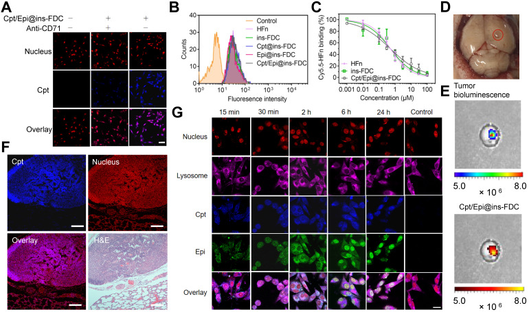
Figure 3.
Tumor targeting property and internalization of Cpt/Epi@ins-FDC in cells. (A) Cpt/Epi@ins-FDC binds to CD71 in tumor cells. CLSM imaging of U87MG cells treated with Cpt/Epi@ins-FDC in the presence or absence of anti-CD71 mAbs. Red, propidium iodide (PI), Excitation/emission wavelength (λex/λem) = 535 nm/615 nm; blue, Cpt, λex/λem = 365 nm/500 nm. Scale bar = 20 µm. (B) Flow cytometry histograms of U87MG cells after incubation with different formulations for 2 h. (C) Competitive binding assay of Cpt/Epi@ins-FDC, ins-FDC and HFn. (D) Digital image of brain from U87MG tumor xenografted mouse. (E) Following i.v. injection of Cpt/Epi@ins-FDC, in vivo NIRF imaging indicated the Cpt/Epi@ins-FDC accumulated specifically in the tumor area. λex/λem = 365 nm/500 nm. (F) Cpt/Epi@ins-FDC based fluorescence staining, and H&E staining of paraffin-embedded lung slices from mice with lung metastasis tumors of HepG2. Red, PI, λex/λem = 535 nm/615 nm; blue, Cpt, λex/λem = 365 nm/500 nm. Scale bar = 200 µm. (G) Internalization of Cpt/Epi@ins-FDC by U87MG cells and the sequential released behaviour of Cpt and Epi at indicated times revealed by CLSM. Purple, Cy5 labled Lysosomal Associated Membrane Protein 1 (LAMP1), λex/λem = 650 nm/700 nm; Red, PI, λex/λem = 535 nm/615 nm; blue, Cpt, λex/λem = 365 nm/500 nm; Green, Epi, λex/λem = 485 nm/575 nm. Scale bar = 20 µm.

