ABSTRACT
Guillain-Barre syndrome (GBS) is an acute immune-mediated disease of the peripheral nerves and nerve roots (polyradiculoneuropathy) that is usually elicited by various infections. We present a case of GBS after receiving the second dose of Pfizer-COVID 19 vaccine. Diagnosis was made after performing an accurate clinical examination, electromyoneurography and laboratory tests. In particular, anti-ganglioside antibodies have tested positive. During this pandemic with ongoing worldwide mass vaccination campaign, it is critically important for clinicians to rapidly recognize neurological complications or other side effects associated with COVID-19 vaccination.
KEYWORDS: Pfizer-BioNTech COVID-19 vaccine, neurological complications, guillain-barre syndrome, electromyoneurography, anti-ganglioside antibodies
Introduction
After spreading to multiple countries, coronavirus disease 2019 (COVID-19) was declared a pandemic by the World Health Organization (WHO) on March 11, 2020. Since then, interdisciplinary research has consistently aimed at developing strategies to limit its diffusion. The median incubation period for SARS-CoV-2 is estimated to be 5.1 days, and the majority of patients will develop symptoms within 11.5 days of infection.1 A range of clinical manifestations related to COVID-19 has been demonstrated, from asymptomatic or paucisymptomatic forms to clinical diseases characterized by acute respiratory distress, septic shock and multi-organ dysfunction.1 Multiple neurological complications have been associated with COVID-19 infection.2,3 The first mass vaccination program started in early December 2020. In the clinical trials of the vaccine, multiple side effects have been reported ranging from mild symptoms including but not limited to injection site pain, myalgia, fatigue, and fever to more serious side effects including anaphylactic shock.4,5 A recent article published on February 20216 reported the first case of Guillan Barrè syndrome (GBS) after the first dose of Pfizer COVID-19 vaccine. Here, we describe, for the first time in the vaccination scientific panorama, GBS occurred after receiving the second dose of the Pfizer vaccine.
Case presentation
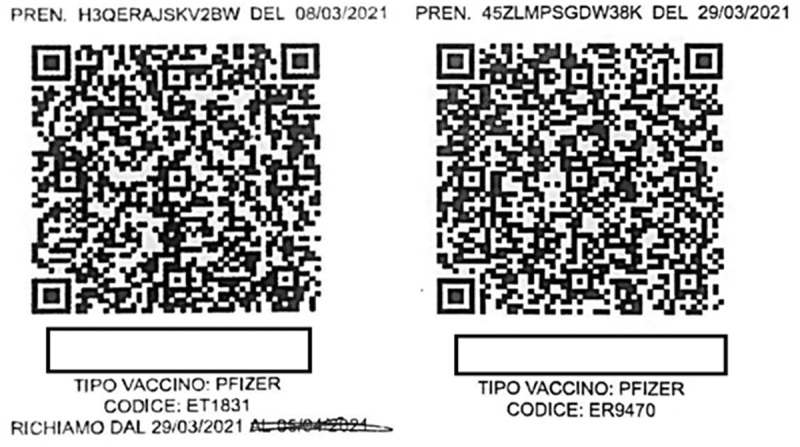
We present the case of an Italian 82-year-old woman, nonsmoker, suffering from permanent atrial fibrillation and arterial hypertension, on pharmacological treatment with rivaroxaban and with a preexisting walking disorder due to bilateral hip prosthesis for which she walked with a walker. Her height and body weight were, respectively, 1.75 meters (m) and 85 kilograms (Kg), with a body mass index of 28 Kg/m2 (overweight). She received her second dose of the Pfizer COVID-19 vaccine (Figure 1 and Table 1) according to the instructions provided by the manufacturer for the administration of the vaccine (Manufacturer: Pfizer, Inc., and BioNTech, Type of Vaccine: mRNA, Number of Shots: 2 shots, 21 days apart).
Figure 1.

Patient’s COVID-19 vaccination QR codes related to the the 1st dose of March 8th and to the 2nd dose of March 29th.
Table 1.
Vaccine characteristics of the two shots
| First dose | Second dose | |
|---|---|---|
| Vaccine lot number | ET1831 | ER9470 |
| Vaccine route of administration | Intramuscular | Intramuscular |
| Anatomic site of injection | Deltoid muscle of the right arm | Deltoid muscle of the left arm |
| Timetable of administration | 6 p.m. | 6 p.m. |
Two weeks later she presented a progressive worsening of walking, so she went to the emergency department. The patient began to experience a sudden worsening of walking associated with a lack of strength and sensitivity in the lower limbs. In the following days the appearance of similar symptoms in the upper limbs was associated with greater involvement of the proximal arm than the distal. The walking disturbance had become so severe that the patient was completely bedridden.
For this reason, she was taken to the emergency room. Her physical examination revealed normal mental status and speech. She had an unremarkable cranial nerve examination and no visible facial weakness or asymmetry was appreciated. Motor examination demonstrated normal bulk and tone in bilateral upper and lower extremities, strength in bilateral upper extremities was noted to be 1/5 in proximal muscles and 3/5 in distal muscles, according to the Medical Research Council [MRC] scale. The examination of muscle group strength testing showed diffuse weakness in the lower limbs quantifiable in 1/5.
The patient presented with superficial tactile hypoesthesia of the 4 limbs distally and she had areflexia in both upper and lower extremities. CT of the brain and cervical spine resonance were normal.
Standard laboratory tests and special blood tests (HbA1c, ANA, ENA, anti‐DNA, c‐ANCA, p‐ANCA, HIV, HBV, HCV, serum vitamin B12‐level, and serum protein electrophoresis) were also within the normal range. Campylobacter jejuni antibodies were tested as negative. Two nasopharyngeal swabs for SARS-COV2 were negative. A lumbar puncture was performed and cerebrospinal fluid (CSF) analysis showed albumin cytologic dissociation (protein of 5,7 gr/l and 2 cells), consistent with the diagnosis of GBS. CSF cytology was negative and the analysis of all neurotropic viruses gave negative results in the same fluid.
Taking the medical history, the patient had denied any symptoms (including respiratory and gastrointestinal ones) in the 2–4 weeks prior to the onset of neurological symptoms. The only trigger identified was the vaccine given 15 days earlier.
To complete the diagnostic framework, anti-ganglioside antibodies tests and electromyoneurography were carried out (Tables 2 and 3). Our patient received 0.4 g/kg/day intravenous immunoglobulin (IVIG) for five consecutive days, with improvement of the strength.
Table 2.
Nerve conduction study parameters in the patient
| Nerve stimulated | Stimulation site | Amplitude | Latency (ms) | Conduction velocity (m/s) | F wave (ms) | ||||
|---|---|---|---|---|---|---|---|---|---|
| RT | LT | RT | LT | RT | LT | RT | LT | ||
| Median (s) | Wrist | / | / | / | / | ||||
| Ulnar (s) | Wrist | / | / | / | / | ||||
| Sural (s) | Calf | / | / | / | / | ||||
| Radial (s) | Back of hand | / | / | / | / | ||||
| Median (m) | Wrist | 0,1 | 1,6 | 18,3 | 14,4 | / | 31 | / | 50,5 |
| AF | 1,4 | ||||||||
| Ulnar (m) | Wrist | 5,3 | 5 | 42 | 46,2 | ||||
| BE | 4,4 | ||||||||
| Peroneal (m) | Ankle | / | 1,5 | / | 10,2 | / | 25 | / | / |
| BF | 1 | ||||||||
Amplitude motor = mV, Amplitude sensory = µV; m = motor study; s = sensory study; RT = right; LT = left; AF = antecubital fossa; BE = below elbow; BF = below fibula; “/“ = absent.
Table 3.
Electromyography parameters in the patient
| Spontaneous activity | Voluntary activity (recruitment) | |||
|---|---|---|---|---|
| DELTOIDEUS | / | / | ↓↓ | ↓↓ |
| INTEROSSEUS DORSALIS I | / | / | ↓ | ↓ |
| VASTUS MEDIALIS | / | / | ↓↓ | ↓↓ |
| TIBIALIS ANTERIOR | / | / | ↓↓ | ↓↓ |
| EXTENSOR DIGITORUM LONGUS | + | + | 3–4 MUAPs | 3–4 MUAPs |
| EXTENSOR DIGITORUM BREVIS | + | + | 1–2 MUAPs | 1–2 MUAPs |
| EXTENSOR HALLUCIS LONGUS | + | + | 3–4 MUAPs | 3–4 MUAPs |
| ABDUCTOR HALLUCIS | + | + | 1–2 MUAPs | 1–2 MUAPs |
Spontaneous activity = fibrillation potentials and positive sharp waves.
↓ intermediate pattern; ↓↓ discrete activity.
“/” = absent. “+” = present.
MUAP: motor unit action potential.7
Electroneurography results (performed on the eleven day of hospitalization) showed acute sensory-motor neuropathy of the demyelinating type (Table 2). Electromyography showed decreased recruitment to the analysis of voluntary muscle activity, with signs of spontaneous activity (Table 3). Anti-ganglioside antibodies, in particular anti-sulfatide IgG (+) and IgM (++), anti-GM2 IgM (+) and anti-GM4 IgM (+) antibodies, were positive.
The patient did not show any signs of respiratory compromise. After therapy with IVIG, the patient presented a slight improvement in strength in the upper and lower limbs (upper limbs, according to the MRC scale 3/5 proximally, 4/5 distally; lower limbs 2/5). The patient received physical therapy during the hospital stay and was discharged to acute rehabilitation facility. The case was pointed out to Italian Medicines Agency (AIFA).
Discussion
GBS is an acute immune-mediated disease of the peripheral nerves and nerve roots (polyradiculoneuropathy) that is usually elicited by various infections. The classic clinical manifestations of GBS are progressive, ascending, symmetrical flaccid limbs paralysis, along with areflexia or hyporeflexia and with or without cranial nerve involvement, which can progress over the course of a few days to several weeks.
Antiganglioside antibodies are principally associated with autoimmune peripheral neuropathies. In these disorders, immune attack is inadvertently directed at peripheral nerve by autoantibodies that target glycan structures borne by glycolipids, particularly gangliosides concentrated in nerve myelin and axons. GM1, GD1b, GD1a, and GQ1b are important antigens, but many other gangliosides have also been identified as antibody targets.8,9 Usually, the trigger of the GBS is Campylobacter jejuni enteritis,10 therefore in the diagnostic process the serology for Campylobacter enteritidis should be checked.
Several vaccines against SARS-CoV-2 have been developed: messenger RNA (mRNA) vaccines (by Pfizer-BioNTech, Moderna and CureVac), and adenoviral vector vaccines (by Janssen-Johnson & Johnson, Astra-Zeneca, Sputnik-V, and CanSino).11 The mRNA vaccines introduce mRNA into cells, usually via a lipid nanoparticle (LNP). Inside the human body, mRNA enters the human cell and instructs the cells to identify the spike protein found on the surface of SARS-COV-2, the virus that causes COVID-19. Our bodies then recognize the spike protein as an invader and produce antibodies against it. Later, if these antibodies encounter the actual virus, they are ready to recognize and kill the virus before it can cause illness. In some patients, this immune response can trigger autoimmune processes that lead to the production of antibodies against the myelin, causing in this way GBS. In the Vaccine Adverse Event Reporting System (VAERS) database have been reported other autoimmune diseases, such as acute disseminated encephalomyelitis (6 cases), transverse myelitis (9 cases), facial palsy (190 cases), GBS (32 cases).12 Unlike the recent article6 in which antibodies to gangliosides were not analyzed, in our case the antibodies were positive, demonstrating the autoimmune process that caused the syndrome.
Therefore, the definitive diagnosis of post-vaccination GBS was made after meeting all clinical, laboratory and instrumental criteria, excluding viral infection as a trigger. The correlation with the vaccine was also supported by the timing of symptom onset, reported exactly after two weeks of administration and in line with the clinical onset of a GBS.
Given the positivity of antibodies to glycolipids, is it conceivable that the same LNPs trigger the autoimmune disease? Further studies are needed to confirm this hypothesis, setting out from the assumption that at present it has been shown that in some subjects the vaccine can cause an important immune response and in a good percentage of cases this reaction goes against both central and peripheral nervous system targets, with adverse neurological outcomes.
Conclusions
During this pandemic with ongoing worldwide mass vaccination campaign, it is critically important for clinicians to rapidly recognize neurological complications or other side effects associated with COVID-19 vaccination. We would like to highlight that the risk of neurological complications or any other adverse effect associated with COVID-19 vaccination is low and the benefits of the vaccination outweigh any potential risks or side effects, both at the individual and society levels.
However, it is certainly important to carry out a complete panel of laboratory and instrumental diagnostic tests on patients who present neurological symptoms after COVID-19 vaccination, to ensure an adequate pharmacovigilance and to develop a complete understanding of the potential complications that we do not know enough about at present.
Funding Statement
This paper did not receive any specific grant from funding agencies in the public, commercial, or not-for-profit sectors.
Disclosure of potential conflicts of interest
The authors reiterate that there are no conflicts of interest.
References
- 1.Cascella M, Rajnik M, Cuomo A, Dulebohn SC, Di Napoli R. Features, evaluation and treatment coronavirus (COVID-19). In StatPearls. Treasure Island, FL: StatPearls Publishing; 2020 [accessed 2021 May 25]. http://www.ncbi.nlm.nih.gov/books/NBK554776/ [PubMed] [Google Scholar]
- 2.Espinosa PS, Rizvi Z, Sharma P, Hindi F, Filatov A.. Neurological complications of coronavirus disease (COVID- 19): encephalopathy, MRI brain and cerebrospinal fluid findings: case 2. Cureus. 2020;12:e7930. [DOI] [PMC free article] [PubMed] [Google Scholar]
- 3.Filatov A, Sharma P, Hindi F, Espinosa PS.. Neurological complications of coronavirus disease (COVID- 19): encephalopathy. Cureus. 2020;12:e7352. [DOI] [PMC free article] [PubMed] [Google Scholar]
- 4.Kim JH, Marks F, Clemens JD. Looking beyond COVID-19 vaccine phase 3 trials. Nat Med. 2021;27:205–11. doi: 10.1038/s41591-021-01230-y. [DOI] [PubMed] [Google Scholar]
- 5.Polack FP, Thomas SJ, Kitchin N, Absalon J, Gurtman A, Lockhart S, Perez JL, Pérez Marc G, Moreira ED, Zerbini C, et al. Safety and efficacy of the BNT162b2 mRNA Covid-19 vaccine. N Engl J Med. 2020;31:2603–15. doi: 10.1056/NEJMoa2034577. [DOI] [PMC free article] [PubMed] [Google Scholar]
- 6.Waheed S, Bayas A, Hindi F, Rizvi Z, Espinosa PS. Neurological Complications of COVID-19: Guillain-Barre syndrome following Pfizer COVID-19 vaccine. Cureu.s. 2021;13:e13426. [DOI] [PMC free article] [PubMed] [Google Scholar]
- 7.Dengler R, de Carvalho M, Shahrizaila N, Nodera H, Vucic S, Grimm A, Padua L, Schreiber S, Kneiser MK, Hobson‐Webb LD, et al. AANEM-IFCNGlossary of terms in neuromuscular electrodiagnostic medicine and ultrasound. Muscle Nerve. 2020;62:10–12. doi: 10.1002/mus.26868. [DOI] [PubMed] [Google Scholar]
- 8.Willison HJ, Yuki N. Peripheral neuropathies and anti-glycolipid antibodies. Brain J Neurol. 2002;125(Pt 12):2591–625. doi: 10.1093/brain/awf272. [DOI] [PubMed] [Google Scholar]
- 9.Kusunoki S. Anti-ganglioside antibodies in Guillain-Barré syndrome; useful diagnostic markers as well as possible pathogenetic factors. Intern Med. 2003;42(6):457–58. doi: 10.2169/internalmedicine.42.457. [DOI] [PubMed] [Google Scholar]
- 10.Ho TW, Hsieh ST, Nachamkin I, Willison HJ, Sheikh K, Kiehlbauch J, Flanigan K, McArthur JC, Cornblath DR, McKhann GM, et al. Motor nerve terminal degeneration provides a potential mechanism for rapid recovery in acute motor axonal neuropathy after Campylobacter infection. Neurology. 1997;48(3):717–24. doi: 10.1212/WNL.48.3.717. [DOI] [PubMed] [Google Scholar]
- 11.Drugs and lactation database (LactMed) [Internet]. Bethesda (MD): National Library of Medicine (US); 2006. [Updated 2021 May 17]. COVID-19 vaccines. [Google Scholar]


