



Abstract
Nuclear endocrinology is the main ignitor for founding the Korean Society of Nuclear Medicine (KSNM) in the early 1960s by outstanding pioneering medical doctors. Management of thyroid diseases required nuclear medicine technology in the early days of the KSNM and was rapidly developed by advancements in nuclear medicine technology. Nuclear thyroidology remains one of the main clinical applications in nuclear medicine worldwide. Nuclear medicine technology provides essential information for diagnosing and assessing diseases of the parathyroid glands, pituitary gland, and neuroendocrine tumors (NETs). In addition, therapeutic nuclear medicine is essential for managing nonresectable NETs. Nuclear endocrinology remains a major section in clinical nuclear medicine, and members of the KSNM have contributed to progressing better management of benign and malignant endocrine diseases. This review summarizes the historical activities and milestone contributions to nuclear endocrinology made by the members of the KSNM over the past 60 years to congratulate the KSNM on its 60-year anniversary.
Keywords: Nuclear endocrinology, Thyroid gland, Parathyroid gland, Pituitary gland, Neuroendocrine tumor
Introduction
Nuclear endocrinology is the ignitor for the Korean Society of Nuclear Medicine (KSNM) which was launched by outstanding pioneering medical doctors. Diagnosis and treatment of thyroid diseases were rapidly developed by advancing nuclear medicine technology and have been the most prominent interests in nuclear medicine since the early 1960s. In particular, it is a representative area that realizes theranosis with a “magic bullet” in real clinical practice. Nuclear endocrinology has grown along with the long history of nuclear medicine, and a new movement has recently been emerging. In particular, new radiopharmaceuticals have been introduced in Korea, expanding the scope of diagnosis and treatment in nuclear medicine. Clinical nuclear medicine provides essential information for diagnosing and assessing diseases of the parathyroid glands, pituitary gland, and neuroendocrine tumors (NETs). In addition, therapeutic nuclear medicine is essential for managing nonresectable NETs.
In this article, historical events involving nuclear endocrinology and the KSNM—focused on the thyroid and parathyroid glands, pituitary gland, and NETs—will be discussed.
Thyroid Disease and Radioiodine Therapy
Radioiodine: the Beginning of Nuclear Medicine
The history of nuclear medicine in Korea began with the usage of I-131. Usage of I-131 was first implemented at Seoul National University Hospital and Dongsan Presbyterian Hospital around the same time (Fig. 1). In August 1959, Professor Lee MH started to use radioiodine to measure thyroid uptake, excretion, and therapy in Seoul National University Hospital. On May 30, 1960, a radioisotope clinic opened in Seoul National University Hospital, and patients began receiving treatment there [1]. Doctor Toh SH installed equipment for using radioiodine at Dongsan Presbyterian Hospital in 1959. However, laws for using radioisotopes in humans were not well established yet. While waiting for legislation, he opened a radioisotope clinic in March 1960; started using I-131 on March 30, 1960; and administered I-131 for the treatment of hyperthyroidism on May 15, 1960 [2, 3].
Fig. 1.
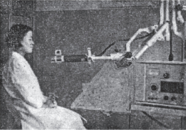
Radioactive iodine uptake measurement in the 1960s
In 1961, the United States (US) Atomic Energy Commission donated rectilinear dot scanner, well type scintillation counter, survey meter, and uptake system to Seoul National University, Kyungpook National University, Chonnam National University, and Pusan National University. This equipment contributed a lot to the nuclear medicine clinical practices in South Korea (Fig. 2).
Fig. 2.

a Image acquisition of a thyroid gland with a dot scanner in the 1970s and b thyroid scintigraphic images
Radioiodine therapy gradually increased until 2000. However, the trend of using radioiodine for hyperthyroidism seems to have decreased. In a survey of experts on thyroid disease in 1992, antithyroid drug treatment was the first choice for 81% of responders, 11% of responders chose radioiodine for first-line therapy, and 8% chose surgery as the initial treatment modality [4]. In 2013, 97.1% of experts reported choosing antithyroid drugs, and the remaining 2.9% reported choosing radioiodine for the initial treatment modality [5].
As the number of newly diagnosed patients with thyroid cancer rapidly increased from 3414 patients (2000) to 44,718 patients (2012), the number of patients receiving radioiodine therapy also rapidly increased from 5588 cases (2000) to 30,971 cases (2012) (Fig. 3) [6, 7]. There were concerns about the overdetection of small thyroid cancer with widespread use of ultrasonography as a health screening, and there were debates about the diagnosis and treatment of thyroid cancer [8]. After publication of the 2015 American Thyroid Association management guidelines [9], diagnosis of thyroid cancer rapidly decreased to 25,482 patients (2015) owing to the change in the indications for pathologic confirmation using fine-needle aspiration for thyroid nodules. As the proportion of lobectomies significantly increased, the number of cases of radioiodine therapy also decreased to 9848 cases (2020).
Fig. 3.

Numbers of patients receiving radioiodine therapy and incidences of newly diagnosed thyroid cancer
Thyroid Research of The Korean Society of Nuclear Medicine
Dr. Toh SH reported on the I-131 uptake of the thyroid glands in 150 healthy Koreans with euthyroid status, 73 patients with various thyroid diseases, and 25 patients with various conditions that influence I-131 uptake. He also reported on the experience of using I-131 treatment for nine patients with Graves’ disease in 1961 [2]. In the same year, Professor Lee MH reported 144 cases (including two patients with thyroid cancer) of patients receiving I-131 therapy [10]. About 4–7 mCi of I-131 were usually administered for hyperthyroidism, and 10–36 mCi were administered for thyroid cancer [2, 10]. After that, high-dose radioiodine was widely used to treat thyroid cancers. There were conflicts with the regulatory agency related to medical radioisotope use, but starting then, shielded isolation rooms for radioiodine therapy were installed [11].
As Professor Mazzaferri demonstrated the beneficial effects of radioiodine therapy in differentiated thyroid cancer in 1994 [12], radioiodine therapy was considered one of the main therapeutic tools for differentiated thyroid cancer. After the cloning of sodium/iodide symporter (NIS) in 1996 [13], the relationship between thyroid cancer and radioiodine was studied more in depth. Since the first use of radioiodine therapy for thyroid cancer in Korea [10], radioiodine therapy has been widely applied for managing patients with thyroid cancer. Various nuclear medicine modalities were widely used for assessing thyroid cancers, and many clinical studies were performed by members of the KSNM to visualize the characteristics of thyroid cancers using various nuclear medicine modalities. The concordance of immunohistochemical staining of NIS and positivity on I-131 scans in humans was elucidated [14]. F-18 fluorodeoxyglucose (FDG) positron emission tomography (PET) localized metastatic sites in I-131 scan-negative thyroid carcinoma [15], and F-18 FDG uptake of untreated papillary thyroid carcinoma has an inverse correlation with NIS expression [16]. There were flip-flop phenomena of I-131 and F-18 FDG in metastatic lesions of differentiated thyroid cancer and differences in survival depending on I-131 or F-18 FDG avidity of thyroid cancers [17].
Technological advancements of the gamma camera were widely applied in imaging studies of thyroid cancers. I-131 single positron emission computed tomography (SPECT)/computed tomography (CT) could provide useful information for detecting hidden metastases of differentiated thyroid cancers and provide excellent diagnostic accuracy [18, 19]. SPECT/CT was also used for a dosimetric approach and revealed differences in radioiodine biokinetics between thyroid remnants and lymph node (LN) metastases [20]. Recent deep learning technology was applied for automated localization of the metastatic LNs of thyroid cancer on postablation whole-body planar scans [21].
F-18 FDG avidity of thyroid nodules was assessed, and the relationship with cancer was validated [16, 22, 23]. The role of preoperative F-18 FDG PET/CT in thyroid cancer was also assessed, and high F-18 FDG avidity of the primary tumor was related to LN metastasis in patients with papillary thyroid cancer [24–26]. A multicenter, retrospective study was performed by members of the KSNM and revealed that high F-18 FDG uptake of primary lesions on preoperative PET/CT has prognostic value according to the extent of metastatic LNs [27]. F-18 FDG PET/CT has preoperative diagnostic value for LN staging in thyroid cancer and has a complementary role compared with ultrasonography in meta-analyses [28, 29].
Because major concerns about radioiodine refractoriness of thyroid cancers are related to the de-differentiation of cancer, many studies were performed to overcome the radioiodine refractoriness. The maximum safe dose of I-131 was applied for treating differentiated patients with thyroid cancer [30]. Retinoic acid was also used to increase the therapeutic effect of I-131 in patients with radioiodine-refractory papillary thyroid cancer [31]. Many studies were performed to find pharmacological candidates for increasing I-131 uptake in thyroid cancer [32, 33]. It was revealed that glycosylation of NIS regulates its membrane translocation and enhanced radioiodine uptake [34]. Codon optimization of NIS was accomplished by members of the KSNM, although NIS was cloned by American scientists [35]. The high-throughput screening system was established for detecting pharmacological NIS enhancers with a molecular imaging technique using a molecular imaging technology [36], and a new tyrosine kinase inhibitor was newly validated for the re-differentiation of radioiodine-refractory thyroid cancer [37].
Salivary gland damage is a common adverse effect of radioiodine therapy, and many studies were performed to understand and overcome it. Salivary gland dysfunction was clearly demonstrated after radioiodine therapy [38] and was well evaluated using salivary gland scintigraphy 5 years after radioiodine therapy [39]. Salivary gland massage during radioiodine therapy showed the protective effect of salivary gland function [40–44].
Thyroid Study Group and the Clinical Trials Network
Thyroid clinics and research were closely linked with the establishment of the KSNM. However, the Thyroid Study Group of the KSNM was established relatively late on October 30, 2009 (Fig. 4). As many clinical specialties were managing patients with thyroid cancers, the Thyroid Study Group interacted with other medical academic societies for the establishment of consensus and guidelines. The Thyroid Study Group held symposiums and gathered opinions about nuclear thyroidology to make suggestions and guidelines. The Thyroid Study Group engaged in creating many Korean guidelines for managing thyroid diseases [45–47]. Members of the Thyroid Study Group actively interacted with the Korean Thyroid Association and published “Radioiodine Therapy Guidance for Patients” [48].
Fig. 4.

Ten-year anniversary of the Thyroid Study Group of the Korean Society of Nuclear Medicine
To provide the right answers to certain clinical questions that impact the real world, data from one hospital may not be enough, and big clinical data from multiple hospitals are needed to support the answers. The first Clinical Trials Network (CTN) for thyroid cancer was initiated in 2014, and nine institutions participated. Three articles were published about the role of preoperative F-18 FDG PET/CT [27, 49, 50]. The second CTN was started in 2016 to creating evidence for clinical values of radioactive iodine treatment through the analysis of radioiodine treatment–related clinical data for differentiated thyroid cancer. The data were collected from more than 4000 patients in 25 hospitals, and these data were used for publications in the European Journal of Nuclear Medicine and Molecular Imaging in 2020 [51].
Radioimmunoassay
Yalow and Berson developed radioimmunoassay (RIA) for the measurement of plasma insulin in humans in 1959 [52]. After developing this method using radionuclide and antigen–antibody reactions, RIA was widely applied for the measurement of peptide hormones and small molecules because the low concentration of these substances required the remarkable sensitivity and specificity of RIA. RIA was first introduced in Korea in 1969 [53], and a new scintillation gamma counter was installed in 1970 for using RIA in clinical application (Fig. 5). In 1974, T4 measurements using kits were initiated; since then, researchers have been able to measure hormones, cancer markers, hepatitis antigens/antibodies, vitamins, and other small molecules using RIA.
Fig. 5.

Radioimmunoassay in the 1970s
Members of the KSNM have also contributed to the standardization and the quality control of the RIA methods since 1971. The Scientific Division of the KSNM published “Standardization of Thyroidal Radioiodine (I-131) Uptake Study in Korea” in 1971 [54]. From 1984 to 1986, external quality assessment in RIA was performed in 15 hospitals in Korea and was supported by the International Atomic Energy Agency (IAEA) [55]. In 1987, the data procession course of RIA was held by the KSNM and IAEA for nuclear medicine technicians. External quality assurance began in September 1989, and 28 institutions participated. Owing to the need for and importance of quality control, a new Director of Quality Assurance was established in 1993, and a Committee of Quality Assurance was established in 2010. In 2019, 59 institutions were certified for RIA by the KSNM.
Parathyroid Diseases
Parathyroid hormone (PTH) is secreted by the chief cells of the parathyroid gland, and modulates calcium and phosphorus homeostasis by affecting bones, small intestine, and kidneys [56]. Parathyroid diseases cause an abnormally increased or decreased level of calcium in blood, resulting in various symptoms such as osteoporosis, renal stones, heart disease, high blood pressure, muscle weakness, fatigue, and depression [57]. Research conducted by members of the KSNM has revealed the important roles of nuclear medicine imaging in evaluating parathyroid diseases over the past 60 years.
Primary hyperparathyroidism is caused by autonomous hyperfunction of a parathyroid adenoma or, less frequently, four-gland hyperplasia [57]. Nuclear medicine imaging can show hyperfunctioning parathyroid glands, allowing for differential diagnosis, decision-making on the treatment direction, and accurate localization for surgical removal [58]. In especially, parathyroid imaging is useful for patients who have no parathyroid abnormalities on CT or MR images and who have persistent hypercalcemia even after parathyroidectomy. Members of the KSNM have made parathyroid scintigraphy a routine part of the preoperative workup of patients with the clinical diagnosis of hyperparathyroidism through the following clinically significant studies. The uptake grade and washout on Tc-99 m sestamibi (MIBI) dual-phase parathyroid scintigraphy, along with age and PTH serum concentration, were reported to be useful diagnostic criteria for differentiating benign from malignant parathyroid lesions [59]. Also, it was reported that more parathyroid lesions could be visualized on SPECT/CT compared with planar scans, showing the usefulness of SPECT/CT for detecting hidden parathyroid lesions in patients with thyroid parenchymal Tc-99 m MIBI retention [60]. It was reported that quantitative indicators of parathyroid SPECT/CT can be used to assess the underlying functional status and disease severity of parathyroid adenomas [61]. The usefulness of maximum standardized uptake value (SUVmax) at the delayed phase of Tc-99 m MIBI SPECT/CT was demonstrated for the identification of parathyroid adenomas and hyperplasia [62]. It was shown that a SUVmax of 3.2 at the delayed phase SPECT/CT was accurate enough to identify parathyroid lesions that cause hyperparathyroidism, and a SUVmax of 7.1 was efficient to differentiate between parathyroid adenomas and hyperplasia. Recently, it was reported that dual-phase SPECT/CT have higher sensitivity than single-phase SPECT/CT in detecting hyperfunctioning parathyroid lesions [63]. In addition to Tc-99 m MIBI scintigraphy, researchers at the KSNM have shown that nuclear medicine imaging with other radiopharmaceuticals is useful for evaluating parathyroid diseases. In a systematic review and meta-analysis, F-18 fluorocholine PET/CT was demonstrated to have the high sensitivity and specificity for localization of hyperfunctioning parathyroid glands [64]. C-11 methionine PET/CT was suggested to be a useful diagnostic modality for detecting parathyroid adenomas and hyperplasia, showing that the sensitivity of C-11 methionine PET/CT is 91.7% and that of Tc-99 m MIBI scintigraphy is 41.7% [65].
Pituitary Tumors
Members of the KSNM have also revealed that nuclear medicine imaging is helpful for evaluating pituitary tumors. The majority of pituitary tumors are benign adenomas arising from adenohypophyseal cells of the anterior pituitary [66]. Excessive secretion of pituitary hormones from these tumors can lead to endocrine disorders such as hyperprolactinemia, acromegaly, Cushing’s syndrome, and hyperthyroidism [66]. Although nuclear medicine imaging is not used as a routine tool to evaluate pituitary tumors, focal uptake is occasionally observed in the pituitary gland on F-18 FDG PET/CT images [58]. Members of the KSNM also have revealed that F-18 FDG PET is helpful for evaluating pituitary tumors through the following studies. One study reported that incidental pituitary uptake was found in 0.8% of the study population [67]. This study showed that 40.8% of pituitary F-18 FDG uptake was pathologic lesions; pituitary adenomas (89.7%) were the most common cause of these pathologic lesions. Another study reported that the incidence of pituitary uptake was 0.073% in 40,967 patients and that the F-18 FDG uptake for macroadenomas was higher than that for microadenomas [68]. Furthermore, the study revealed a strong positive correlation between pituitary F-18 FDG uptake and serum thyroid-stimulating hormone, suggesting that pituitary F-18 FDG uptake seems to hold an important clue about the functional thyroid statuses of subjects with diffuse thyroid FDG uptake.
Neuroendocrine Tumors
Diagnosis of Neuroendocrine Tumors
MIBG imaging
Radioiodinated metaiodobenzylguanidine (MIBG) is the first radiopharmaceutical for imaging of some NETs, particularly catecholamine-secreting tumors, medullary thyroid carcinomas (MTCs), and carcinoid tumors [69]. It was first introduced into clinical practice in 1981 by Sisson from the University of Michigan [70]. Since the late 1980s, it has been used to diagnose NETs in Korea and has spread further as domestic production has become possible. During this period, diagnostic approaches using I-123/I-131 MIBG scans were performed on various diseases in Korea, and case reports and some original articles were published that provided a theoretical and clinical basis. Many case reports have shown that NETs were well visualized by MIBG scans even in unusual or difficult cases and that the scans had helped clinical decision-making. In one study, compared with biochemical and anatomical imaging tests, I-123/I-131 MIBG scans showed better diagnostic performance for NETs, demonstrating that it is an effective diagnostic tool [71]. Based on this clinical evidence in Korea, I-123/I-131 MIBG has been reimbursed by the National Health Insurance System (NHIS) since 2004. As shown in Fig. 6, diagnostic MIBG scans have been continuously conducted until recently, but the domestic supply of radioiodinated MIBG is not consistent owing to the unstable operation of the production facility.
Fig. 6.

Number of nuclear medicine imaging tests for neuroendocrine tumors in Korea over the past 8 years. MIBG, metaiodobenzylguanidine; PET, positron emission tomography
Several radiopharmaceuticals for PET imaging have the same functional properties as MIBG, such as C-11 hydroxyephedrine, C-11 epinephrine, C-11 phenylephrine, and F-18 fluoropropylbenzylguanidine. Although PET images using these radiopharmaceuticals have been reported to show higher sensitivity and specificity compared with I-123/I-131 MIBG SPECT images, these radiotracers are still used, but only for research purposes [72].
Somatostatin receptor imaging
Another diagnostic imaging test used for NETs is somatostatin receptor (SSTR) imaging. The rationale for performing SSTR imaging is based on the overexpression of SSTRs by the majority of NETs and provides information about their presence throughout the body, revealing additional metastases compared with conventional anatomical imaging. The In-111 pentetreotide was the first peptide-based radiopharmaceutical ever approved, and the US Food and Drug Administration (FDA) approved it as an imaging radiopharmaceutical in 1994. In Korea, the In-111 pentetreotide scan was approved in 2001, and, although not in large numbers, it is steadily being implemented (Fig. 6). In the 2010s, many changes occurred in how NETs were diagnosed using PET scans in Korea. Ga-68 DOTATOC and F-18 fluorodihydroxyphenylalanine (FDOPA), radiopharmaceuticals for PET, have become clinically available in Korea since 2014. Subsequently, Ga-68 DOTATOC and F-18 FDOPA PET have been reimbursed by the NHIS since 2016 and 2018, respectively.
In many studies by members of the KSNM, Ga-68 DOTATOC PET showed superior diagnostic performance for NETs compared with other nuclear medicine and anatomical imaging tests in common [73–77]. Recently, a multidisciplinary research group in Korea reported the current consensus on the diagnosis of gastroenteropancreatic NETs (GEP-NETs). In the consensus report, they recommended SSTR-targeting imaging for newly diagnosed GEP-NET cases because it may provide vital complementary information and, thus, influence therapeutic management [73].
Because NETs are a heterogeneous group of malignancies originating from the neuroendocrine cells of various organs, they show varying degrees of SSTR expression. Therefore, PET scans using other radiopharmaceuticals can show better diagnostic performance than SSTR PET imaging in some NETs. The amine precursor, DOPA, can also be labeled with F-18 to image NETs. In a comparative study of diagnostic performance for several PET radiopharmaceuticals (i.e., F-18 FDG, F-18 FDOPA, Ga-68 SSTR, and C-11 methionine), F-18 DOPA PET clearly performed the best in detecting recurrent MTC [74].
In Korea, the change in the number of NET imaging studies can be seen in Fig. 6. The In-111 pentetreotide scan has been continuously performed until recently. However, since the introduction of Ga-68 DOTATOC PET in 2016, it has gradually decreased, and use of Ga-68 DOTATOC PET scans has been increasing very rapidly. Usage of F-18 FDOPA has also been gradually increasing since 2016. Overall, diagnostic imaging for NETs is growing in the number of tests in total owing to the increase in SSTR imaging. As treatment using peptide receptor radionuclide therapy (PRRT), such as Lu-177 DOTATATE, gradually expands, it is expected that the use of such SSTR imaging before and after the treatment will expand. Recently, the Korean Pheochromocytoma and Paraganglioma Task Force has developed guidelines for the diagnosis of the tumors regarding controversial issues in Korea [75]. They suggest using I-123 MIBG scan, Ga-68 SSTR PET, and F-18 FDOPA PET as the functional imaging modalities for these tumors based on the genotype and location of tumors, availability of radiopharmaceuticals, and clinical situation.
As mentioned previously, Ga-68 DOTATOC and F-18 FDOPA PET are excellent diagnostic tools for NET evaluation, but there are hurdles to overcome in order for these modalities to spread nationwide. Ga-68 DOTATOC can only be synthesized in hospitals using an expensive gallium generator, and F-18 FDOPA is still being distributed limitedly by region in Korea. These issues need to be solved to allow for nationwide availability.
Therapy for Neuroendocrine Tumors
I-131 MIBG therapy
A highly specific nuclear medicine technique for NETs is being applied for treatment and diagnosis. Eligible patients for effective radionuclide therapy can be prescreened using an I-123 MIBG scan or Ga-68 DOTATOC PET prior to treatment. This pair of molecular targeted treatment and companion diagnostics is an excellent example of theranosis and precision medicine.
I-131 MIBG therapy is a relatively old treatment method for NETs using the same intratumoral drug absorption mechanism as a diagnostic scan and has been used to treat NETs worldwide since the 1980s. In Korea, the Korea Atomic Energy Research Institute (KAERI) has been producing I-131 MIBG since 2001; in the same year, the NHIS began reimbursing high-dose I-131 MIBG therapy. This therapy has been used to treat neuroblastomas, pheochromocytomas, and MTCs. In particular, more than 3000 children in Korea were treated with I-131 MIBG for neuroblastomas. The number of patients receiving I-131 MIBG therapy has increased gradually from the 1990s to the present and has maintained a similar number of cases over the past 5 years (Fig. 7).
Fig. 7.

Number of radionuclide therapy for neuroendocrine tumors in Korea. MIBG, metaiodobenzylguanidine
I-131 MIBG therapy is a clinically well-established treatment method, and many treatment guidelines suggest this therapeutic approach. Among them, the European Society for Medical Oncology–European Reference Network for rare adult solid cancers stated in the clinical guidelines for malignant pheochromocytoma that radionuclide therapy is an effective treatment and that I-131 MIBG therapy is one of the most frequent therapeutic approaches for the disease [76]. The guidelines suggest that I-131 MIBG therapy could be considered a first-line approach in patients displaying avid uptake of I-123 MIBG in all tumoral lesions. Recently, I-131 MIBG has gone beyond monotherapy and has been applied in combination with other therapies to enhance the therapeutic effect. These clinical trials showed that high-dose I-131 MIBG therapy in combination with chemotherapy or autologous stem cell transplantation could be a feasible option for high-risk pediatric neuroblastoma [78, 79].
However, owing to the low production compared with the demand and the unstable supply of I-131 MIBG in Korea, the waiting time for patients in need of treatment is very long and remains an urgent problem.
Peptide receptor radionuclide therapy
An alternative emerging therapeutic approach is PRRT, which uses radiolabeled somatostatin analogs to bind with SSTRs expressed on tumor cells, thereby delivering a cytotoxic radionuclide to the tumor cells. In PRRT, Lu-177 DOTATATE (Lutathera®) has recently become a global hot topic, including Korea, and is considered a game changer for NETs. PRRT has been used for therapeutic purposes since the mid-1990s, but it has only been studied in several single-arm clinical trials. NETTER-1 was the first multicenter, stratified, open, randomized, controlled, two-arm, phase III clinical trial using Lu-177 DOTATATE [80]. The NETTER-1 study demonstrated that Lu-177 DOTATATE showed an improved progression-free survival (PFS) (20-month PFS rates, 65.2% vs. 10.8%) and higher response rates (18% vs. 3%) than high-dose octreotide treatment [81]. Based on the results of this clinical trial, Lutathera was approved by the European Medicines Agency (EMA) and the US FDA in 2017 and 2018, respectively. Lutathera is the first US FDA- and EMA-approved radiopharmaceutical for PRRT. Although Lu-177 DOTATATE is not yet reimbursed by the NHIS in Korea, it has been available through the Korea Orphan and Essential Drug Center since November 2019. The Korean Ministry of Food and Drug Safety approved the drug for domestic use in July 2020 and has expanded treatment opportunities for domestic patients with NETs to include a support program for 1 year in March 2021. The NET patient society and the KSNM put in a lot of effort and led these policy changes [82]. In 2020, the first year Lutathera therapy became available in Korea, PRRT and I-131 MIBG were performed in similar numbers of cases. As a result, the number of NET treatment cases in 2020 more than doubled from that of the previous year (Fig. 7).
As PRRT has recently become available in Korea, clinical experiences with this treatment have been reported. The results of PRRT in 25 patients with NETs using Y-90 DOTATATE and Lu-177 DOTATATE were reported. More than half of the lesions responded to PRRT, and a higher tumor remission rate was correlated with a high baseline SUVmax on Ga-68 DOTANOC PET imaging [83]. A meta-analysis study of overall therapeutic efficacy showed that Lu-177-labeled PRRT is an effective treatment option for patients with inoperable or metastatic NETs [84].
Following the NETTER-1 trial, the NETTER-2 trial began in 2020, and Korea also participated. The aim of NETTER-2 was to determine if Lutathera in combination with long-acting octreotide prolongs PFS in patients with GEP-NET when given as a first-line treatment. As Korean hospitals participated in this study, the clinical experience and data of PRRT for NETs in Korea can also contribute to international research.
After efforts to localize Lu-177 production, the KAERI started to produce Lu-177 in earnest in 2020 and continues to work hard to localize the production of Lu-177 DOTATATE. It is hoped that the localization of the radiopharmaceutical for PRRT will benefit many patients in Korea at a lower price. In addition, the case of Lutathera provides us with another hopeful message in addition to the academic evidence discussed previously. The fact that Novartis recognized the value of Lutathera and acquired Advanced Accelerator Applications for $3.9 billion confirms that nuclear medicine is still a valuable medical technology and that new breakthroughs and advancements are possible in the future.
Author Contribution
The study was designed by Byeong-Cheol Ahn. Material preparation and data collection were performed by Chae Moon Hong, Young Jin Jeong, and Hae Won Kim. The data analysis was performed by Chae Moon Hong, Young Jin Jeong, and Hae Won Kim. The first draft of the manuscript was written by Chae Moon Hong, Young Jin Jeong, and Hae Won Kim and all authors commented on previous versions of the manuscript. All authors read and approved the final manuscript.
Data Availability
Contact the corresponding author for data requests.
Declarations
Competing Interests
Chae Moon Hong, Young Jin Jeong, Hae Won Kim, and Byeong-Cheol Ahn declare that they have no competing interests.
Ethics Approval and Consent to Participate
Not applicable.
Consent for Publication
Not applicable.
Footnotes
Publisher's Note
Springer Nature remains neutral with regard to jurisdictional claims in published maps and institutional affiliations.
Chae Moon Hong, Young Jin Jeong and Hae Won Kim contributed equally to this work
Contributor Information
Chae Moon Hong, Email: cmhong@knu.ac.kr.
Young Jin Jeong, Email: nmjeong@gmail.com.
Hae Won Kim, Email: hwkim.nm@gmail.com.
Byeong-Cheol Ahn, Email: abc2000@knu.ac.kr.
References
- 1.50 Year Department of Nuclear Medicine Seoul National University. 2010.
- 2.Toh SH. Thyroid and radioactive I131. J Korean Med Assoc. 1961;4:72–83. [Google Scholar]
- 3.Chun MH, Kim YS, Suh DS, Bae SH, Hong SC, Toh SH. A long follow-up study on Graves’ disease after I131 Treatment. Korean J Med. 1969;12:641–647. [Google Scholar]
- 4.Cho BY, Koh CS. Current trends in the diagnosis and treatment of Graves’ disease in Korea. Endocrinol Metab. 1992;7:216–227. [Google Scholar]
- 5.Moon JH, Yi KH. The diagnosis and management of hyperthyroidism in Korea: consensus report of the Korean Thyroid Association. Endocrinol Metab (Seoul) 2013;28:275–279. doi: 10.3803/EnM.2013.28.4.275. [DOI] [PMC free article] [PubMed] [Google Scholar]
- 6.Statistics of Nuclear Medicine in Korea. In. https://www.ksnm.or.kr///education/sub2_5.php? Accessed 18 Jul 2021.
- 7.Cancer Registry Statistics in Korea (KOSIS). In. https://kosis.kr/statHtml/statHtml.do?orgId=117&tblId=DT_117N_A00023. Accessed 18 Jul 2021.
- 8.Lee J, Jo I. Differentiated thyroid cancer and radioactive Iodine: past, present and future. Int J Thyroidol. 2019;12:71–78. [Google Scholar]
- 9.Haugen BR, Alexander EK, Bible KC, Doherty GM, Mandel SJ, Nikiforov YE, et al. 2015 American Thyroid Association management guidelines for adult patients with thyroid nodules and differentiated thyroid cancer: the American Thyroid Association guidelines task force on thyroid nodules and differentiated thyroid cancer. Thyroid. 2016;26:1–133. doi: 10.1089/thy.2015.0020. [DOI] [PMC free article] [PubMed] [Google Scholar]
- 10.Lee MH, Kang SS, Koh CS, Lee JK, Nam KY, Jin BH, et al. Investigation and treatment of disease of the thyroid gland with radioactive iodine. Korean J Med. 1961;4:29–48. [Google Scholar]
- 11.The Korean Society of Nuclear Medicine: 50 years. The Korean Society of Nuclear Medicine; 2011.
- 12.Mazzaferri EL, Jhiang SM. Long-term impact of initial surgical and medical therapy on papillary and follicular thyroid cancer. Am J Med. 1994;97:418–428. doi: 10.1016/0002-9343(94)90321-2. [DOI] [PubMed] [Google Scholar]
- 13.Dai G, Levy O, Carrasco N. Cloning and characterization of the thyroid iodide transporter. Nature. 1996;379:458–460. doi: 10.1038/379458a0. [DOI] [PubMed] [Google Scholar]
- 14.Min JJ, Chung JK, Lee YJ, Jeong JM, Lee DS, Jang JJ, et al. Relationship between expression of the sodium/iodide symporter and 131I uptake in recurrent lesions of differentiated thyroid carcinoma. Eur J Nucl Med. 2001;28:639–645. [PubMed] [Google Scholar]
- 15.Chung JK, So Y, Lee JS, Choi CW, Lim SM, Lee DS, et al. Value of FDG PET in papillary thyroid carcinoma with negative 131I whole-body scan. J Nucl Med. 1999;40:986–992. [PubMed] [Google Scholar]
- 16.Moon SH, Oh YL, Choi JY, Baek CH, Son YI, Jeong HS, et al. Comparison of 18F-fluorodeoxyglucose uptake with the expressions of glucose transporter type 1 and Na+/I- symporter in patients with untreated papillary thyroid carcinoma. Endocr Res. 2013;38:77–84. doi: 10.3109/07435800.2012.713426. [DOI] [PubMed] [Google Scholar]
- 17.Hong CM, Ahn BC, Jeong SY, Lee SW, Lee J. Distant metastatic lesions in patients with differentiated thyroid carcinoma Clinical implications of radioiodine and FDG uptake. Nuklearmedizin. 2013;52(121):9. doi: 10.3413/Nukmed-0541-12-11. [DOI] [PubMed] [Google Scholar]
- 18.Oh JR, Byun BH, Hong SP, Chong A, Kim J, Yoo SW, et al. Comparison of 131I whole-body imaging, 131I SPECT/CT, and 18F-FDG PET/CT in the detection of metastatic thyroid cancer. Eur J Nucl Med Mol Imaging. 2011;38:1459–1468. doi: 10.1007/s00259-011-1809-x. [DOI] [PubMed] [Google Scholar]
- 19.Jeong SY, Lee SW, Kim HW, Song BI, Ahn BC, Lee J. Clinical applications of SPECT/CT after first I-131 ablation in patients with differentiated thyroid cancer. Clin Endocrinol (Oxf) 2014;81:445–451. doi: 10.1111/cen.12460. [DOI] [PubMed] [Google Scholar]
- 20.Hong CM, Kim CY, Son SH, Jung JH, Lee CH, Jeong JH, et al. I-131 biokinetics of remnant normal thyroid tissue and residual thyroid cancer in patients with differentiated thyroid cancer: comparison between recombinant human TSH administration and thyroid hormone withdrawal. Ann Nucl Med. 2017;31:582–589. doi: 10.1007/s12149-017-1188-x. [DOI] [PubMed] [Google Scholar]
- 21.Kavitha M, Lee CH, Shibudas K, Kurita T, Ahn BC. Deep learning enables automated localization of the metastatic lymph node for thyroid cancer on (131)I post-ablation whole-body planar scans. Sci Rep. 2020;10:7738. doi: 10.1038/s41598-020-64455-w. [DOI] [PMC free article] [PubMed] [Google Scholar]
- 22.Chun AR, Jo HM, Lee SH, Chun HW, Park JM, Kim KJ, et al. Risk of malignancy in thyroid incidentalomas identified by fluorodeoxyglucose-positron emission tomography. Endocrinol Metab (Seoul) 2015;30:71–77. doi: 10.3803/EnM.2015.30.1.71. [DOI] [PMC free article] [PubMed] [Google Scholar]
- 23.Kwon SY, Kim J, Jung SH, Chong A, Song HC, Bom HS, et al. Preablative stimulated thyroglobulin levels can predict malignant potential and therapeutic responsiveness of subcentimeter-sized, 18F-fluorodeoxyglucose-avid cervical lymph nodes in patients with papillary thyroid cancer. Clin Nucl Med. 2016;41:e32–e38. doi: 10.1097/RLU.0000000000000889. [DOI] [PubMed] [Google Scholar]
- 24.Cho SG, Kwon SY, Kim J, Cho DH, Na MH, Kang SR, et al. Risk factors of malignant fluorodeoxyglucose-avid lymph node on preablation positron emission tomography in patients with papillary thyroid cancer undergoing radioiodine ablation therapy. Medicine (Baltimore) 2019;98:e14858. doi: 10.1097/MD.0000000000014858. [DOI] [PMC free article] [PubMed] [Google Scholar]
- 25.Jung JH, Kim CY, Son SH, Kim DH, Jeong SY, Lee SW, et al. Preoperative prediction of cervical lymph node metastasis using primary tumor SUVmax on 18F-FDG PET/CT in patients with papillary thyroid carcinoma. PLoS One. 2015;10:e0144152. doi: 10.1371/journal.pone.0144152. [DOI] [PMC free article] [PubMed] [Google Scholar]
- 26.Kang SY, Bang JI, Kang KW, Lee HY, Chung JK. FDG PET/CT for the early prediction of RAI therapy response in patients with metastatic differentiated thyroid carcinoma. PLoS One. 2019;14:e0218416. doi: 10.1371/journal.pone.0218416. [DOI] [PMC free article] [PubMed] [Google Scholar]
- 27.Kwon SY, Choi EK, Kong EJ, Chong A, Ha JM, Chun KA, et al. Prognostic value of preoperative 18F-FDG PET/CT in papillary thyroid cancer patients with a high metastatic lymph node ratio: a multicenter retrospective cohort study. Nucl Med Commun. 2017;38:402–406. doi: 10.1097/MNM.0000000000000657. [DOI] [PubMed] [Google Scholar]
- 28.Kim DH, Kim SJ. Diagnostic role of F-18 FDG PET/CT for preoperative lymph node staging in thyroid cancer patients; a systematic review and metaanalysis. Clin Imaging. 2020;65:100–107. doi: 10.1016/j.clinimag.2020.04.030. [DOI] [PubMed] [Google Scholar]
- 29.Kim K, Shim SR, Lee SW, Kim SJ. Diagnostic values of F-18 FDG PET or PET/CT, CT, and US for preoperative lymph node staging in thyroid cancer: a network meta-analysis. Br J Radiol. 2021;94:20201076. doi: 10.1259/bjr.20201076. [DOI] [PMC free article] [PubMed] [Google Scholar]
- 30.Lee JJ, Chung JK, Kim SE, Kang WJ, Park DJ, Lee DS, et al. Maximal safe dose of I-131 after failure of standard fixed dose therapy in patients with differentiated thyroid carcinoma. Ann Nucl Med. 2008;22:727–734. doi: 10.1007/s12149-007-0179-8. [DOI] [PubMed] [Google Scholar]
- 31.Oh SW, Moon SH, Park DJ, Cho BY, Jung KC, Lee DS, et al. Combined therapy with 131I and retinoic acid in Korean patients with radioiodine-refractory papillary thyroid cancer. Eur J Nucl Med Mol Imaging. 2011;38:1798–1805. doi: 10.1007/s00259-011-1849-2. [DOI] [PubMed] [Google Scholar]
- 32.Singh TD, Jeong SY, Lee SW, Ha JH, Lee IK, Kim SH, et al. Inverse agonist of estrogen-related receptor γ enhances sodium iodide symporter function through mitogen-activated protein kinase signaling in anaplastic thyroid cancer cells. J Nucl Med. 2015;56:1690–1696. doi: 10.2967/jnumed.115.160366. [DOI] [PubMed] [Google Scholar]
- 33.Choi YJ, Lee JE, Ji HD, Lee BR, Lee SB, Kim KS, et al. Tunicamycin as a novel redifferentiation agent in radioiodine therapy for anaplastic thyroid cancer. Int J Mol Sci. 2021;22:1077. doi: 10.3390/ijms22031077. [DOI] [PMC free article] [PubMed] [Google Scholar]
- 34.Chung T, Youn H, Yeom CJ, Kang KW, Chung JK. Glycosylation of sodium/iodide symporter (NIS) regulates its membrane translocation and radioiodine uptake. PLoS One. 2015;10:e0142984. doi: 10.1371/journal.pone.0142984. [DOI] [PMC free article] [PubMed] [Google Scholar]
- 35.Kim YH, Youn H, Na J, Hong KJ, Kang KW, Lee DS, et al. Codon-optimized human sodium iodide symporter (opt-hNIS) as a sensitive reporter and efficient therapeutic gene. Theranostics. 2015;5:86–96. doi: 10.7150/thno.10062. [DOI] [PMC free article] [PubMed] [Google Scholar]
- 36.Oh JM, Kalimuthu S, Gangadaran P, Baek SH, Zhu L, Lee HW, et al. Reverting iodine avidity of radioactive-iodine refractory thyroid cancer with a new tyrosine kinase inhibitor (K905–0266) excavated by high-throughput NIS (sodium iodide symporter) enhancer screening platform using dual reporter gene system. Oncotarget. 2018;9:7075–7087. doi: 10.18632/oncotarget.24159. [DOI] [PMC free article] [PubMed] [Google Scholar]
- 37.Oh JM, Baek SH, Gangadaran P, Hong CM, Rajendran RL, Lee HW, et al. A novel tyrosine kinase inhibitor can augment radioactive iodine uptake through endogenous sodium/iodide symporter expression in anaplastic thyroid cancer. Thyroid. 2020;30:501–518. doi: 10.1089/thy.2018.0626. [DOI] [PubMed] [Google Scholar]
- 38.Lim SM, Hong SW, Lee JO, Kang TW. The change of the salivary function after the high dobe radioiodine treatment in the patients with differentiated thyroid dancer. Korean J Nucl Med. 1989;23:7–12. [Google Scholar]
- 39.Jeong SY, Kim HW, Lee SW, Ahn BC, Lee J. Salivary gland function 5 years after radioactive iodine ablation in patients with differentiated thyroid cancer: direct comparison of pre- and postablation scintigraphies and their relation to xerostomia symptoms. Thyroid. 2013;23:609–616. doi: 10.1089/thy.2012.0106. [DOI] [PMC free article] [PubMed] [Google Scholar]
- 40.Son SH, Hong CM, Jeong SY, Lee J, Ahn B-C. Clinical outcome of parotid gland massage for preventing parotid gland dysfunction in patients treated with radioiodine therapy for differentiated thyroid cancer: a prospective longitudinal follow-up study. Int J Thyroidol. 2021;14:6–17. [Google Scholar]
- 41.Kim HW, Ahn BC, Lee SW, Lee J. Effect of parotid gland massage on parotid gland Tc-99m pertechnetate uptake. Thyroid. 2012;22:611–616. doi: 10.1089/thy.2011.0188. [DOI] [PMC free article] [PubMed] [Google Scholar]
- 42.Hong CM, Son SH, Kim CY, Kim DH, Jeong SY, Lee SW, et al. Emptying effect of massage on parotid gland radioiodine content. Nucl Med Commun. 2014;35:1127–1131. doi: 10.1097/MNM.0000000000000176. [DOI] [PubMed] [Google Scholar]
- 43.Son SH, Lee CH, Jung JH, Kim DH, Hong CM, Jeong JH, et al. The preventive effect of parotid gland massage on salivary gland dysfunction during high-dose radioactive iodine therapy for differentiated thyroid cancer: a randomized clinical trial. Clin Nucl Med. 2019;44:625–633. doi: 10.1097/RLU.0000000000002602. [DOI] [PubMed] [Google Scholar]
- 44.Ahn BC. Reduction of salivary gland damage during radioiodine therapy for differentiated thyroid cancers. Nucl Med Mol Imaging. 2020;54:126–127. doi: 10.1007/s13139-020-00643-w. [DOI] [PMC free article] [PubMed] [Google Scholar]
- 45.Yi KH, Moon JH, Kim I-J, Bom H-S, Lee J, Chung WY, et al. The diagnosis and management of hyperthyroidism consensus - report of the Korean Thyroid Association. Int J Thyroidol. 2013;6:1–11. doi: 10.3803/EnM.2013.28.4.275. [DOI] [PMC free article] [PubMed] [Google Scholar]
- 46.Yi KH, Park YJ, Koong S-S, Kim J-H, Na DG, Ryu J-S, et al. Revised Korean Thyroid Association management guidelines for patients with thyroid nodules and thyroid cancer. Int J Thyroidol. 2010;3:65–96. [Google Scholar]
- 47.Yi KH, Lee EK, Kang H-C, Koh Y, Kim SW, Kim IJ, et al. 2016 Revised Korean Thyroid Association management guidelines for patients with thyroid nodules and thyroid cancer. Int J Thyroidol. 2016;9:59–126. [Google Scholar]
- 48.Ahn BC, Kang H-C, Oh S, Kim BH, Lee KH, Park WS, et al. Radioiodine therapy guidance for patients. 2nd ed.: Korean Thyroid Association; 2019.
- 49.Choi EK, Chong A, Ha JM, Jung CK, O JH, Kim SH, Clinicopathological characteristics including BRAF V600E mutation status and PET/CT findings in papillary thyroid carcinoma. Clin Endocrinol (Oxf) 2017;87:73–9. doi: 10.1111/cen.13335. [DOI] [PubMed] [Google Scholar]
- 50.Chong A, Ha JM, Han YH, Kong E, Choi Y, Hong KH, et al. Preoperative lymph node staging by FDG PET/CT with contrast enhancement for thyroid cancer: a multicenter study and comparison with neck CT. Clin Exp Otorhinolaryngol. 2017;10:121–128. doi: 10.21053/ceo.2015.01424. [DOI] [PMC free article] [PubMed] [Google Scholar]
- 51.Kwon SY, Lee SW, Kong EJ, Kim K, Kim BI, Kim J, et al. Clinicopathologic risk factors of radioactive iodine therapy based on response assessment in patients with differentiated thyroid cancer: a multicenter retrospective cohort study. Eur J Nucl Med Mol Imaging. 2020;47:561–571. doi: 10.1007/s00259-019-04634-8. [DOI] [PubMed] [Google Scholar]
- 52.Yalow RS, Berson SA. Assay of plasma insulin in human subjects by immunological methods. Nature. 1959;184:1648–1649. doi: 10.1038/1841648b0. [DOI] [PubMed] [Google Scholar]
- 53.Kim DJ, Min BS, Bahk YW, Kim BS. Determination of serum thyroxine levels in normal Korean subjects and various thyroid diseases. Korean J Nucl Med. 1969;3:33–38. [Google Scholar]
- 54.Scientific division of the Korean Society of Nuclear Medicine. Standardization of thyroidal radioiodine (131I) uptake study in Korea. Korean J Nucl Med. 1971;5:71–6.
- 55.Bahk Y-W, Kim W-I, Chung S-K. Progress report on “external quality assessment in radioimmunoassay of thyroid-related hormones in the Republic of Korea, 1986”. Korean J Nucl Med. 1987;21:1–3. [Google Scholar]
- 56.Brewer HB, Jr, Fairwell T, Ronan R, Sizemore GW, Arnaud CD. Human parathyroid hormone: amino-acid sequence of the amino-terminal residues 1–34. Proc Natl Acad Sci U S A. 1972;69:3585–3588. doi: 10.1073/pnas.69.12.3585. [DOI] [PMC free article] [PubMed] [Google Scholar]
- 57.Suliburk JW, Perrier ND. Primary hyperparathyroidism. Oncologist. 2007;12:644–653. doi: 10.1634/theoncologist.12-6-644. [DOI] [PubMed] [Google Scholar]
- 58.Ziessman HA, O’Malley JP, Thrall JH. Nuclear medicine: the requisites. Elsevier Health Sciences; 2013.
- 59.Cheon M, Choi JY, Chung JH, Lee JY, Cho SK, Yoo J, et al. Differential findings of Tc-99m sestamibi dual-phase parathyroid scintigraphy between benign and malignant parathyroid lesions in patients with primary hyperparathyroidism. Nucl Med Mol Imaging. 2011;45:276–284. doi: 10.1007/s13139-011-0103-y. [DOI] [PMC free article] [PubMed] [Google Scholar]
- 60.Hwang SH, Rhee Y, Yun M, Yoon JH, Lee JW, Cho A. Usefulness of SPECT/CT in parathyroid lesion detection in patients with thyroid parenchymal (99m)Tc-sestamibi retention. Nucl Med Mol Imaging. 2017;51:32–39. doi: 10.1007/s13139-016-0438-5. [DOI] [PMC free article] [PubMed] [Google Scholar]
- 61.Im HJ, Lee IK, Paeng JC, Lee KE, Cheon GJ, Kang KW, et al. Functional evaluation of parathyroid adenoma using 99mTc-MIBI parathyroid SPECT/CT: correlation with functional markers and disease severity. Nucl Med Commun. 2014;35(6):649–654. doi: 10.1097/MNM.0000000000000102. [DOI] [PubMed] [Google Scholar]
- 62.Suh HY, Na HY, Park SY, Choi JY, So Y, Lee WW, et al. The usefulness of maximum standardized uptake value at the delayed phase of Tc-99m sestamibi single-photon emission computed tomography/computed tomography for identification of parathyroid adenoma and hyperplasia. Medicine (Baltimore) 2020;99:e21176. doi: 10.1097/MD.0000000000021176. [DOI] [PMC free article] [PubMed] [Google Scholar]
- 63.Lee SH, Shin E, Ha S, Oh JS, Song DE, Ryu JS. Is dual-phase SPECT/CT with 99mTc-sestamibi better than single-phase SPECT/CT for lesion localization in patients with hyperparathyroidism? Medicine (Baltimore) 2020;99(19):e19989. doi: 10.1097/MD.0000000000019989. [DOI] [PMC free article] [PubMed] [Google Scholar]
- 64.Kim SJ, Lee SW, Jeong SY, Pak K, Kim K. Diagnostic performance of F-18 fluorocholine PET/CT for parathyroid localization in hyperparathyroidism: a systematic review and meta-analysis. Horm Cancer. 2018;9:440–447. doi: 10.1007/s12672-018-0347-4. [DOI] [PMC free article] [PubMed] [Google Scholar]
- 65.Chun IK, Cheon GJ, Paeng JC, Kang KW, Chung JK, Lee DS. Detection and characterization of parathyroid adenoma/hyperplasia for preoperative localization: comparison between (11)C-methionine PET/CT and (99m)Tc-sestamibi scintigraphy. Nucl Med Mol Imaging. 2013;47:166–172. doi: 10.1007/s13139-013-0212-x. [DOI] [PMC free article] [PubMed] [Google Scholar]
- 66.Shimon I, Melmed S. Genetic basis of endocrine disease: pituitary tumor pathogenesis. J Clin Endocrinol Metab. 1997;82:1675–1681. doi: 10.1210/jcem.82.6.3987. [DOI] [PubMed] [Google Scholar]
- 67.Hyun SH, Choi JY, Lee KH, Choe YS, Kim BT. Incidental focal 18F-FDG uptake in the pituitary gland: clinical significance and differential diagnostic criteria. J Nucl Med. 2011;52:547–550. doi: 10.2967/jnumed.110.083733. [DOI] [PubMed] [Google Scholar]
- 68.Jeong YH, Kim D, Lee JW, Rhee Y, Nam KH, Yun M, et al. Pituitary 18F-FDG uptake correlates with serum TSH levels in subjects with diffuse thyroid 18F-FDG uptake. Clin Nucl Med. 2015;40:632–636. doi: 10.1097/RLU.0000000000000793. [DOI] [PubMed] [Google Scholar]
- 69.Rufini V, Calcagni ML, Baum RP. Imaging of neuroendocrine tumors. Semin Nucl Med. 2006;36:228–247. doi: 10.1053/j.semnuclmed.2006.03.007. [DOI] [PubMed] [Google Scholar]
- 70.Sisson J, Shapiro B, Beierwaltes WH, Nakajo M, Glowniak J, Mangner T, et al. Treatment of malignant pheochromocytoma with a new radiopharmaceutical. Trans Assoc Am Physicians. 1983;96:209–217. [PubMed] [Google Scholar]
- 71.Moon EH, Lim ST, Jeong YJ, Kim DW, Jeong HJ, Sohn MH. Efficacy of I-123/I-131 metaiodobenzylguanidine scan as a single initial diagnostic modality in pheochromocytoma: comparison with biochemical test and anatomic imaging. Nucl Med Mol Imaging. 2009;43:436–442. [Google Scholar]
- 72.Lee EJ, Lee KH. PET application in neuroendocrine tumors. Endocrinol Metab (Seoul) 2007;22:397–406. [Google Scholar]
- 73.Yoo C, Oh CR, Kim ST, Bae WK, Choi HJ, Oh DY, et al. Systemic treatment of advanced gastroenteropancreatic neuroendocrine tumors in Korea: literature review and expert opinion. Cancer Res Treat. 2021;53:291–300. doi: 10.4143/crt.2020.1233. [DOI] [PMC free article] [PubMed] [Google Scholar]
- 74.Lee SW, Shim SR, Jeong SY, Kim SJ. Comparison of 5 different PET radiopharmaceuticals for the detection of recurrent medullary thyroid carcinoma: a network meta-analysis. Clin Nucl Med. 2020;45:341–348. doi: 10.1097/RLU.0000000000002940. [DOI] [PubMed] [Google Scholar]
- 75.Ku EJ, Kim KJ, Kim JH, Kim MK, Ahn CH, Lee KA, et al. Diagnosis for pheochromocytoma and paraganglioma: a joint position statement of the Korean pheochromocytoma and paraganglioma task force. Endocrinol Metab (Seoul) 2021;36:322–338. doi: 10.3803/EnM.2020.908. [DOI] [PMC free article] [PubMed] [Google Scholar]
- 76.Fassnacht M, Assie G, Baudin E, Eisenhofer G, de la Fouchardiere C, Haak HR, et al. Adrenocortical carcinomas and malignant phaeochromocytomas: ESMO-EURACAN clinical practice guidelines for diagnosis, treatment and follow-up. Ann Oncol. 2020;31:1476–1490. doi: 10.1016/j.annonc.2020.08.2099. [DOI] [PubMed] [Google Scholar]
- 77.Lee DY, Lee SH, Kim BJ, Kim W, Yoon PW, Lee SJ, et al. Usefulness of 68Ga-DOTATOC PET/CT to localize the culprit tumor inducing osteomalacia. Sci Rep. 2021;11:1819. doi: 10.1038/s41598-021-81491-2. [DOI] [PMC free article] [PubMed] [Google Scholar]
- 78.Lee JW, Lee S, Cho HW, Ma Y, Yoo KH, Sung KW, et al. Incorporation of high-dose (131)I-metaiodobenzylguanidine treatment into tandem high-dose chemotherapy and autologous stem cell transplantation for high-risk neuroblastoma: results of the SMC NB-2009 study. J Hematol Oncol. 2017;10:108. doi: 10.1186/s13045-017-0477-0. [DOI] [PMC free article] [PubMed] [Google Scholar]
- 79.Suh JK, Koh KN, Min SY, Kim YS, Kim H, Im HJ, et al. Feasibility and effectiveness of treatment strategy of tandem high-dose chemotherapy and autologous stem cell transplantation in combination with (131) I-MIBG therapy for high-risk neuroblastoma. Pediatr Transplant. 2020;24:e13658. [DOI] [PubMed]
- 80.Marco M, Antonella C, Ettore S. Peptide receptor radionuclide therapy after NETTER-1 clinical trial: what should not be left behind. Clin Trans Imaging. 2019;7:155–157. [Google Scholar]
- 81.Strosberg J, El-Haddad G, Wolin E, Hendifar A, Yao J, Chasen B, et al. Phase 3 trial of (177)Lu-Dotatate for midgut neuroendocrine tumors. N Engl J Med. 2017;376:125–135. doi: 10.1056/NEJMoa1607427. [DOI] [PMC free article] [PubMed] [Google Scholar]
- 82.Kang KW. Functional imaging and peptide receptor radionuclide therapy for pancreatic neuroendocrine tumor. Korean J Pancreas Biliary Tract. 2021;26:10–14. [Google Scholar]
- 83.Oh S, Prasad V, Lee DS, Baum RP. Effect of peptide receptor radionuclide therapy on somatostatin receptor status and glucose metabolism in neuroendocrine tumors: intraindividual comparison of Ga-68 DOTANOC PET/CT and F-18 FDG PET/CT. Int J Mol Imaging. 2011;2011:524130. [DOI] [PMC free article] [PubMed]
- 84.Kim SJ, Pak K, Koo PJ, Kwak JJ, Chang S. The efficacy of (177)Lu-labelled peptide receptor radionuclide therapy in patients with neuroendocrine tumours: a meta-analysis. Eur J Nucl Med Mol Imaging. 2015;42:1964–1970. doi: 10.1007/s00259-015-3155-x. [DOI] [PubMed] [Google Scholar]
Associated Data
This section collects any data citations, data availability statements, or supplementary materials included in this article.
Data Availability Statement
Contact the corresponding author for data requests.