Abstract
Background
Lateral end clavicle fractures can be challenging due to the small and often comminuted lateral fragment, problems with union and stability and implant morbidity. We retrospectively reviewed outcomes of Tightrope device in isolation to treat lateral end clavicle fractures.
Methods
Subjective and objective measures were assessed for 29 patients. The subjective comprised of functional clinical scores: Oxford shoulder score and EuroQoL5D. The objective measures were maintenance of fracture reduction, bone healing and complications.
Results
Median age was 36 years and 72% of cases were male patients. Average clinical follow up time was 21 months. Evaluation of latest radiographs showed that all reductions were maintained post-operatively. Twenty-two fractures had united and one patient had established non-union. Functional outcomes showed predominantly good results with Oxford shoulder score average of 41, EuroQoL5D index score of 0.78 and EuroQol Visual Analogue Scale 76. The overall post-operative complication rate was 10%; only one case requiring a secondary procedure.
Discussion
In our series, using the Tightrope as the sole device to treat displaced lateral end of clavicle fractures resulted in good radiological and functional outcomes, with minimal complications requiring secondary procedures. We believe the Tightrope device is a good method of fixing these challenging fractures and advocate its use.
Keywords: Lateral clavicle fracture, Tightrope
Introduction
Lateral end clavicle fractures account for 10–20%1–4 of all clavicle fractures, with the majority occurring in the elderly after simple falls.5,6 These fractures tend to be minimally displaced, with non-operative management being the treatment of choice for the elderly, resulting in a good overall functional outcome. Management is more controversial for the displaced lateral end of clavicle fracture in younger patients with higher functional demand. Although these fractures are less common than shaft fractures, they account for nearly 50% of overall clavicle fracture non-unions.7,8
Neer describes a classification of lateral clavicle fractures, defined by the relation of the fracture site to, and the continuity of, the coracoclavicular ligament. 7
In Neer type 1 fractures, the fracture line is lateral to the coracoclavicular ligament, which remains intact, stabilising the medial fragment and preventing significant displacement. This results in a fracture pattern amenable to satisfactory bone healing, without need for surgical intervention. Neer type 2 configurations have a fracture medial to the coracoclavicular ligament, disrupting the stability of the acromioclavicular joint. These type 2 fractures are subdivided into 2a injuries, whereby the fracture lies medial to the entire coracoclavicular ligament complex, which remains intact, and 2b, where either the conoid or the entire coracoclavicular ligament complex is disrupted. Several opposing anatomical forces act on this fracture pattern. 7 These forces result in significant displacement of the fragments thus reducing the likelihood of fracture healing in conservatively treated type 2 distal clavicle fractures.
With the knowledge of these anatomical factors and with the literature demonstrating high (>20%) non-union and mal-union rate after non-operative treatment,7,9,10 Neer type 2 distal clavicle fractures are generally surgically managed. These can be challenging fractures to fix due to the often small comminuted fragment laterally and the marked displacement of the medial shaft. Many surgical techniques are described in the literature, all with their own merits and complications. Operative approaches can be split broadly into direct osteosynthesis or indirect coracoclavicular stabilisation, as well as techniques combining both.11,12 Direct approaches include, but are not limited to, hook plate, 13 contoured distal locking plates and transacromial Knowles pin. 14 A 2013 meta-analysis 15 demonstrated all these methods have good rates of fracture union with no significant difference in functional outcome (level 3 or 4 evidence papers). Hook plates have an increased risk in complications including irritation, shoulder stiffness and infection, as well as the need for routine further operation for removal of metalwork. Locking plates, although have a low complication rate in comparison to other methods, 16 can be technically difficult due to small comminuted distal fragments and may still need to be removed.
The aim of this study is to assess the functional and radiological outcomes for patients undergoing tightrope fixation in isolation to treat acute displaced lateral end clavicle fractures. We employed an operative protocol of open reduction and fixation using an AC Tightrope implant (Arthrex) as a single device to achieve reduction of the medial clavicle fragment to allow fracture healing.
Materials and methods
This was a single centre retrospective study. A cohort of patients was compiled. A database was formed to collect information regarding demographics; age and gender, follow up time, surgery, post-operative complications, clinical outcomes, bone healing, fracture reduction and functional scores. This was obtained through review of the operation note, clinic letters, radiographs, and subsequent functional clinical scores were obtained over the telephone. The inclusion criterion was isolated acute displaced lateral end clavicle fractures (within 3 weeks of injury) in an active individual where the deemed surgery was more appropriate. We excluded open fractures, those with concomitant scapula fractures, a history of previous clavicular surgery and patients with neurovascular compromise.
All procedures were carried out using a standardised technique. The majority of cases were performed as day case procedures. Some patients were done as in patients due to other associated injuries. The patients were in the beach chair position on a shoulder table. A vertical “bra-strap” type incision was used extending from the dorsal ridge of the clavicle and extending distally till the tip of the coracoid. Skin flaps were raised and the fascia split longitudinally in line with the clavicle with use of a distal T-shape extension if necessary for better access. The coracoid was adequately exposed. A 3.5 mm drill was used to drill separate holes in the clavicle and the coracoid, and the drill was wiggled to slightly increase the diameter of the hole to accommodate the 4 mm button passing through. We recommend leaving a 2 cm gap between the drill hole medially in the clavicle shaft and the lateral fracture site on the clavicle. The coracoid drill position is near the base of the coracoid and centrally to reduce the risk of lateral blow out fracture. The Arthrex endo-button system was used. A guide wire was used to pass the button through the clavicle and subsequently through the coracoid. It was retrieved and then pulled back to ensure it has flipped and is secure against the coracoid. The clavicle was reduced as best as possible, ensuring the optimal distance between the under surface of the clavicle and the coracoid. The desired tension was finalised and button secured further with knots. As a secondary measure, the threads which are attached to the coracoid button are then tied over with the threads from the tied clavicle button to reduce the bulkiness of the knot and to reinforce the repair. Depending on surgeon preference, an image intensifier was used intra-operatively in some of the cases. However, in most of our case series this was not deemed necessary and was not performed.
The outcome was based on subjective and objective measures. The subjective comprised of functional clinical scores: Oxford shoulder score (OSS) and EuroQoL5D (EQ5D). The objective measures were maintenance of fracture reduction, bone healing and complications.
Results
A total of 29 patients were identified. We obtained functional clinical scores and radiological assessment for 26 and 28 patients, respectively. The procedures were carried out between June 2015 and October 2019. The demographics are detailed in Table 1. The median length of radiological follow-up was based on the time elapsed between the operation date and the latest available radiograph on the system. Clinical follow up time was defined as the period between surgery and either the latest clinic letter or scoring interview date, which ever was the later. Only one patient had no post-op radiographs as they failed to attend follow up appointments, but we were able to score them virtually. Another patient had one-month follow-up, both radiologically and clinically. Unsuccessful attempts were then made to score this individual virtually. There were three other patients with only one month radiological follow-up, with no further subsequent imaging, but further clinical appointments. These patients subsequently underwent virtual follow-up scoring.
Table 1.
Demographics and follow-up.
| Gender | 72% Male | 28% Female |
|---|---|---|
| Average | Range | |
| Age | 36 years | 14–73 years |
| Radiological follow-up | 6 months | 1–22 months |
| Clinical follow-up | 21 months | 1–54 months |
At time of data analysis 26 patients had been discharged, while 2 were still under active review. We were unable to record the outcome of one patient as no scores or clinic letters were available. During this period we report no intra-operative or immediate post-operative complications.
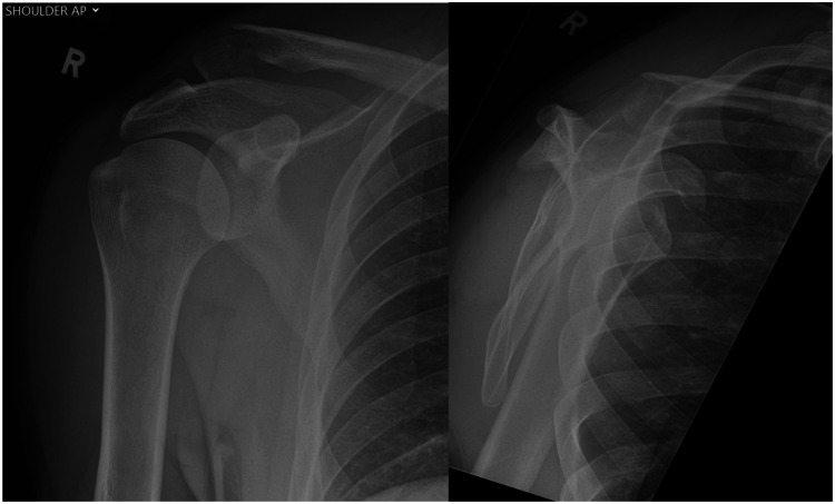
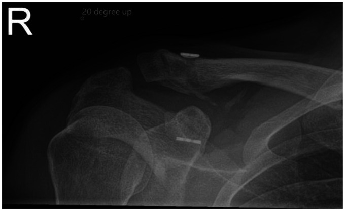
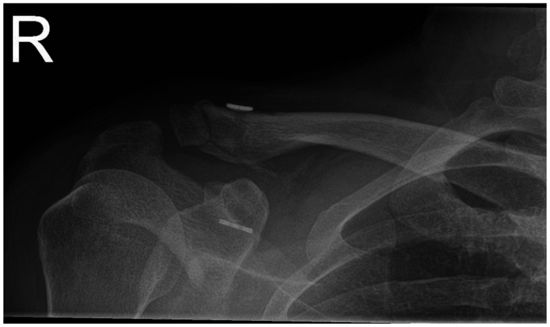
Radiologically, the reduction was maintained in all cases when evaluating serial and final radiographs, and there were no fixation failures within the follow up period thus far (see Figures 1 to 3).
Figure 1.
Pre-op radiographs.
Figure 3.
Final radiograph (3 months post-op).
Assessing bony healing: 22 (79 %) cases had united fully radiologically. There were five (17%) cases of progressive healing at the time of last radiograph, within the eight-week post-operative period; four patients did not attend for repeat imaging but functional scores and clinical review indicated a good outcome, and one case did not have repeat imaging nor clinical scoring and functional review. There was one (4%) case of established non-union, confirmed with a CT scan at the five-month mark; this was in a patient who had persistent pain secondary to adhesive capsulitis.
Clinically, there were good post-operative functional scores for OSS (41 points), EQ5D (0.78) and EuroQol Visual Analogue Scale (EQVAS) (76).
The total complication rate was 10% (a total of 3 cases), although there were no intra-operative or early complications. We have one case where the patient had persistent pain and developed adhesive capsulitis of the shoulder, necessitating treatment in its own right. The patient did recover and improve with clinical scores of 33 for OSS, 0.5 for EQ5D and 75 for EQVAS. We also had one patient reporting symptomatic prominence of the tightrope and persistent pain which resulted in subsequent removal of the tightrope implant with good resolution. There was one case of coracoid button migration, although reduction was maintained and union achieved, with no further intervention or treatment required.
Discussion
We believe that this is the largest series to date of Tightrope fixation device being used as the sole surgical technique for treating lateral end of clavicle fractures. Our results show positive clinical functional scores and maintenance of fracture reduction in all cases. We have also achieved high union rates and low complication rates.
Indirect stabilisation has been described using both open and arthroscopic approaches, 17 using different suture materials and with tendon graft reconstructions (both synthetic or allo/autografts). Small case series show these techniques appear to have good functional and radiological outcomes in the short and medium term17–20 with the advantage of the lower profile implants causing less irritation than hook plates or pins, reducing re-operation rate, as well as not relying on the need to obtain bony purchase on small, possibly comminuted lateral fragments as in locking plates.
This technique offers significant advantage over the hook plates; as it does not necessitate a second procedure for removal. It is advantageous to lateral end clavicle plates; that may be prominent and again require removal, or may fail to maintain reduction of the coracoclavicular ligaments.15,21,22 By securing the reduction of the medial clavicle segment directly and independently of the lateral fragment or acromion, we are able to confidently maintain reduction. The additional benefit of Tightrope device is the cost, which is significantly less than locking plates or hook plates.
Whilst there have been arthroscopic techniques described for this procedure, 17 we feel it is safer to do this under direct vision with better positioning of the drill hole at base of coracoid reducing the risk of coracoid fracture. This technique also allows for two separate drill holes, each directed appropriately for the coracoid and clavicle, in contrary to the arthroscopic single with a combined drill hole. It is paramount that the drill hole is central or medial on the coracoid base to prevent cortical blow out fracture laterally. The medial coracoid provides better bone stock.
These procedures have an excellent track record of being performed in a day surgery unit setting. The cost of the tightrope compared to the lateral end clavicle plates is significantly lower. Day surgery setting is cost effective and has high patient satisfaction. The additional benefits to such an approach include improved outcomes and dedicated care which in turn leads to better value and is supported by the value based healthcare agenda. Such patient driven outcome and costs improvement agendas are of relevance to all healthcare systems, especially at this time. 23 We acknowledge that this is a retrospective case series and not all cases have confirmed radiological union as some patients were unavailable for further radiological follow-up; as our cohort was mainly a young dynamic population, and only had functional scores carried out at the final review. A further prospective study with a standardised protocol post-operatively, with clinical and radiological review at fixed points, may be useful. Additionally, in the future a randomised study, comparing this technique to other methods may be helpful in establishing the optimum procedure to achieve good outcomes whilst reducing both complications and costs. 23 The positive subjective and objective outcome measures, in the setting of a displaced lateral clavicle fracture with traditionally high rates of non-union, support the use of Tightrope device as the sole method of fixation in these cases.
Figure 2.
Post-op radiograph (6 weeks post-op).
Acknowledgements
We would like to thank the business invention unit and medical records at Kings College Hospital for their assistance and guidance in this research.
Footnotes
Declaration of Conflicting Interests: The author(s) declared no potential conflicts of interest with respect to the research, authorship, and/or publication of this article.
Funding: The author(s) received no financial support for the research, authorship, and/or publication of this article.
Ethical Review and Patient Consent: Kings college Hospital London does not require ethical approval for reporting individual cases or case series. No consent was required as there is no patient identifiable information. Verbal consent was taken for this series due to the Coronavirus situation.
Contributorship: KAT, MG and TA contributed to the data collection and write up. KK, RT, TCS and AT contributed to supervision, review and editing. All authors approved the final version of the manuscript.
ORCID iDs: K Al-Tawil https://orcid.org/0000-0001-6105-6698
T Colegate-Stone https://orcid.org/0000-0001-6847-2205
References
- 1.Edwards DJ, Kavanagh TG, Flannery MC. Fractures of the distal clavicle: a case for fixation. Injury 1992; 23: 44–46. [DOI] [PubMed] [Google Scholar]
- 2.van der Meijden OA, Gaskill TR, Millett PJ. Treatment of clavicle fractures: current concepts review. J Shoulder Elbow Surg 2012; 21: 423–429. [DOI] [PubMed] [Google Scholar]
- 3.Nordqvist A, Petersson C. The incidence of fractures of the clavicle. Clin Orthop Relat Res 1994; 300: 127–132. [PubMed] [Google Scholar]
- 4.Robinson CM. Fractures of the clavicle in the adult. Epidemiology and classification. J Bone Joint Surg Br 1998; 80: 476–484. [DOI] [PubMed] [Google Scholar]
- 5.Goldberg JA, Bruce WJ, Sonnabend DH, et al. Type 2 fractures of the distal clavicle: a new surgical technique. J Shoulder Elbow Surg 1997; 6: 380–382. [DOI] [PubMed] [Google Scholar]
- 6.Robinson CM, Cairns DA. Primary nonoperative treatment of displaced lateral fractures of the clavicle. J Bone Joint Surg Am 2004; 86: 778–782. [DOI] [PubMed] [Google Scholar]
- 7.Neer CS, 2nd. Fractures of the distal third of the clavicle. Clin Orthop Relat Res 1968; 58: 43–50. [PubMed] [Google Scholar]
- 8.Neer CS, 2nd. Fracture of the distal clavicle with detachment of the coracoclavicular ligaments in adults. J Trauma 1963; 3: 99–110. [DOI] [PubMed] [Google Scholar]
- 9.Nordqvist A, Redlund-Johnell I, von Scheele A, et al. Shortening of clavicle after fracture. Incidence and clinical significance, a 5-year follow-up of 85 patients. Acta Orthop Scand 1997; 68: 349–351. [DOI] [PubMed] [Google Scholar]
- 10.Nordqvist A, Petersson C, Redlund-Johnell I. The natural course of lateral clavicle fracture. 15 (11–21) year follow-up of 110 cases. Acta Orthop Scand 1993; 64: 87–91. [DOI] [PubMed] [Google Scholar]
- 11.Rieser GR, Edwards K, Gould GC, et al. Distal-third clavicle fracture fixation: a biomechanical evaluation of fixation. J Shoulder Elbow Surg 2013; 22: 848–855. [DOI] [PubMed] [Google Scholar]
- 12.Madsen W, Yaseen Z, LaFrance R, et al. Addition of a suture anchor for coracoclavicular fixation to a superior locking plate improves stability of type IIB distal clavicle fractures. Arthroscopy 2013; 29: 998–1004. [DOI] [PubMed] [Google Scholar]
- 13.Haidar SG, Krishnan KM, Deshmukh SC. Hook plate fixation for type II fractures of the lateral end of the clavicle. J Shoulder Elbow Surg 2006; 15: 419–423. [DOI] [PubMed] [Google Scholar]
- 14.Kao FC, Chao EK, Chen CH, et al. Treatment of distal clavicle fracture using Kirschner wires and tension-band wires. J Trauma 2001; 51: 522–525. [DOI] [PubMed] [Google Scholar]
- 15.Stegeman SA, Nacak H, Huvenaars KH, et al. Surgical treatment of Neer type-II fractures of the distal clavicle: a meta-analysis. Acta Orthop 2013; 84: 184–190. [DOI] [PMC free article] [PubMed] [Google Scholar]
- 16.Zhang C, Huang J, Luo Y, et al. Comparison of the efficacy of a distal clavicular locking plate versus a clavicular hook plate in the treatment of unstable distal clavicle fractures and a systematic literature review. Int Orthop 2014; 38: 1461–1468. [DOI] [PMC free article] [PubMed] [Google Scholar]
- 17.Motta P, Bruno L, Maderni A, et al. Acute lateral dislocated clavicular fractures: arthroscopic stabilization with TightRope. J Shoulder Elbow Surg 2014; 23: e47–52. [DOI] [PubMed] [Google Scholar]
- 18.Robinson CM, Akhtar MA, Jenkins PJ, et al. Open reduction and endobutton fixation of displaced fractures of the lateral end of the clavicle in younger patients. J Bone Joint Surg Br 2010; 92: 811–816. [DOI] [PubMed] [Google Scholar]
- 19.Zheng YR, Lu YC, Liu CT. Treatment of unstable distal-third clavicule fractures using minimal invasive closed-loop double endobutton technique. J Orthop Surg Res 2019; 14: 37–37. [DOI] [PMC free article] [PubMed] [Google Scholar]
- 20.Yagnik GP, Porter DA, Jordan CJ. Distal clavicle fracture repair using cortical button fixation with coracoclavicular ligament reconstruction. Arthrosc Tech 2018; 7: e411–e415. [DOI] [PMC free article] [PubMed] [Google Scholar]
- 21.Sajid S, Fawdington R, Sinha M. Locking plates for displaced fractures of the lateral end of clavicle: potential pitfalls. Int J Shoulder Surg 2012; 6: 126–129. [DOI] [PMC free article] [PubMed] [Google Scholar]
- 22.Martetschläger F, Kraus TM, Schiele CS, et al. Treatment for unstable distal clavicle fractures (Neer 2) with locking T-plate and additional PDS cerclage. Knee Surg Sports Traumatol Arthrosc 2013; 21: 1189–1194. [DOI] [PubMed] [Google Scholar]
- 23.Colegate-Stone T, Tavakkolizadeh A, Moxham J, et al. Increasing value: The King’s College Hospital experience. Br J Healthc Manag 2016; 22: 225–233. [Google Scholar]


