Abstract
Long non-coding RNAs (lncRNAs) are an emerging group of ncRNAs that can modulate gene expression at the transcriptional or translational levels. In the present work, previously published transcriptomic data were used to identify lncRNAs expressed in gilthead sea bream skeletal muscle, and their transcription levels were studied under different physiological conditions. Two hundred and ninety lncRNAs were identified and, based on transcriptomic differences between juveniles and adults, a total of seven lncRNAs showed potential to be important for muscle development. Our data suggest that the downregulation of most of the studied lncRNAs might be linked to increased myoblast proliferation, while their upregulation might be necessary for differentiation. However, with these data, as it is not possible to propose a formal mechanism to explain their effect, bioinformatic analysis suggests two possible mechanisms. First, the lncRNAs may act as sponges of myoblast proliferation inducers microRNAs (miRNAs) such as miR-206, miR-208, and miR-133 (binding energy MEF < −25.0 kcal). Secondly, lncRNA20194 had a strong predicted interaction towards the myod1 mRNA (ndG = −0.17) that, based on the positive correlation between the two genes, might promote its function. Our study represents the first characterization of lncRNAs in gilthead sea bream fast skeletal muscle and provides evidence regarding their involvement in muscle development.
Keywords: lncRNA, muscle development, gilthead sea bream
1. Introduction
Gilthead sea bream (Sparus aurata) is one of the most cultivated species in the Mediterranean area, whose production has already exceeded wild-fish captures [1]. Although this species has been extensively studied due to its commercial interest, more research is still needed to understand some physiological aspects better, such as the molecular networks involved in muscle growth control and development. As with most teleosts, gilthead sea bream shows indeterminate growth with continuous muscle accretion throughout its life thanks to processes of hyperplasia (i.e., recruitment of new muscle fibers) and hypertrophy (i.e., increase in fiber size) [2,3]. These two processes are the result of increased myogenesis, a complex process involving the coordination of several molecular networks, including the myogenic regulatory factors (MRFs), a group of transcription factors that play a key role in the control of the myogenesis [3,4]. In the onset of myogenesis, the myogenic factor 5 (Myf5) and the myogenic determination factors (Myods) are required for the determination of the myogenic lineage and the proliferation of the muscle satellite cells. Then, the myogenin (Myog) and the myocyte enhancer factor 2 (Mef2) lead to myoblasts fusion and differentiation, and the myogenic factor 6 (Myf6/Mrf4) is responsible for myotubes maturation. In the myoblasts fusion process, different membrane proteins are also involved [5,6], including the cadherins (Cdhs), which mediate cell–cell adhesions [7]; the caveolins (Cavs), implicated in vesicular trafficking and signal transduction [8]; and a recently discovered protein duo, Myomaker (Mymk) and Myomixer (Mymx), involved in plasma membrane hemifusion or fusion pore formation, respectively [9,10]. Besides, the Crk adaptor proteins (Crk and Crkl) and the dedicator of cytokinesis (Docks) are crucial members of the intracellular signaling networks that trigger myotubes formation [11].
Although it has traditionally been supposed that most genetic information is transacted by proteins and that RNA only plays an intermediary role, in the last decades, it was demonstrated that a majority of genomes from complex organisms are in fact transcribed into non-coding RNAs (ncRNAs) [12]. The term ncRNA refers to RNAs that are not translated into a protein but have a wide spectrum of functions in many cellular processes, both in physiological and pathological situations and are crucial in controlling transcription of other genes [12,13,14].
The long ncRNAs (lncRNAs) are a class of ncRNAs characterized by having a length ranging from 200 nt to 100 kb [15]. They can exhibit a wide or tissue-specific expression, and their transcription is usually lower than those of protein-coding genes [15]. Although the lncRNAs do not normally code for proteins, a fraction of putative small open reading frames (sORFs) were found in some lncRNAs that are translated into micropeptides [16,17], as in the case of Mymx [10,17,18]. Besides, lncRNAs play a fundamental role in the fine control of gene expression, acting at different levels: modulating chromatin structure, acting upon transcription factors binding, controlling RNA splicing, regulating translation, modifying mRNA stability, or interacting directly with proteins [13,19,20]. LncRNAs can also interact with microRNAs (miRNAs), another type of ncRNAs involved in posttranscriptional regulation of protein-coding genes by mRNA cleavage, translational repression, or mRNA destabilization [21]. LncRNAs can act as miRNAs precursors or as miRNAs sponges, thus altering their regulatory effect on mRNAs and introducing an additional layer of complexity in the miRNA-target interaction network [21,22,23]. All the specific interactions of lncRNAs are sometimes based on sequence complementarity, but it is suggested that, in many cases, the lncRNAs’ function is defined by their three-dimensional structure [24]. This might be the reason why the evolutionary conservation of the nucleotide sequence of lncRNAs is very low, and it is proposed that the secondary structure is conserved [24]. In some cases, where lncRNAs codify for micropeptides, such as the mymx gene, their sequence is conserved between species, but that only happens on very rare occasions [10,18].
Currently, most of the studies in the field of lncRNAs are limited to humans and model species showing that lncRNAs have key roles in glucose and lipid metabolism [25], the immune response [26,27], the occurrence and development of cancers [28], and the neural development [29], among many others. Moreover, several lncRNAs were described as important molecules that can also regulate myogenesis in mammals, although the function of the vast majority is not yet well defined [30,31]. They may be implicated in the maintenance of satellite cells pool, their activation, proliferation, differentiation, and self-renewal [32,33]. For example, Linc-RAM is a lncRNA that is specifically expressed in skeletal muscle tissue and promotes myogenic differentiation by interacting with Myod [34]. The lncRNA Irm regulates the expression of myogenic genes by binding to Mef2d, which in turn promotes the assembly of Myod/Mef2d on the regulatory elements of target genes [30]. The lncRNA Munc is also required for optimal myogenic differentiation since it induces the expression of myod, myog, and myosin heavy chain 3 [35,36]. The fact that lncRNAs have such important roles in muscle development involves them not only in physiological conditions but also in pathological ones, such as dystrophy, atrophy, aberrant hypertrophy, or even in the recovery after an injury, in the process of necrosis, and muscle regeneration [16,32,33].
While the study of lncRNAs is progressing fast in humans and model species, information on teleost fish is scarce, and most studies are based on high-through sequencing platforms [37,38,39], providing valuable information on the molecular interactions between lncRNAs, mRNAs, and miRNAs. However, RNA-Seq studies can give us a limited picture of the lncRNAs functions due to RNA-Seq inherent limitations, such as the relatively small number of animals used and physiological conditions able to test. In addition, these studies of lncRNAs are of very little utility unless they are conducted in the same species of interest since the low conservation of lncRNAs sequences, even between closely related species, makes it difficult to translate findings on one species to another [24]. There are some recent studies in fish where the lncRNAs were reported to participate in many biological processes, including immune response [39,40,41], sex differentiation [42,43], smoltification process [44], intestinal homeostasis [45], and lipid metabolism [46]. Regarding the role of lncRNAs in fish muscle development and growth, there are very few studies that address this issue [37,47], and there is still much to explore in this field.
In gilthead sea bream, the mechanisms orchestrating the myogenesis and the molecular basis of muscle plasticity have only been studied from the perspective of protein-coding genes [4,48,49,50,51,52,53,54]. Hence, building on this research, the present work aims to identify lncRNAs with potential functions on the development and growth of fast skeletal muscle of gilthead sea bream. Therefore, existing transcriptomic data from the white muscle of this species were used to find expressed lncRNAs, and then the transcriptional profile of a subgroup was examined under different experimental conditions in which white muscle development and remodeling are expected.
2. Materials and Methods
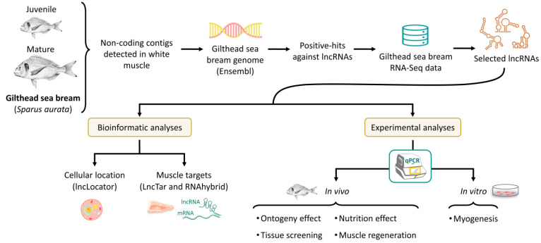
Figure 1 shows the main steps followed for the identification and characterization of gilthead sea bream fast skeletal muscle lncRNAs. Specific databases and software used for each step are indicated in brackets.
Figure 1.
Workflow for the detection and analysis of gilthead sea bream fast skeletal muscle lncRNAs. Gilthead sea bream figure was extracted from https://www.fao.org/fishery/culturedspecies/Sparus_aurata/es (accessed on 15 November 2021).
2.1. Identification of lncRNAs in Gilthead Sea Bream
In order to detect lncRNAs expressed in the gilthead sea bream fast skeletal muscle, existent transcriptomic data obtained from publicly available GS FLX 454 normalized libraries were used [55]. The GS FLX 454 transcriptomes were blasted (BLASTn) against all lncRNAs annotated in the gilthead sea bream genome (http://www.ensembl.org/ (accessed on 20 November 2021)) using the BLAST2GO software (part of OmicsBox package v.1.0) [56]. The threshold for a contig to be considered a positive hit for a lncRNA was set at an e-value lower than 1 × 10−90 and similarity over 98%. The expression of identified lncRNAs was further investigated using fast skeletal muscle RNA-Seq data from mature and immature male gilthead sea breams (Figure 1).
2.2. RNA-Seq
The transcriptomic analysis was performed at the Institute of Marine Biology, Biotechnology, and Aquaculture of the Hellenic Centre of Marine Sciences (HCMR, Crete, Greece). The use of animals used for the transcriptomic analysis was approved by the relevant Greek authorities (National Veterinary Services) under the license No 32356 (AΔA: 984I7ΛK-K65). All procedures involving animals were conducted following the “Guidelines for the treatment of animals in behavioral research and teaching” [57], the Ethical justification for the use and treatment of fishes in research: an update [58], and the “Directive 2010/63/EU of the European Parliament and the council of 22 September 2010 on the protection of animals used for scientific purposes” (EU, 2010).
Briefly, the white muscle was sampled from non-mature (juveniles) and mature male gilthead sea breams. The total RNA was extracted with NuceloZOL (Macherey-Nagel, Duren, Germany) following the manufacturer’s instructions. RNA quantity was assessed by Nano-Drop ND-1000 spectrophotometer (NanoDrop Technologies, Wilmington, DE, USA). The quality of the extracted RNA was evaluated by agarose (1%) gel electrophoresis and by RNA Pico Bioanalysis chip (Agilent 2100 Bioanalyzer, Agilent, CA, USA).
Library construction and paired-end (PE) sequencing was carried out by Novogen (Novogene, UK). Muscle reference transcriptome and count matrix was generated applying Trinity software Trinity v.2.13.2 [59]. Differential expression was assessed using Bioconductor packages, including DESeq2 [60].
The RNA-Seq data were deposited in the European Nucleotide Archive (ENA) under the accession number PRJEB50017.
2.3. Prediction of Target mRNAs, miRNAs, and Cell Location of lncRNAs
LncRNAs were compared against the full cDNA sequences of 149 protein-coding genes and miRNAs known to be involved in muscle regulation (http://www.ensembl.org/ (accessed on 20 November 2021)) (Supplementary File S1). The mRNA target predictions were made using LncTar software v.1.0 (https://www.cuilab.cn/lnctar (accessed on 20 November 2021)) [61] with a normalized interaction threshold of ndG < −0.08. The potential interactions with mature miRNAs were investigated using RNAhybrid v.2.2.1 (https://bibiserv2.cebitec.uni-bielefeld.de/rnahybrid (accessed on 20 November 2021)) [62] with a minimal free energy (MFE) threshold of <−20.0 kcal. The subcellular location of lncRNAs was predicted using the lncLocator software v.1.0 (http://www.csbio.sjtu.edu.cn/bioinf/lncLocator/ (accessed on 20 November 2021)) [63] (Figure 1). MiRNAs sequences were obtained from the Ensembl database, and their mature sequences were predicted by alignment with known mature forms from zebrafish (Danio rerio) and tilapia (Oreochromis niloticus), downloaded from the miRbase database (https://www.mirbase.org (accessed on 20 November 2021)).
2.4. Experimental Trials
All animal-handling procedures were conducted following the Directive 2010/63/EU of the European Parliament and the council of 22 September 2010 on the protection of animals used for scientific purposes, the guidelines of the Spanish and Catalan governments, and with the approval of the Ethics and Animal Care Committee of the University of Barcelona.
Gilthead sea breams used in all the experiments were obtained from a commercial hatchery (Piscimar, Burriana, Castellón, Spain) and were acclimatized to the facilities at the University of Barcelona (Barcelona, Spain) for a minimum of two weeks prior to sampling or experimental manipulations. Fish were fed ad libitum twice a day with commercial pellets (Skretting, Burgos, Spain) and held at 23 ± 1 °C, a salinity of 35–37‰ and a photoperiod of 12 h light/12 h dark in a semi-closed recirculation system with a weekly renewal of 20–30%.
In order to infer the functions of the lncRNAs in the muscle, their transcriptional profile was analyzed in different in vivo and in vitro experimental conditions.
2.4.1. In Vivo Experiments
For the exploration of the ontogeny effect on the expression of the lncRNAs, the epaxial white muscle tissue was collected from groups of 8 fish each of fingerlings (6.0 ± 0.5 g), juveniles (122.4 ± 2.3 g), and matures/adults (387.1 ± 41.9 g). For lncRNAs screening, the following tissues were extracted from 4 fish of 214.0 ± 12.1 g: white muscle, red muscle, skin, gills, eye, heart, adipose tissue, bone, brain, pituitary, spleen, stomach, proximal and distal intestine, liver, head kidney, pyloric caeca, and gonads. In order to analyze the effects of fasting and refeeding on lncRNA transcription, fish with an initial body weight of 50.0 ± 3.0 g were fasted for 21 days and then refed for 7 days. Samples of white muscle tissue were taken from 6 fish at the beginning of fasting and at the end of it at 0, 2, 5, and 24 h and 7 days after refeeding (as previously described in [64]). A muscle regeneration experiment was conducted to study the possible role of the lncRNAs in myogenesis after a muscle injury. Fish of 15.4 ± 3.5 g were used, and an injury was performed with a 2.108 mm diameter needle inserted vertically into the left epaxial muscle below the sixth radius to a depth of 1 cm. Samplings were performed at days 0, 1, 2, 4, 8, 16, and 30 after the injury. Each day, from 10 injured fish, a section of the muscle was removed from the left side (injured) and the right side, as self-control for each fish (for more details of this experiment, see Perelló-Amorós et al. [18]).
2.4.2. In Vitro Myogenesis: Primary Myocyte Cell Culture
In order to infer the function of the lncRNAs during the process of myogenesis, their expression was analyzed in the different phases of a myocyte cell culture. This culture consists of a short period where they remain quiescent satellite cells (day 1), then a proliferative stage of the myoblasts (until day 4 of the culture), followed by a fusion phase of the myoblast to form early myotubes and their final maturation (from day 4 onwards) [4,65]. Six different cell cultures were performed following the protocol previously described by Montserrat et al. (2007) [65]. Cells were seeded at a density of 2 × 10⁶ cells per well in 6-well plastic plates (9.6 cm2/well) (Nunc, Labclinics, Barcelona, Spain). Cells were maintained at 23 °C and 2.5% CO2 in Dulbecco’s Modified Eagle Medium supplemented with 10% fetal bovine serum and 1% antibiotic–antimycotic solution. All media and reagents were obtained from Sigma-Aldrich (Tres Cantos, Madrid, Spain). The medium was renewed every 2 days during the culture. Samples were taken on days 2, 4, 6, 8, 10, and 12 after satellite cells seeding.
2.5. Primer Design
Primers for qPCR were designed from Ensembl lncRNAs sequences using Primer3 software v.0.4.0 [66] with a melting temperature of 60 °C. Primers, possible hairpins, or non-desirable primer-dimers were investigated using NetPrimer (http://www.premierbiosoft.com/netprimer/netprlaunch/netprlaunch.html (accessed on 13 November 2021)). Primers used in the present study are summarized in Supplementary File S2.
2.6. Gene Expression
2.6.1. RNA Extraction and cDNA Synthesis
For RNA extraction from tissue, 40 to 500 mg of tissue (depending on tissue yield) were used, and total RNA was extracted with 1 mL of TRI Reagent® Solution (Applied Biosystems, Alcobendas, Madrid, Spain). For RNA extraction from cells, the cells seeded in 3 replicate wells were pooled together at each sampling point during the culture, and total RNA was extracted with 1 mL of TRI Reagent® Solution. The RNA concentration and purity of the samples were determined using the Nanodrop 2200TM (Thermo Scientific, Alcobendas, Madrid, Spain). The RNA integrity was checked in a 1% (w/v) agarose gel stained with SYBR-Safe® DNA Gel Stain (Life Technologies, Alcobendas, Madrid, Spain). For cDNA synthesis, 1.1 µg of total RNA was treated with DNase I Amplification Grade (Life Technologies, Alcobendas, Barcelona, Spain) and retrotranscribed with the Transcriptor First Strand cDNA Synthesis Kit® (Roche, Sant Cugat del Vallès, Spain) [49].
2.6.2. Quantitative Real-Time PCR (qPCR)
The qPCRs were performed following the MIQE guidelines [67] in a CFX384TM Real-Time System (Bio-Rad, El Prat de Llobregat, Barcelona, Spain) using iTAQ Universal SYBR® Green Supermix (Bio-Rad, El Prat de Llobregat, Barcelona, Spain). The analyses were carried out in triplicate, using for each reaction: 2.5 µL of iTAQ Universal SYBR® Green Supermix, 1 µL of cDNA, 250 nM (final concentration) of forward and reverse primers, and 1.25 µL of DEPC water. The qPCR program consisted of 3 min at 95 °C, 39 × (10 s at 95 °C, 30 s at the annealing temperature of the primers and fluorescence detection), followed by an amplicon dissociation analysis from 55 to 95 °C with an increase of 0.5 °C each 30 s [49].
All the primers were first validated using a dilution curve with a pooled sample made before the analyses to confirm reaction specificity, efficiency of the primer pairs, absence of primer-dimers, and to determine the appropriate cDNA dilution to work with.
The mRNA transcript level of each studied gene was calculated relative to the geometric mean of the combination of the two most stable reference genes (ef1a: elongation factor 1 alpha, rps18: ribosomal protein s18, and rpl27a: ribosomal protein l27a) (confirmed by the geNorm algorithm) using the Bio-Rad CFX Manager™ software v.3.1, and following the Pfaffl method [68].
2.7. Statistics
All statical analyses and graphs were conducted using R-Studio v.1.1.419 [69] and ggplot2 [70]. Data normality and homogeneity of variance were estimated using Shapiro–Wilk and Levene’s tests. A Box-Cox transformation approach was used to transform non-normally distributed data and tested again on normality and homogeneity of variance assumptions. Differences between measurements were analyzed using a t-test or an ANOVA model followed by a Tukey’s post hoc test when homogeneity of variance was achieved. Pearson’s test was used to estimate the correlation between the transcription of the different genes studied.
Unless otherwise indicated, values are shown as mean ± SD. The signification threshold was established as the p-value (p) < 0.05.
3. Results
3.1. lncRNAs Selection
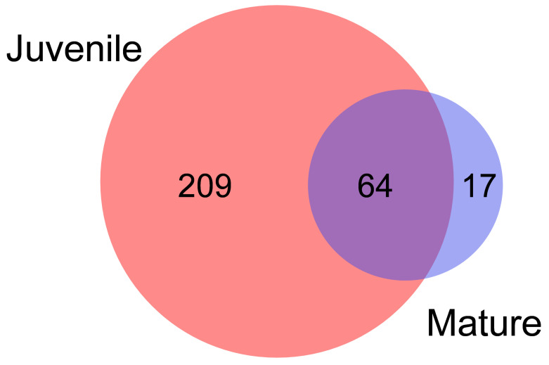
Because many lncRNA can show low levels of the transcriptome, normalized GS FLX 454 transcriptomic data from juvenile and adult gilthead sea bream were used to detect as many as possible in the gilthead sea bream fast skeletal muscle [55]. A total of 290 lncRNAs were identified, with 209 found only in juveniles, 64 shared between juveniles and adults, and 17 expressed only in adults (Figure 2; Supplementary File S3).
Figure 2.
LncRNAs detected in fast skeletal muscle GS FLX 454 transcriptomes. Venn diagram representing LncRNAs identified in the GS FLX 454 transcriptomes of juvenile and mature gilthead sea breams from Garcia de la serrana et al. (2012) [55]. The number of lncRNAs found only in juveniles or matures is shown inside the individual circles, while lncRNAs shared are shown in the intersection.
LncRNAs of interest were ranked using their relative expression in the GS FLX 454 transcriptomes (data not shown). However, as it is a normalized transcriptome expression, data are not accurate; therefore, non-normalized RNA-Seq data from fast skeletal muscle of juveniles and adults of gilthead sea bream was used to estimate their abundance initially. The number of counts mapped for each of the lncRNAs was extracted and selected the 20 first lncRNAs in which at least one count was present in all the samples (Supplementary File S4). From those 20 selected, at least eight of them were already showing significant differences in the expression between juveniles and matures (Supplementary File S4). Primers were designed to amplify the selected 20 lncRNAs, with only 12 successfully amplified by qPCR. These 12 lncRNAs were named throughout the text as “lncRNA” plus the last five numbers of their Ensembl Transcript ID.
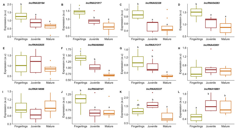
Our interest was focused on those lncRNAs that might have an active role in muscle development; therefore, the expression of the 12 lncRNAs candidates was studied in the fast skeletal muscle of gilthead sea bream fingerlings, juveniles, and matures (Figure 3). The lncRNA20194, lncRNA21817, lncRNA02328, lncRNA54283, lncRNA60660, lncRNA31317, and lncRNA40141 were significantly more expressed in the fingerlings compared to matures and/or juveniles (Figure 3A–D,F–G,J). The lncRNA16861 showed significantly lower expression in fingerlings compared to juveniles and matures (Figure 3L). The lncRNA62925, lncRNA43061, lncRNA14696, and lncRNA05337 had similar transcription in the three ontogenetic stages (Figure 3E,H–I,K).
Figure 3.
Expression of selected lncRNAs in gilthead sea bream fast skeletal muscle at different ontogenetic stages. Boxplots showing lncRNAs expression in gilthead fast skeletal muscle obtained from fingerlings (green box), juveniles (dark red box) or adults (orange box) (A–L). Gene expression is indicated as arbitrary units (a.u). Significant differences between ontogenetic stages are indicated as different letters when p < 0.05. Outliers are presented as points.
Since it was intended to study lncRNAs with relevant roles in muscle growth and development, those lncRNAs more expressed in the stages in which the fish growth rates are higher (fingerling and juvenile stages) had to be selected. Hence, lncRNA20194, lncRNA21817, lncRNA54283, lncRNA60660, and lncRNA40141 were prioritized and selected to study their transcriptional profile under different physiological conditions further. Moreover, the lncRNA16861 that had lower transcription in fingerlings compared to juveniles and matures, and the lncRNA14696 that showed no differences between stages were selected to explore the transcription of lncRNAs with different patterns of expression. In addition, for the selection of lncRNAs, the expression level of each lncRNA was taken into account, and those with the highest expression were selected.
3.2. lncRNAs Subcellular Location and Targets
In order to elucidate the possible functions of the selected lncRNAs, their sequences were analyzed to determine their predicted subcellular location and their possible interactions with protein-coding genes mRNA and miRNAs related to muscle growth and development in other vertebrates (Table 1).
Table 1.
Bioinformatic predictions of lncRNA interactions with muscle-related mRNAs and miRNAs and subcellular location.
| LncRNAs | mRNA | ndG | miRNA | MEF (kcal) | Subcellular Location | Probability |
|---|---|---|---|---|---|---|
| lncRNA14696 | lamtor2 (ENSSAUG00010013916) | −0.087 | miR-133a1/2 | −25.9 | Nucleus | 0.93 |
| miR-133b | −29.2 | |||||
| miR-499 | −20.6 | |||||
| miR-1 | −21.9 | |||||
| miR-206 | −28.3 | |||||
| miR-208 | −22.3 | |||||
| lncRNA16861 | lamtor1 (ENSSAUG00010010852) | −0.087 | miR-133b | −23.2 | Cytoplasm | 0.89 |
| miR-488 | −21.3 | |||||
| miR-1 | −21.4 | |||||
| miR-206 | −23.3 | |||||
| miR-208 | −21.3 | |||||
| lncRNA20194 | myod1 (ENSSAUG00010008630) | −0.170 | miR-133a1/2 | −23.4 | Cytoplasm | 0.82 |
| miR-133b | −26.2 | |||||
| miR-499 | −22.5 | |||||
| miR-206 | −21.1 | |||||
| miR-208 | −28.3 | |||||
| lncRNA21817 | NA | NA | miR-133a1/2 | −23.3 | Cytoplasm | 0.70 |
| miR-133b | −27.3 | |||||
| miR-499 | −23.4 | |||||
| miR-206 | −27.2 | |||||
| miR-208 | −21.6 | |||||
| lncRNA40141 | eif4ebp1 (ENSSAUG00010019173) | −0.085 | miR-133a1/2 | −22.4 | Cytoplasm | 0.46 |
| miR-133b | −25.8 | Nucleus | 0.26 | |||
| miR-499 | −20.5 | |||||
| miR-1 | −22.0 | |||||
| miR-206 | −25.2 | |||||
| miR-208 | −26.1 | |||||
| lncRNA54283 | lamtor1 (ENSSAUG00010010852) | −0.082 | miR-133b | −23.4 | Nucleus | 0.49 |
| pax7b (ENSSAUG00010013590) | −0.080 | miR-206 | −24.3 | Cytoplasm | 0.37 | |
| miR-208 | −21.9 | |||||
| lncRNA60660 | NA | NA | miR-206 | −25.1 | Nucleus | 0.67 |
| Cytoplasm | 0.27 |
The strength of the predicted interactions with muscle-related mRNAs is indicated as length normalized free energy (ndG). The strength of the predicted interactions between lncRNAs against muscle-related miRNAs is indicated as minimal free energy (MFE). The probability of predicted subcellular cell location is also indicated. NA: not analyzed. lamtor1 (late endosomal/lysosomal adaptor, MAPK and MTOR activator 1), lamtor2 (late endosomal/lysosomal adaptor, MAPK and MTOR activator 2), myod1 (myogenic determination factor 1), eif4ebp1 (eukaryotic translation initiation factor 4E-binding protein 1), pax7b (paired box 7b).
The lncRNA16861, lncRNA20194, and lncRNA21817 were predicted to be in the cytoplasm, while lncRNA14696 was predicted to be in the nucleus, all of them with a probability over 0.70 (Table 1). Three of the selected lncRNAs, lncRNA40141, lncRNA54283, and lncRNA60660, appeared to have a relatively low probability of being in the cytoplasm and the nucleus (Table 1).
Regarding the possible interactions between selected lncRNAs and genes known to be involved in muscle growth and development, it was observed that several lncRNAs showed some interaction with these genes (Table 1). However, only lncRNA20194 appeared to have strong interaction with myod1 (ndG < −0.170) (Table 1).
The analysis of possible interactions between selected lncRNAs and miRNAs revealed that all the lncRNAs selected in the present work interact with several miRNAs with an MFE lower than −20 kcal (Table 1). Some of the lncRNAs showed relatively strong interactions (MFE < −25 kcal) with different miRNAs, as in the case of lncRNA14696 (miR-133a1/2, miR-133b and miR-206 with MFE of −25.9, −29.2 and −28.3 kcal), lncRNA20194 (miR-133b and miR-208 with MFE of −26.2 and −28.3 kcal, respectively), lncRNA21817 (miR-133b and miR-206 with MFE of −27.3 and −27.2 kcal, respectively), lncRNA40141 (miR-133b, miR-206 and miR-208 with MFE of −25.8, −25.2 and −26.1 kcal), and lncRNA60660 (miR-206 with MFE of −25.1 kcal).
3.3. lncRNAs Expression in Gilthead Sea Bream Muscle
3.3.1. lncRNAs Expression in Fast Muscle of Fingerlings, Juveniles, and Adults of Gilthead Sea Bream
In order to identify the possible roles of the selected lncRNAs in the development of the fast skeletal muscle of gilthead sea bream, their expression levels were studied under different in vivo and in vitro conditions. First, lncRNAs expression was analyzed on fast skeletal muscle from fish in different ontogenetic stages, expanding from fast-growing (fingerlings and juveniles) to slow-growing stages (mature/adult) (Figure 3). The majority of lncRNAs studied had a lower expression during the mature stage (lncRNA20194, lncRNA21817, lncRNA02328, lncRNA54283, lncRNA60660, lncRNA31317, and lncRNA40141) (Figure 3A–D,F–G,J) with a significant reduction between 77 and 34%. It was also found that lncRNA62925, lncRNA43061, lncRNA14696, and lncRNA05337 showed no significant changes in expression between stages (Figure 3E,H–I,K). Only lncRNA16861 had a significantly higher expression during juvenile and mature stages with an increase of 112 and 91%, respectively (Figure 3L).
3.3.2. lncRNAs Tissue Screening
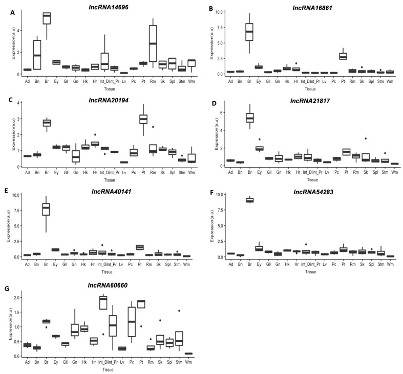
The analysis of lncRNAs expression in 18 different tissues showed that they were not exclusive of fast skeletal muscle (Wm) but detected in all tissues analyzed (Figure 4). Interestingly, except for lncRNA14696, the fast skeletal muscle was among the tissues with lower lncRNAs expression. In all cases, the slow muscle (Rm) showed a significantly higher expression than the fast skeletal muscle (Figure 4). It is also interesting that while lncRNA14696, lncRNA20194, and lncRNA60660 (Figure 4A,C,G) had variable expression among tissues, lncRNA16861, lncRNA21817, lncRNA40141, and lncRNA54283 had a much higher expression (5- to 7-fold change compared to fast muscle) in the brain (Figure 4B,D–F).
Figure 4.
Boxplots showing lncRNAs expression in different gilthead sea bream tissues (n = 4) (A–G): Ad (Adipose tissue), Bn (Bone), Br (Brain), Ey (Eye), Gl (Gills), Hk (Head kidney), Hr (Heart), Int_Di (Distal Intestine), Int_Pr (Proximal Intestine), Lv (Liver), Pc (Pyloric caeca), Pt (Pituitary), Rm (Slow muscle), Sk (Skin), Spl (Spleen), Stm (Stomach), and Wm (Fast muscle). Gene expression is indicated as arbitrary units (a.u). Outliers are presented as points.
3.3.3. lncRNAs Expression in Response to Nutrition
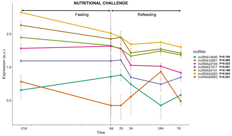
The regulation of lncRNAs transcription by the gilthead sea bream fast skeletal muscle in response to changes in the nutritional status was also analyzed. Therefore, fish were fasted for 21 days and then refed to satiation for 7 days (Figure 5). The lncRNA16861, lncRNA54283, lncRNA60660, and lncRNA40141 decreased their expression between 40 and 25% after 21 days of food deprivation, while lncRNA20194 and lncRNA21817 did not change, and lncRNA14696 increased a 32% (Figure 5). Almost all lncRNAs, except for lncRNA16861, decreased their expression and kept a low transcription during the 7 days of refeeding, ranging between 50 and 30% reduction compared to pre-fasting values. Only lncRNA16861 showed a significantly high transcription 24 h after refeeding started (23% increase compared to pre-fasting values), with a non-significant reduction of 35% by the end of the refeeding period.
Figure 5.
LncRNAs transcription in response to a nutritional challenge. Plot showing gene expression of 7 lncRNAs in response to 21 days of fasting followed by 7 days of refeeding. Gene expression is expressed as average ± SD (n = 6) of arbitrary units (a.u). The statistical effect of nutrition is indicated for each lncRNA analyzed.
3.3.4. lncRNAs Expression during Myogenesis
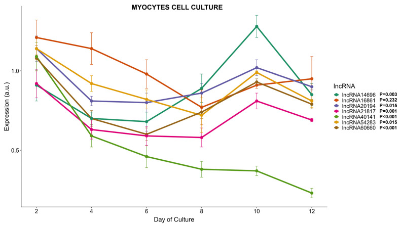
In order to study the role of the lncRNAs in the myogenesis process in more detail, their expression was analyzed during the course of a primary myocyte culture, from the proliferative stage (days 0 to 4) and through the complete differentiation process (day 4 to 12). All lncRNAs analyzed except lncRNA16861 (which showed a more stable expression during the whole culture) had a 20–35% decrease in their transcription between days 4 and 6 (Figure 6), concomitant with the myoblast proliferation phase. Until day 6, lncRNAs lncRNA20194, lncRNA60660, lncRNA21817, and lncRNA54283 kept low transcription profiles (15–35% lower compared to the beginning of the culture). Between days 8 and 12, when myoblasts fuse to form myotubes and their maturation, several lncRNAs started to increase their transcription again (Figure 6). LncRNA20194, lncRNA60660, lncRNA21817, and lncRNA54283 recovered 90% of the transcription compared to day 2; lncRNA16861, which recovered 85% of the day 2 transcription; and lncRNA14696, which even had an increase of 40% compared to day 2 values (Figure 6). The lncRNA40141 did not follow any of those patterns and steadily continued reducing its transcription until day 12, when it was 80% lower than at the beginning of the cell culture (Figure 6).
Figure 6.
LncRNAs transcription during myocytes cell culture development. Plot showing gene expression of 7 lncRNAs during gilthead sea bream fast skeletal muscle myocytes cell culture. Gene expression is expressed as average ± SD (n = 6) of arbitrary units (a.u). The statistical effect of fasting and refeeding is indicated for each lncRNA analyzed.
3.3.5. lncRNAs Expression during Muscle Regeneration
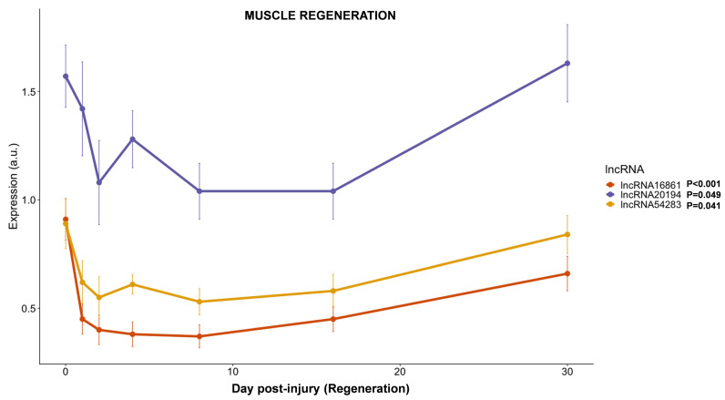
The expression of three of the selected lncRNAs, the one predicted to interact with myod1 (lncRNA20194), one of the lncRNA whose expression was higher in fingerlings (lncRNA54283), and the only lncRNA with higher expression in adults and juveniles (lncRNA16861), was studied during 30 days of regeneration after an induced injury in the epaxial fast skeletal muscle of juvenile gilthead sea breams. All three lncRNAs reduced their transcription by around 10 to 50% between days 1 and 2 after the injury (Figure 7). Except for a spike in the transcription of lncRNA20194 at day 4 of regeneration, all lncRNAs analyzed kept a low transcription level (between 40 and 60% lower than pre-injury levels) for at least 16 days while recovering nearly normal values after 30 days of regeneration (Figure 7).
Figure 7.
LncRNAs transcription in response to induced muscle injury. Plot showing the gene expression of three lncRNAs during 30 days of regeneration after an induced injury in the epaxial fast skeletal muscle of gilthead sea bream juveniles. Gene expression is expressed as average ± SD (n = 6) of arbitrary units (a.u). The statistical effect of muscle regeneration is indicated for each lncRNA analyzed.
3.3.6. Correlation between lncRNAs and Genes Related to Muscle Development
In order to further understand the possible role of the selected lncRNAs on muscle development, their correlation with muscle-related genes in the same samples of the regeneration experiment and the myocytes cell culture was estimated [18,71] (Table 2).
Table 2.
Correlation between gilthead sea bream lncRNAs and fast muscle-related genes.
| lncRNAs | myf5 | myod1 | myod2 | myog | mef2c | myf6 | cdh15 | cav3 | mymx | mymk | dock5 | crk-a | crk-b | crkl |
|---|---|---|---|---|---|---|---|---|---|---|---|---|---|---|
| lncRNA14696 | 0.2 | 0.1 | −0.1 | −0.2 | 0.4 * | −0.1 | −0.1 | 0.3 | −0.1 | 0.1 | 0.5 ** | 0.3 | 0.1 | 0.4 * |
| lncRNA16861 | 0 | −0.1 | −0.1 | 0.1 | −0.1 | 0.1 | 0.1 | −0.4 * | 0.1 | −0.4 *** | 0.1 | −0.02 | 0.3 | −0.1 |
| lncRNA20194 | 0.0 | 0.6 *** | 0.1 | −0.4 * | 0.2 | 0.4 *** | −0.2 | 0.0 | 0.6 *** | 0.3 * | 0.4 ** | 0.4 * | 0.4 * | 0.2 |
| lncRNA21817 | 0 | 0.1 | −0.4 * | −0.5 ** | 0 | 0.1 | −0.4 * | 0.1 | −0.4 * | 0.1 | 0.6 ** | 0.4 * | 0.4 * | 0.3 |
| lncRNA40141 | −0.3 | 0.5 ** | −0.5 ** | −0.2 | −0.2 | −0.2 | −0.1 | −0.1 | −0.2 | −0.3 | 0.7 *** | 0.6 *** | 0.8 *** | 0.4 * |
| lncRNA54283 | −0.1 | 0.3 * | 0 | −0.1 | 0 | 0.1 | 0 | −0.6 ** | 0.3 * | −0.1 | 0.5 *** | 0.5** | 0.6 *** | 0.4 * |
| lncRNA60660 | 0.2 | 0.2 | −0.2 | −0.4 * | 0.0 | −0.4 * | −0.2 | −0.1 | −0.3 | 0.1 | 0.7 *** | 0.7 *** | 0.4 ** | 0.6 *** |
Pearson correlation index between lncRNA and muscle-related genes expression. Significant correlations are indicated as follow: p < 0.05 *, p < 0.01 **, and p < 0.001 ***. myf5 (myogenic factor 5), myod1 (myogenic determination factor 1), myod2 (myogenic determination factor 2), myog (myogenin), mef2c (myocyte enhancer factor 2c), myf6 (myogenic factor 6), cdh15 (cadherin 15), cav3 (caveolin 3), mymx (myomixer), myk (myomaker), dock5 (dedicator of cytokinesis 5), crk (Crk adaptor protein), crkl (Crk-like adaptor protein).
Interestingly, the great majority of lncRNAs analyzed (except for lncRNA16861) were positively correlated to dock5 (r = 0.40–0.70) (Table 2). Beyond dock5, several correlations between lncRNAs and muscle related genes were found: lncRNA14696 correlated to mef2c (r = 0.4) and crkl (r = 0.4); lncRNA16861 correlated to cav3 (r = −0.4) and mymk (r = −0.4); lncRNA20194 correlated to myod1 (r = 0.6), myog (r = −0.4), myf6 (r = 0.4), mymx (r = 0.6), mymk (r = 0.3), crk-a (r = 0.4), and crk-b (r = 0.4); lncRNA21817 correlated to myod2 (r = −0.4), myog (r = −0.5), cdh15 (r = −0.4), mymx (r = −0.4), crk-a (r = 0.4), and crk-b (r = 0.4); lncRNA40141 correlated to myod1 (r = 0.5), myod2 (r = −0.5), crk-a (r = 0.6), crk-b (r = 0.8), and crkl (r = 0.4); lncRNA54283 correlated to myod1 (r = 0.3), cav3 (r = −0.6,) mymx (r = 0.3), crk-a (r = 0.5), crk-b (r = 0.6), and crkl (r = 0.4); and lncRNA60660 correlated to myog (r = −0.4), myf6 (r = −0.4), crk-a (r = 0.7), crk-b (r = 0.4), and crkl (r = 0.6). The gene expression of myod1 in the regeneration experiment and the myocytes cell culture is presented in the Supplementary File S5.
4. Discussion
In the present work, we took advantage of the existing transcriptomic data of gilthead sea bream fast skeletal muscle to focus on the non-coding genes, which have traditionally been overshadowed by the protein-coding genes. Because of the low expression of many lncRNAs, we used the normalized data published by Garcia de la serrana et al. (2012) from juvenile and adult gilthead sea breams’ fast skeletal muscle [55] to detect low expressed lncRNAs. By using this approach, 290 lncRNAs were identified in the fast skeletal muscle and were differently distributed between juveniles and adults. A greater number of lncRNAs was found in the juvenile gilthead sea bream transcriptome than in adults (209 compared to 17), which is a suggestion that the vast majority of lncRNAs detected play an important role in regulating muscle growth during that stage.
Primer design for the selected lncRNAs proved to be a challenge, with great difficulties finding primers with no self-dimer and cross-dimmer interactions or forming hairpins with themselves. These problems in primer design are likely to be the result of the different evolutionary pressures that act over the lncRNAs. While mRNAs are linear in structure to be translated, most of lncRNAs functions are derived from their three-dimensional structure; therefore, lncRNAs tend to form double strands within their sequence [24,72]. This increases the probability of two primers forming dimers and hairpins, increasing the difficulty of primer design. In addition, some lncRNAs were so poorly expressed that it was not possible to amplify them even from pure cDNA. Due to both obstacles mentioned, 8 of the 20 pre-selected lncRNAs could not be amplified since it was not possible to design suitable primers for them, or their expression was so low that it was not correct to analyze it by qPCR.
It is interesting to notice that four of the lncRNAs studied had a much higher transcription in the brain than in any other tissue, and even in those cases where lncRNAs were also expressed in many other tissues, the brain appeared to be one with a higher level of transcription. This should not be a surprise since previous studies reported that lncRNAs seem to be very important for the development of the neural system [29], with some researchers pointing out that around 40% of lncRNAs can be found to be highly expressed in the brain [73]. The expression in skeletal muscle was always very low, which might be in contradiction to an important role in muscle development. However, this is not necessarily the case for lncRNAs that are, in general, very low expressed [15]. Besides, all lncRNAs showed higher expression levels in slow muscle than in fast muscle. The difference between these two types of muscles could be because slow muscle is a metabolically and functionally more active tissue than fast muscle [74], and the role of lncRNAs in this tissue is likely to be more relevant. Nevertheless, further investigation will be necessary to understand the differences between the two tissues in the context of lncRNAs functions better.
The majority of lncRNAs showed similar patterns of regulation in response to different physiological contexts. For instance, most of them showed few changes in response to fasting but decreased shortly after refeeding started. Similarly, the three lncRNAs analyzed in the fish injury model were also downregulated shortly after muscle regeneration started. The correspondence in the regulation between lncRNAs is likely the results of the process of selection that was followed, in which it was prioritized those lncRNAs with higher expression in fingerlings and juveniles. The rationale behind this decision was to find lncRNAs with relevant roles in muscle growth and development. Then, those most expressed in the stages in which the fish growth rates are higher (fingerling and juvenile stages) had to be selected. Therefore, it is not unusual that those lncRNAs with the highest expression in the fingerling stage appeared to have similar patterns of transcription, while the only lncRNA selected with the lowest expression in fingerling (lncRNA16861) tended to have a different expression profile during fasting-refeeding but not muscle regeneration. The role played by these lncRNAs can be hypothesized from their expression profile during in vitro culture of myocytes. All lncRNAs, except for lncRNA16861, significantly changed their expression during the cell culture. Most of the lncRNAs were downregulated in the proliferative phase while increased again during the differentiation. These results suggest that their downregulation is important to maintain the proliferation of the myoblast, while their increase might be necessary for the transition to differentiation. Only lncRNA40141 appeared to continuously decrease during myogenesis, suggesting that its inhibition might be important for the progression of the myogenic program. The downregulation of lncRNAs to promote proliferation and their increase to trigger the differentiation seems to fit with the results obtained in the in vivo experiments since the majority of lncRNAs were also decreased during proliferative stages while increased during fusion events. For instance, several studies in teleost fish demonstrated that after a period of food deprivation, when food intake is restored, there is an increase in proliferative muscle markers followed much later by an increase in differentiation [48,75,76,77]. Likewise, after a muscle injury, there is early activation of the satellite cells that are attracted to the injured zone and stimulated to proliferate, while later, during the regeneration of the injury, they fuse to form new fibers [78,79]. Similar roles in regulating muscle development were described for lncRNAs in humans, such as Neat1 and Lnc-31 [31,80] promoting proliferation, and Myoparr, Munc, and LncMyod promoting differentiation [35,81,82]. Our correlation analysis suggests the possibility that lncRNAs action is performed regulating the transcription of, at least, some of the genes controlling myogenesis progression, such as myog, myod1, or myf5 [83,84], whose expression correlated positively with some of the lncRNAs analyzed. It is also interesting that all lncRNAs, except for lncRNA16861 (only lncRNA with high expression in adult fish), correlate positively with dock5 and, more than half of them to crk, both genes known to be involved in myoblast fusion [11]. These results also fit with the hypothesis that the lncRNAs identified are necessary to increase their transcription during differentiation, where myoblast fusion is a crucial step.
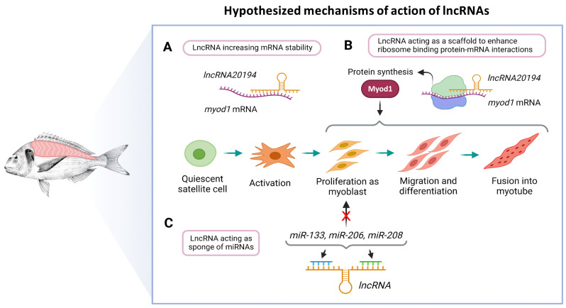
The lncRNAs have multiple mechanisms to regulate gene transcription at very different levels [20]. Therefore, with the present data, it is not possible to establish the exact mechanisms of action. However, we studied two possible mechanisms of regulation, their interaction with mRNAs and miRNAs. The great majority of lncRNAs have weak interactions with mRNAs from genes known to be related to muscle development (ndG < −0.10). Only lncRNA20194 showed a strong interaction with myod1 (ndG = −0.17), as well as had a positive correlation with myod1 (r = 0.6; p < 0.001). Although no formal mechanism of action can be proposed with the present data, one possible way of regulation could be a direct interaction of lncRNA20194 with myod1 increasing its function, for instance, stabilizing its mRNA or acting as a scaffold to enhance ribosome binding protein-mRNA interactions (Figure 8A,B) [85,86]. This hypothesis is also consistent with the cellular location of lncRNA20194 since it was predicted to be in the cytoplasm (probability of 0.82) [87]. These results strongly suggest that the lncRNA20194 is a promising candidate for further research.
Figure 8.
Hypothesized mechanisms of action of lncRNAs in the fast skeletal muscle of gilthead sea bream. The lncRNA20194 might increase myod1 mRNA stability (A) or act as a scaffold to enhance ribosome binding protein-mRNA interaction and thus promote Myod1 synthesis and function (B). The lncRNAs could also act as a sponge of miRNAs that are known to enhance myoblast proliferation, thereby regulating this step of myogenesis (C). These hypotheses are derived from the bioinformatic analyses.
The second mechanism of action could be the interaction with miRNA acting as sponges. Sponges lncRNAs can act as a decoy for one or multiple miRNAs, preventing them from binding to their targets [22]. All lncRNAs were able to bind some of the best-known miRNAs that regulate muscle development (miR-1, miR-133a, miR-133b, miR-499, miR-206, and miR-208). The miRNAs used to study their interaction with lncRNAs can either promote proliferation (miR-206, miR-208, and miR-133), differentiation (miR-1), or fiber contractile phenotype (miR-499) [88,89,90]. Some of the predicted interactions between lncRNAs and miRNAs were relatively strong (MFE < −25 kcal), but miR-133, miR-206, and miR-208 were found in all cases. These results and the proposed hypothesis also fit with the results in the expression profiles observed in the in vivo experiments and throughout the myocytes cell culture since the lncRNAs downregulation happened during proliferation phases. If acting as sponges, their downregulation would lead to an increase in miR-133, miR-206, and miR-208, thus promoting proliferation (Figure 8C).
In order to confirm these hypothesized mechanisms of action of lncRNAs in the fast skeletal muscle of gilthead sea bream, further studies would be necessary. The suppression or overexpression of the lncRNAs would help to unravel their specific role in the regulation of myogenesis. However, transfection of primary myoblast cultures has yet to be developed in this species. Another drawback would be that there are no fish skeletal muscle cell lines, which for the study of lncRNAs would even have to be a specific gilthead sea bream cell line. In order to verify the interaction between lncRNA20194 and Myod1 protein, it would be useful to perform, for example, a crosslinking immunoprecipitation combined with high-throughput sequencing (CLIP-seq) [91], which needs an antibody that works to immunoprecipitate the Myod1 protein. The investigation of the lncRNA mRNA and lncRNA miRNAs interactions will also pose a great challenge since the methods to decode RNA-RNA interactions, such as the ligation of interacting RNA and high-throughput sequencing (LIGR-seq), the psoralen analysis of RNA interactions and structures (PARIS), or the sequencing of psoralen-crosslinked, ligated, and selected hybrids (SPLASH), have never been tested in fish [92,93]. Finally, regarding the cellular localization of lncRNAs, RNA in situ hybridization [94] would be a valuable tool to confirm the results obtained from the bioinformatic analyses. Therefore, these subsequent functional studies on the lncRNAs will increase our understanding of the complex networks involved in regulating gilthead sea bream muscle growth and development.
5. Conclusions
The present work represents a first attempt to identify relevant lncRNAs for the development and growth of gilthead sea bream fast skeletal muscle. We found 290 lncRNAs expressed in fast skeletal muscle and identified seven that were differentially regulated according to the physiological context. The majority of lncRNAs studied were downregulated in those stages in which myoblast proliferation was more active and increased during fusion. The bioinformatic analysis suggested two possible mechanisms of action: first, by acting as sponges of miR-133, miR-206, and miR-208, important in promoting proliferation; secondly, by interacting with myod1 mRNA, one of the myogenic regulatory factors regulating myogenesis. These results will serve as important resources for future studies that further investigate their ways of action and roles in muscle growth and development of gilthead sea bream.
Acknowledgments
The authors would like to thank the personnel from the facilities at the School of Biology (CCtUB) for the maintenance of the fish and Piscimar for providing the fish.
Supplementary Materials
The following are available online at https://www.mdpi.com/2073-4409/11/3/428/s1. Supplementary File S1: Full cDNA sequences of 149 protein-coding genes and miRNAs known to be involved in muscle regulation; Supplementary File S2: Primers used in the Real-Time quantitative PCR analyses; Supplementary File S3: lncRNAs Ensembl IDs; Supplementary File S4: Figure showing the expression of a subgroup of lncRNAs in fast skeletal muscle from adults and juveniles sequenced by RNA-Seq; Supplementary File S5: Figures showing the expression of myod1 along the regeneration experiment and the primary culture of myocytes.
Author Contributions
Conceptualization: D.G.S. and I.G.-P.; methodology: D.G.S.; software: D.G.S.; validation: I.G.-P. and A.M.-S.; experiments performance: M.P.-A.; formal analysis: I.G.-P., A.M.-S., and D.G.S.; investigation: I.G.-P., A.M.-S., and D.G.S.; resources: D.G.S. and E.S.; data curation: D.G.S., I.G.-P., and A.M.-S.; writing—original draft preparation: I.G.-P. and D.G.S.; writing—review and editing: I.G.-P., D.G.S., J.G., J.B., and E.S.; visualization: I.G.-P. and D.G.S.; supervision: D.G.S.; project administration: J.G. and J.B.; funding acquisition: J.G. and J.B. All authors have read and agreed to the published version of the manuscript.
Funding
This study was supported by the projects from the “Ministerio de Economia y Competitividad” (MINECO) AGL2015-70679-R and the “Ministerio de Ciencia, Innovación y Universidades” (MICIUN) RTI2018-100757-B-I00 to J.G. and J.B.; and by the European Union’s Horizon 2020 Research and Innovation Programme under grant agreement No 81792 (AQUA-FAANG). D.G.S. is a Tenure-Track Serra Húnter Lecturer. I.G.-P. and M.P.-A. were supported by predoctoral fellowships from the “Ministerio de Ciencia e Innovación” (MICINN) and the MINECO, PRE2019-089578 and BES-2016-078697, respectively.
Institutional Review Board Statement
The study was conducted following the Directive 2010/63/EU of the European Parliament and the council of 22 September 2010 on the protection of animals used for scientific purposes, and the guidelines of the Spanish and Catalan governments, and with the approval of the Ethics and Animal Care Committee of the University of Barcelona (permit numbers: CEEA 110/17, DAAM 9488; CEEA OB 72/17).
Informed Consent Statement
Not applicable.
Data Availability Statement
The data presented in this study are available in the current article and its corresponding Supplementary material.
Conflicts of Interest
The authors declare no conflict of interest.
Footnotes
Publisher’s Note: MDPI stays neutral with regard to jurisdictional claims in published maps and institutional affiliations.
References
- 1.APROMAR APROMAR Report AQUACULTURE IN SPAIN. [(accessed on 29 November 2021)]. Available online: http://www.apromar.es/content/informes-anuales.
- 2.Mommsen T.P. Paradigms of growth in fish. Comp. Biochem. Physiol.—B Biochem. Mol. Biol. 2001;129:207–219. doi: 10.1016/S1096-4959(01)00312-8. [DOI] [PubMed] [Google Scholar]
- 3.Johnston I.A. Environment and plasticity of myogenesis in teleost fish. J. Exp. Biol. 2006;209:2249–2264. doi: 10.1242/jeb.02153. [DOI] [PubMed] [Google Scholar]
- 4.García de la serrana D., Codina M., Capilla E., Jiménez-Amilburu V., Navarro I., Du S.J., Johnston I.A., Gutiérrez J. Characterisation and expression of myogenesis regulatory factors during in vitro myoblast development and in vivo fasting in the gilthead sea bream (Sparus aurata) Comp. Biochem. Physiol. A Mol. Integr. Physiol. 2014;167:90–99. doi: 10.1016/j.cbpa.2013.10.020. [DOI] [PubMed] [Google Scholar]
- 5.Johnston I.A., Garcia de la serrana D., Devlin R.H. Muscle fibre size optimisation provides flexibility for energy budgeting in calorie-restricted coho salmon transgenic for growth hormone. J. Exp. Biol. 2014;217:3392–3395. doi: 10.1242/jeb.107664. [DOI] [PMC free article] [PubMed] [Google Scholar]
- 6.Lehka L., Rędowicz M.J. Mechanisms regulating myoblast fusion: A multilevel interplay. Semin. Cell Dev. Biol. 2020;104:81–92. doi: 10.1016/j.semcdb.2020.02.004. [DOI] [PubMed] [Google Scholar]
- 7.Maître J.L., Heisenberg C.P. Three Functions of Cadherins in Cell Adhesion. Curr. Biol. 2013;23:R626. doi: 10.1016/j.cub.2013.06.019. [DOI] [PMC free article] [PubMed] [Google Scholar]
- 8.Pfannkuche K. Cell Fusion: Overviews and Methods. 2nd ed. Springer; New York, NY, USA: 2015. pp. 1–248. [DOI] [Google Scholar]
- 9.Leikina E., Gamage D.G., Prasad V., Goykhberg J., Crowe M., Diao J., Kozlov M.M., Chernomordik L.V., Millay D.P. Myomaker and Myomerger Work Independently to Control Distinct Steps of Membrane Remodeling during Myoblast Fusion. Dev. Cell. 2018;46:767–780.e7. doi: 10.1016/j.devcel.2018.08.006. [DOI] [PMC free article] [PubMed] [Google Scholar]
- 10.Perelló-Amorós M., Rallière C., Gutiérrez J., Gabillard J.-C. Myomixer is expressed during embryonic and post-larval hyperplasia, muscle regeneration and differentiation of myoblasts in rainbow trout (Oncorhynchus mykiss) Gene. 2021;790:145688. doi: 10.1016/j.gene.2021.145688. [DOI] [PubMed] [Google Scholar]
- 11.Moore C.A., Parkin C.A., Bidet Y., Ingham P.W. A role for the Myoblast city homologues Dock1 and Dock5 and the adaptor proteins Crk and Crk-like in zebrafish myoblast fusion. Development. 2007;134:3145–3153. doi: 10.1242/dev.001214. [DOI] [PubMed] [Google Scholar]
- 12.Mattick J.S., Makunin I.V. Non-coding RNA. Hum. Mol. Genet. 2006;15:R17–R29. doi: 10.1093/hmg/ddl046. [DOI] [PubMed] [Google Scholar]
- 13.Statello L., Guo C.-J., Chen L.-L., Huarte M. Gene regulation by long non-coding RNAs and its biological functions. Nat. Rev. Mol. Cell Biol. 2021;22:96–118. doi: 10.1038/s41580-020-00315-9. [DOI] [PMC free article] [PubMed] [Google Scholar]
- 14.Tarifeño-Saldivia E., Valenzuela-Miranda D., Gallardo-Escárate C. In the shadow: The emerging role of long non-coding RNAs in the immune response of Atlantic salmon. Dev. Comp. Immunol. 2017;73:193–205. doi: 10.1016/j.dci.2017.03.024. [DOI] [PubMed] [Google Scholar]
- 15.Bhat S.A., Ahmad S.M., Mumtaz P.T., Malik A.A., Dar M.A., Urwat U., Shah R.A., Ganai N.A. Long non-coding RNAs: Mechanism of action and functional utility. Non-coding RNA Res. 2016;1:43–50. doi: 10.1016/j.ncrna.2016.11.002. [DOI] [PMC free article] [PubMed] [Google Scholar]
- 16.Gonçalves T.J.M., Armand A.S. Non-coding RNAs in skeletal muscle regeneration. Non-coding RNA Res. 2017;2:56–67. doi: 10.1016/j.ncrna.2017.03.003. [DOI] [PMC free article] [PubMed] [Google Scholar]
- 17.Choi S.W., Kim H.W., Nam J.W. The small peptide world in long noncoding RNAs. Brief. Bioinform. 2019;20:1853–1864. doi: 10.1093/bib/bby055. [DOI] [PMC free article] [PubMed] [Google Scholar]
- 18.Perelló-Amorós M., Otero-Tarrazón A., Jorge-Pedraza V., García-Pérez I., Sánchez-Moya A., Gabillard J.C., Moshayedi F., Navarro I., Capilla E., Fernández-Borràs J., et al. Myomaker and Myomixer characterization in gilthead sea bream under different myogenesis conditions. Sci. Rep. 2021. submitted . [DOI] [PMC free article] [PubMed]
- 19.Long Y., Wang X., Youmans D.T., Cech T.R. How do lncRNAs regulate transcription? Sci. Adv. 2017;3:eaao2110. doi: 10.1126/sciadv.aao2110. [DOI] [PMC free article] [PubMed] [Google Scholar]
- 20.Neguembor M.V., Jothi M., Gabellini D. Long noncoding RNAs, emerging players in muscle\ndifferentiation and disease. Skelet. Muscle. 2014;4:8. doi: 10.1186/2044-5040-4-8. [DOI] [PMC free article] [PubMed] [Google Scholar]
- 21.Paraskevopoulou M.D., Hatzigeorgiou A.G. Analyzing MiRNA–LncRNA Interactions. Methods Mol. Biol. 2016;1402:271–286. doi: 10.1007/978-1-4939-3378-5_21. [DOI] [PubMed] [Google Scholar]
- 22.Zhang X., Zhou Y., Chen S., Li W., Chen W., Gu W. LncRNA MACC1-AS1 sponges multiple miRNAs and RNA-binding protein PTBP1. Oncogenesis. 2019;8:73. doi: 10.1038/s41389-019-0182-7. [DOI] [PMC free article] [PubMed] [Google Scholar]
- 23.Chen Y., Wan S., Li Q., Dong X., Diao J., Liao Q., Wang G.Y., Gao Z.X. Genome-Wide Integrated Analysis Revealed Functions of lncRNA–miRNA–mRNA Interaction in Growth of Intermuscular Bones in Megalobrama amblycephala. Front. Cell Dev. Biol. 2021;8:1–15. doi: 10.3389/fcell.2020.603815. [DOI] [PMC free article] [PubMed] [Google Scholar]
- 24.Quinn J.J., Chang H.Y. Unique features of long non-coding RNA biogenesis and function. Nat. Rev. Genet. 2016;17:47–62. doi: 10.1038/nrg.2015.10. [DOI] [PubMed] [Google Scholar]
- 25.Zhang T.N., Wang W., Yang N., Huang X.M., Liu C.F. Regulation of Glucose and Lipid Metabolism by Long Non-coding RNAs: Facts and Research Progress. Front. Endocrinol. 2020;11:457. doi: 10.3389/fendo.2020.00457. [DOI] [PMC free article] [PubMed] [Google Scholar]
- 26.Liu W., Ding C. Roles of LncRNAs in viral infections. Front. Cell. Infect. Microbiol. 2017;7:205. doi: 10.3389/fcimb.2017.00205. [DOI] [PMC free article] [PubMed] [Google Scholar]
- 27.Hadjicharalambous M.R., Lindsay M.A. Long Non-Coding RNAs and the Innate Immune Response. Non-coding RNA. 2019;5:34. doi: 10.3390/ncrna5020034. [DOI] [PMC free article] [PubMed] [Google Scholar]
- 28.Li G., Deng L., Huang N., Sun F. The Biological Roles of lncRNAs and Future Prospects in Clinical Application. Diseases. 2021;9:8. doi: 10.3390/diseases9010008. [DOI] [PMC free article] [PubMed] [Google Scholar]
- 29.Vangoor V.R., Gomes-Duarte A., Pasterkamp R.J. Long non-coding RNAs in motor neuron development and disease. J. Neurochem. 2021;156:777–801. doi: 10.1111/jnc.15198. [DOI] [PMC free article] [PubMed] [Google Scholar]
- 30.Sui Y., Han Y., Zhao X., Li D., Li G. Long non-coding RNA Irm enhances myogenic differentiation by interacting with MEF2D. Cell Death Dis. 2019;10:181. doi: 10.1038/s41419-019-1399-2. [DOI] [PMC free article] [PubMed] [Google Scholar]
- 31.Wang S., Zuo H., Jin J., Lv W., Xu Z., Fan Y., Zhang J., Zuo B. Long noncoding RNA Neat1 modulates myogenesis by recruiting Ezh2. Cell Death Dis. 2019;10:505. doi: 10.1038/s41419-019-1742-7. [DOI] [PMC free article] [PubMed] [Google Scholar]
- 32.Wang S., Jin J., Xu Z., Zuo B. Functions and Regulatory Mechanisms of lncRNAs in Skeletal Myogenesis, Muscle Disease and Meat Production. Cells. 2019;8:1107. doi: 10.3390/cells8091107. [DOI] [PMC free article] [PubMed] [Google Scholar]
- 33.Zhao Y., Chen M., Lian D., Li Y., Li Y., Wang J., Deng S., Yu K., Lian Z. Non-Coding RNA Regulates the Myogenesis of Skeletal Muscle Satellite Cells, Injury Repair and Diseases. Cells. 2019;8:988. doi: 10.3390/cells8090988. [DOI] [PMC free article] [PubMed] [Google Scholar]
- 34.Yu X., Zhang Y., Li T., Ma Z., Jia H., Chen Q., Zhao Y., Zhai L., Zhong R., Li C., et al. Long non-coding RNA Linc-RAM enhances myogenic differentiation by interacting with MyoD. Nat. Commun. 2017;8:14016. doi: 10.1038/ncomms14016. [DOI] [PMC free article] [PubMed] [Google Scholar]
- 35.Cichewicz M.A., Kiran M., Przanowska R.K., Sobierajska E., Shibata Y., Dutta A. MUNC, an Enhancer RNA Upstream from the MYOD Gene, Induces a Subgroup of Myogenic Transcripts in trans Independently of MyoD. Mol. Cell. Biol. 2018;38:e00655-17. doi: 10.1128/MCB.00655-17. [DOI] [PMC free article] [PubMed] [Google Scholar]
- 36.Mueller A.C., Cichewicz M.A., Dey B.K., Layer R., Reon B.J., Gagan J.R., Dutta A. MUNC, a Long Noncoding RNA That Facilitates the Function of MyoD in Skeletal Myogenesis. Mol. Cell. Biol. 2015;35:498–513. doi: 10.1128/MCB.01079-14. [DOI] [PMC free article] [PubMed] [Google Scholar]
- 37.Ali A., Al-Tobasei R., Kenney B., Leeds T.D., Salem M. Integrated analysis of lncRNA and mRNA expression in rainbow trout families showing variation in muscle growth and fillet quality traits. Sci. Rep. 2018;8:12111. doi: 10.1038/s41598-018-30655-8. [DOI] [PMC free article] [PubMed] [Google Scholar]
- 38.Luo M., Wang L., Yin H., Zhu W., Fu J., Dong Z. Integrated analysis of long non-coding RNA and mRNA expression in different colored skin of koi carp. BMC Genomics. 2019;20:515. doi: 10.1186/s12864-019-5894-8. [DOI] [PMC free article] [PubMed] [Google Scholar]
- 39.Wei S., Chen Y., Huang L., Ma H., Qi L., Wang Q., Sun M., Zhang X., Sha Z. Analysis of lncRNA and mRNA expression profiles in peripheral blood leukocytes of the half-smooth tongue sole (Cynoglossus semilaevis) treated with chitosan oligosaccharide. Dev. Comp. Immunol. 2021;120:104043. doi: 10.1016/j.dci.2021.104043. [DOI] [PubMed] [Google Scholar]
- 40.Jiang L., Liu W., Zhu A., Zhang J., Zhou J., Wu C. Transcriptome analysis demonstrate widespread differential expression of long noncoding RNAs involve in Larimichthys crocea immune response. Fish Shellfish Immunol. 2016;51:1–8. doi: 10.1016/j.fsi.2016.02.001. [DOI] [PubMed] [Google Scholar]
- 41.Wang M., Jiang S., Wu W., Yu F., Chang W., Li P., Wang K. Non-coding RNAs function as immune regulators in teleost fish. Front. Immunol. 2018;9:2801. doi: 10.3389/fimmu.2018.02801. [DOI] [PMC free article] [PubMed] [Google Scholar]
- 42.Cai J., Li L., Song L., Xie L., Luo F., Sun S., Chakraborty T., Zhou L., Wang D. Effects of long term antiprogestine mifepristone (RU486) exposure on sexually dimorphic lncRNA expression and gonadal masculinization in Nile tilapia (Oreochromis niloticus) Aquat. Toxicol. 2019;215:105289. doi: 10.1016/j.aquatox.2019.105289. [DOI] [PubMed] [Google Scholar]
- 43.Yuan W., Jiang S., Sun D., Wu Z., Wei C., Dai C., Jiang L., Peng S. Transcriptome profiling analysis of sex-based differentially expressed mRNAs and lncRNAs in the brains of mature zebrafish (Danio rerio) BMC Genomics. 2019;20:830. doi: 10.1186/s12864-019-6197-9. [DOI] [PMC free article] [PubMed] [Google Scholar]
- 44.Valenzuela-Muñoz V., Váldes J.A., Gallardo-Escárate C. Transcriptome Profiling of Long Non-coding RNAs During the Atlantic Salmon Smoltification Process. Mar. Biotechnol. 2021;23:308–320. doi: 10.1007/s10126-021-10024-9. [DOI] [PubMed] [Google Scholar]
- 45.Núñez-Acuña G., Détrée C., Gallardo-Escárate C., Gonçalves A.T. Functional Diets Modulate lncRNA-Coding RNAs and Gene Interactions in the Intestine of Rainbow Trout Oncorhynchus mykiss. Mar. Biotechnol. 2017;19:287–300. doi: 10.1007/s10126-017-9750-z. [DOI] [PubMed] [Google Scholar]
- 46.Xu H., Cao L., Sun B., Wei Y., Liang M. Transcriptomic analysis of potential “lncRNA-mRNA” interactions in liver of the marine teleost Cynoglossus semilaevis fed diets with different DHA/EPA ratios. Front. Physiol. 2019;10:331. doi: 10.3389/fphys.2019.00331. [DOI] [PMC free article] [PubMed] [Google Scholar]
- 47.Paneru B., Ali A., Al-Tobasei R., Kenney B., Salem M. Crosstalk among lncRNAs, microRNAs and mRNAs in the muscle ‘degradome’ of rainbow trout. Sci. Rep. 2018;8:8416. doi: 10.1038/s41598-018-26753-2. [DOI] [PMC free article] [PubMed] [Google Scholar]
- 48.Lavajoo F., Perelló-Amorós M., Vélez E.J., Sánchez-Moya A., Balbuena-Pecino S., Riera-Heredia N., Fernández-Borràs J., Blasco J., Navarro I., Capilla E., et al. Regulatory mechanisms involved in muscle and bone remodeling during refeeding in gilthead sea bream. Sci. Rep. 2020;10:184. doi: 10.1038/s41598-019-57013-6. [DOI] [PMC free article] [PubMed] [Google Scholar]
- 49.Perelló-Amorós M., García-Pérez I., Sánchez-Moya A., Innamorati A., Vélez E.J., Achaerandio I., Pujolà M., Calduch-Giner J., Pérez-Sánchez J., Fernández-Borràs J., et al. Diet and Exercise Modulate GH-IGFs Axis, Proteolytic Markers and Myogenic Regulatory Factors in Juveniles of Gilthead Sea Bream (Sparus aurata) Animals. 2021;11:2182. doi: 10.3390/ani11082182. [DOI] [PMC free article] [PubMed] [Google Scholar]
- 50.Sánchez-Moya A., García-Meilán I., Riera-Heredia N., Vélez E.J., Lutfi E., Fontanillas R., Gutiérrez J., Capilla E., Navarro I. Effects of different dietary vegetable oils on growth and intestinal performance, lipid metabolism and flesh quality in gilthead sea bream. Aquaculture. 2020;519:734881. doi: 10.1016/j.aquaculture.2019.734881. [DOI] [Google Scholar]
- 51.Vélez E.J., Azizi S., Millán-Cubillo A., Fernández-Borràs J., Blasco J., Chan S.J., Calduch-Giner J.A., Pérez-Sánchez J., Navarro I., Capilla E., et al. Effects of sustained exercise on GH-IGFs axis in gilthead sea bream (Sparus aurata) Am. J. Physiol.—Regul. Integr. Comp. Physiol. 2016;310:R313–R322. doi: 10.1152/ajpregu.00230.2015. [DOI] [PubMed] [Google Scholar]
- 52.Vélez E.J., Azizi S., Verheyden D., Salmerón C., Lutfi E., Sánchez-Moya A., Navarro I., Gutiérrez J., Capilla E. Proteolytic systems’ expression during myogenesis and transcriptional regulation by amino acids in gilthead sea bream cultured muscle cells. PLoS ONE. 2017;12:e0187339. doi: 10.1371/journal.pone.0187339. [DOI] [PMC free article] [PubMed] [Google Scholar]
- 53.Naya-Català F., Martos-Sitcha J.A., de las Heras V., Simó-Mirabet P., Calduch-Giner J., Pérez-Sánchez J. Targeting the Mild-Hypoxia Driving Force for Metabolic and Muscle Transcriptional Reprogramming of Gilthead Sea Bream (Sparus aurata) Juveniles. Biology. 2021;10:416. doi: 10.3390/biology10050416. [DOI] [PMC free article] [PubMed] [Google Scholar]
- 54.Balbuena-Pecino S., Riera-Heredia N., Vélez E.J., Gutiérrez J., Navarro I., Riera-Codina M., Capilla E. Temperature Affects Musculoskeletal Development and Muscle Lipid Metabolism of Gilthead Sea Bream (Sparus aurata) Front. Endocrinol. 2019;10:173. doi: 10.3389/fendo.2019.00173. [DOI] [PMC free article] [PubMed] [Google Scholar]
- 55.Garcia de la serrana D., Estévez A., Andree K., Johnston I. a Fast skeletal muscle transcriptome of the gilthead sea bream (Sparus aurata) determined by next generation sequencing. BMC Genomics. 2012;13:181. doi: 10.1186/1471-2164-13-181. [DOI] [PMC free article] [PubMed] [Google Scholar]
- 56.Götz S., García-Gómez J.M., Terol J., Williams T.D., Nagaraj S.H., Nueda M.J., Robles M., Talón M., Dopazo J., Conesa A. High-throughput functional annotation and data mining with the Blast2GO suite. Nucleic Acids Res. 2008;36:3420–3435. doi: 10.1093/nar/gkn176. [DOI] [PMC free article] [PubMed] [Google Scholar]
- 57.Anonymous. Guidelines for the treatment of animals in behavioural research and teaching. Anim. Behav. 2000;59:253–257. doi: 10.1006/anbe.1999.1349. [DOI] [PubMed] [Google Scholar]
- 58.Metcalfe J.D., Craig J.F. Ethical justification for the use and treatment of fishes in research: An update. J. Fish Biol. 2011;78:393–394. doi: 10.1111/j.1095-8649.2010.02900.x. [DOI] [PubMed] [Google Scholar]
- 59.Zhao Q.-Y., Wang Y., Kong Y.-M., Luo D., Li X., Hao P. Optimizing de novo transcriptome assembly from short-read RNA-Seq data: A comparative study. BMC Bioinformatics. 2011;12:S2. doi: 10.1186/1471-2105-12-S14-S2. [DOI] [PMC free article] [PubMed] [Google Scholar]
- 60.Love M.I., Huber W., Anders S. Moderated estimation of fold change and dispersion for RNA-seq data with DESeq2. Genome Biol. 2014;15:550. doi: 10.1186/s13059-014-0550-8. [DOI] [PMC free article] [PubMed] [Google Scholar]
- 61.Li J., Ma W., Zeng P., Wang J., Geng B., Yang J., Cui Q. LncTar: A tool for predicting the RNA targets of long noncoding RNAs. Brief. Bioinform. 2015;16:806–812. doi: 10.1093/bib/bbu048. [DOI] [PubMed] [Google Scholar]
- 62.Rehmsmeier M., Steffen P., Höchsmann M., Giegerich R. Fast and effective prediction of microRNA/target duplexes. RNA. 2004;10:1507–1517. doi: 10.1261/rna.5248604. [DOI] [PMC free article] [PubMed] [Google Scholar]
- 63.Cao Z., Pan X., Yang Y., Huang Y., Shen H.-B. The lncLocator: A subcellular localization predictor for long non-coding RNAs based on a stacked ensemble classifier. Bioinformatics. 2018;34:2185–2194. doi: 10.1093/bioinformatics/bty085. [DOI] [PubMed] [Google Scholar]
- 64.Perelló-Amorós M., Vélez E.J., Vela-Albesa J., Sánchez-Moya A., Riera-Heredia N., Hedén I., Fernández-Borràs J., Blasco J., Calduch-Giner J.A., Navarro I., et al. Ghrelin and its receptors in gilthead sea bream: Nutritional regulation. Front. Endocrinol. 2018;9:399. doi: 10.3389/fendo.2018.00399. [DOI] [PMC free article] [PubMed] [Google Scholar]
- 65.Montserrat N., Sánchez-Gurmaches J., García De La serrana D., Navarro I., Gutiérrez J. IGF-I binding and receptor signal transduction in primary cell culture of muscle cells of gilthead sea bream: Changes throughout in vitro development. Cell Tissue Res. 2007;330:503–513. doi: 10.1007/s00441-007-0507-2. [DOI] [PubMed] [Google Scholar]
- 66.Untergasser A., Cutcutache I., Koressaar T., Ye J., Faircloth B.C., Remm M., Rozen S.G. Primer3—new capabilities and interfaces. Nucleic Acids Res. 2012;40:e115. doi: 10.1093/nar/gks596. [DOI] [PMC free article] [PubMed] [Google Scholar]
- 67.Bustin S.A., Benes V., Garson J.A., Hellemans J., Huggett J., Kubista M., Mueller R., Nolan T., Pfaffl M.W., Shipley G.L., et al. The MIQE guidelines: Minimum information for publication of quantitative real-time PCR experiments. Clin. Chem. 2009;55:611–622. doi: 10.1373/clinchem.2008.112797. [DOI] [PubMed] [Google Scholar]
- 68.Pfaffl M.W. A new mathematical model for relative quantification in real-time RT-PCR. Nucleic Acids Res. 2001;29:e45. doi: 10.1093/nar/29.9.e45. [DOI] [PMC free article] [PubMed] [Google Scholar]
- 69.RStudio Team . RStudio: Integrated Development for R. RStudio, PBC; Boston, MA, USA: 2020. [(accessed on 29 November 2021)]. Available online: http://www.rstudio.com/ [Google Scholar]
- 70.Wickham H. Ggplot2: Elegrant graphics for data analysis. 2nd ed. Springer; New York, NY, USA: 2016. [Google Scholar]
- 71.Otero-Tarrazón A., Perelló-Amorós M., Sánchez-Moya A., Jorge-Pedraza V., Moshayedi F., García-Pérez I., Capilla E., Navarro I., Fernández-Borràs J., Garcia de la serrana D., et al. Muscle regeneration in gilthead sea bream: Implications of autocrine GH-IGF axis, the muscle proteolytic systems, novel regulatory factors, and the crosstalk with bone. Front. Endocrinol. 2022. in preparation . [DOI] [PMC free article] [PubMed]
- 72.Chillón I., Marcia M. The molecular structure of long non-coding RNAs: Emerging patterns and functional implications. Crit. Rev. Biochem. Mol. Biol. 2020;55:662–690. doi: 10.1080/10409238.2020.1828259. [DOI] [PubMed] [Google Scholar]
- 73.Zimmer-Bensch G. Emerging Roles of Long Non-Coding RNAs as Drivers of Brain Evolution. Cells. 2019;8:1399. doi: 10.3390/cells8111399. [DOI] [PMC free article] [PubMed] [Google Scholar]
- 74.Altringham J.D., Ellerby D.J. Fish swimming: Patterns in muscle function. J. Exp. Biol. 1999;202:3397–3403. doi: 10.1242/jeb.202.23.3397. [DOI] [PubMed] [Google Scholar]
- 75.Amaral I.P.G., Johnston I.A. Circadian expression of clock and putative clock-controlled genes in skeletal muscle of the zebrafish. AJP Regul. Integr. Comp. Physiol. 2012;302:R193–R206. doi: 10.1152/ajpregu.00367.2011. [DOI] [PubMed] [Google Scholar]
- 76.Garcia de la serrana D., Vieira V.L.A., Andree K.B., Darias M., Estévez A., Gisbert E., Johnston I.A. Development Temperature Has Persistent Effects on Muscle Growth Responses in Gilthead Sea Bream. PLoS ONE. 2012;7:e51884. doi: 10.1371/journal.pone.0051884. [DOI] [PMC free article] [PubMed] [Google Scholar]
- 77.Hagen Ø., Fernandes J.M.O., Solberg C., Johnston I.A. Expression of growth-related genes in muscle during fasting and refeeding of juvenile Atlantic halibut, Hippoglossus hippoglossus L. Comp. Biochem. Physiol.—B Biochem. Mol. Biol. 2009;152:47–53. doi: 10.1016/j.cbpb.2008.09.083. [DOI] [PubMed] [Google Scholar]
- 78.Serrano A.L., Mann C.J., Vidal B., Ardite E., Perdiguero E., Muñoz-Cánoves P. Cellular and Molecular Mechanisms Regulating Fibrosis in Skeletal Muscle Repair and Disease. Curr. Top. Dev. Biol. 2011;96:167–201. doi: 10.1016/B978-0-12-385940-2.00007-3. [DOI] [PubMed] [Google Scholar]
- 79.Yin H., Price F., Rudnicki M.A. Satellite Cells and the Muscle Stem Cell Niche. Physiol. Rev. 2013;93:23–67. doi: 10.1152/physrev.00043.2011. [DOI] [PMC free article] [PubMed] [Google Scholar]
- 80.Ballarino M., Cazzella V., D’Andrea D., Grassi L., Bisceglie L., Cipriano A., Santini T., Pinnarò C., Morlando M., Tramontano A., et al. Novel long noncoding RNAs (lncRNAs) in myogenesis: A miR-31 overlapping lncRNA transcript controls myoblast differentiation. Mol. Cell. Biol. 2015;35:728–736. doi: 10.1128/MCB.01394-14. [DOI] [PMC free article] [PubMed] [Google Scholar]
- 81.Hitachi K., Nakatani M., Takasaki A., Ouchi Y., Uezumi A., Ageta H., Inagaki H., Kurahashi H., Tsuchida K. Myogenin promoter-associated lncRNA Myoparr is essential for myogenic differentiation. EMBO Rep. 2019;20:e47468. doi: 10.15252/embr.201847468. [DOI] [PMC free article] [PubMed] [Google Scholar]
- 82.Gong C., Li Z., Ramanujan K., Clay I., Zhang Y., Lemire-Brachat S., Glass D.J. A long non-coding RNA, LncMyoD, regulates skeletal muscle differentiation by blocking IMP2-mediated mRNA translation. Dev. Cell. 2015;34:181–191. doi: 10.1016/j.devcel.2015.05.009. [DOI] [PubMed] [Google Scholar]
- 83.Tapscott S.J. The circuitry of a master switch: Myod and the regulation of skeletal muscle gene transcription. Development. 2005;132:2685–2695. doi: 10.1242/dev.01874. [DOI] [PubMed] [Google Scholar]
- 84.Hernández-Hernández J.M., García-González E.G., Brun C.E., Rudnicki M.A. The myogenic regulatory factors, determinants of muscle development, cell identity and regeneration. Semin. Cell Dev. Biol. 2017;72:10–18. doi: 10.1016/j.semcdb.2017.11.010. [DOI] [PMC free article] [PubMed] [Google Scholar]
- 85.Abdelmohsen K., Panda A.C., Kang M.-J., Guo R., Kim J., Grammatikakis I., Yoon J.-H., Dudekula D.B., Noh J.H., Yang X., et al. 7SL RNA represses p53 translation by competing with HuR. Nucleic Acids Res. 2014;42:10099–10111. doi: 10.1093/nar/gku686. [DOI] [PMC free article] [PubMed] [Google Scholar]
- 86.Xu H., Jiang Y., Xu X., Su X., Liu Y., Ma Y., Zhao Y., Shen Z., Huang B., Cao X. Inducible degradation of lncRNA Sros1 promotes IFN-γ-mediated activation of innate immune responses by stabilizing Stat1 mRNA. Nat. Immunol. 2019;20:1621–1630. doi: 10.1038/s41590-019-0542-7. [DOI] [PubMed] [Google Scholar]
- 87.Rashid F., Shah A., Shan G. Long Non-coding RNAs in the Cytoplasm. Genomics. Proteomics Bioinformatics. 2016;14:73–80. doi: 10.1016/j.gpb.2016.03.005. [DOI] [PMC free article] [PubMed] [Google Scholar]
- 88.Winbanks C.E., Ooi J.Y., Nguyen S.S., Mcmullen J.R., Bernardo B.C. MicroRNAs differentially regulated in cardiac and skeletal muscle in health and disease: Potential drug targets? Clin. Exp. Pharmacol. Physiol. 2014;41:727–737. doi: 10.1111/1440-1681.12281. [DOI] [PubMed] [Google Scholar]
- 89.Mok G.F., Lozano-Velasco E., Münsterberg A. microRNAs in skeletal muscle development. Semin. Cell Dev. Biol. 2017;72:67–76. doi: 10.1016/j.semcdb.2017.10.032. [DOI] [PubMed] [Google Scholar]
- 90.Horak M., Novak J., Bienertova-Vasku J. Muscle-specific microRNAs in skeletal muscle development. Dev. Biol. 2016;410:1–13. doi: 10.1016/j.ydbio.2015.12.013. [DOI] [PubMed] [Google Scholar]
- 91.Cao M., Zhao J., Hu G. Genome-wide methods for investigating long noncoding RNAs. Biomed. Pharmacother. 2019;111:395–401. doi: 10.1016/j.biopha.2018.12.078. [DOI] [PMC free article] [PubMed] [Google Scholar]
- 92.Schönberger B., Schaal C., Schäfer R., Voß B. RNA interactomics: Recent advances and remaining challenges. F1000Research. 2018;7:F1000. doi: 10.12688/f1000research.16146.1. [DOI] [PMC free article] [PubMed] [Google Scholar]
- 93.Qian X., Zhao J., Yeung P.Y., Zhang Q.C., Kwok C.K. Revealing lncRNA Structures and Interactions by Sequencing-Based Approaches. Trends Biochem. Sci. 2019;44:33–52. doi: 10.1016/j.tibs.2018.09.012. [DOI] [PubMed] [Google Scholar]
- 94.Jensen E. Technical review: In situ hybridization. Anat. Rec. 2014;297:1349–1353. doi: 10.1002/ar.22944. [DOI] [PubMed] [Google Scholar]
Associated Data
This section collects any data citations, data availability statements, or supplementary materials included in this article.
Supplementary Materials
Data Availability Statement
The data presented in this study are available in the current article and its corresponding Supplementary material.


