Abstract
Triadelphia pulvinata, a soil hyphomycete, was found to be the cause of eczematoid, scaly, grey lesions on the skin of both eyelids of a 30-year-old Indian male living in Saudi Arabia. Repeated KOH preparations of the skin scrapings showed presence of sclerotic, branched, septate hyphae. When cultured, skin scrapings from the lesion grew the dematiaceous fungus T. pulvinata. Treatment with topical clotrimazole cured the infection, and no recurrence of the infection was noted in a 5-year follow-up.
Triadelphia pulvinata is a rather rare dematiaceous hyphomycete. It was described as new in 1978 by Maggi et al. (4) following its isolation for the first time from the rhizosphere of the grass Loudetia simplex in the Ivory Coast. The second isolation of the mold was achieved coincidentally by the author in 1981 from soils collected in Saudi Arabia that were contaminated with bat guano. This occurred during a countrywide search in Saudi Arabia for the pathogen Histoplasma capsulatum using a method employing mouse inoculation with soil suspensions from suspicious sites (1). Further studies with experimental mice led to findings and the reporting of T. pulvinata as a potential human pathogen (2). This paper reports the first human infection by T. pulvinata.
(An abstract of this paper was presented as a poster in the Fourteenth Congress of the International Society for Human and Animal Mycology held from 8 to 12 May 2000 in Buenos Aires, Argentina.)
Case report.
The infection involved a 30-year-old Indian male who worked as a porter in one of the hospitals in Riyadh. He made round-trip visits to his country of origin during his annual vacations.
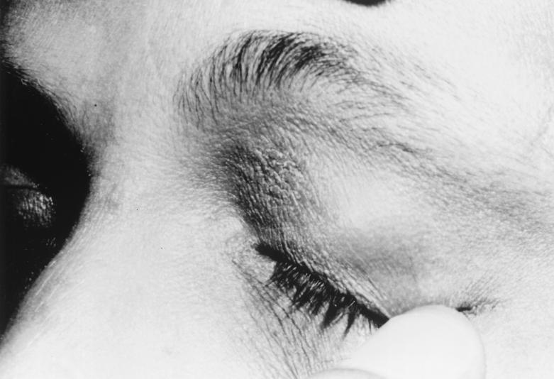
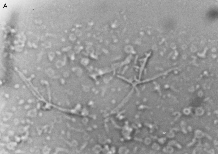
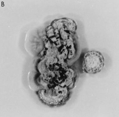
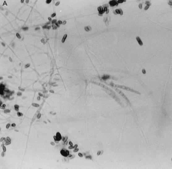
The patient presented with itchy, grey, scaly, eczematoid lesions of 6 months' duration on the skin of both eyelids (Fig. 1). He was seen previously by dermatologists on three different occasions and was put on topical corticosteroids each time. These relieved the itching and quieted the lesions; however, when the treatment course was completed, the symptoms recurred. The patient was healthy otherwise, and the routine chemical and hematological laboratory investigations were within normal limits. Also, his chest X-ray was normal. Skin scrapings from each of the two lesions were collected. Portions of the scrapings were used for direct microscopic examination in 10% KOH and for culture on Sabouraud dextrose agar (SDA) and on SDA supplemented with 50 μg of chloramphenicol ml−1 (Oxoid Ltd., Basingstoke, England), which were incubated at 26 (±1)°C. The direct microscopic examination of the skin scrapings revealed the presence of branched hyphae (Fig. 2A), and the cultures grew an unusual dematiaceous mold which was identified as T. pulvinata (Fig. 2B and 3). Since the organism was very uncommon, repeat skin scrapings from both lesions were taken and their analysis resulted in the same findings by direct microscopy and culture. Therefore, the patient was treated with topical clotrimazole cream (canesten cream containing 0.01 g of clotrimazole/g of cream; Bayer, Leverkusen, Germany) applied to each of the lesions twice daily for 4 weeks. The lesions resolved considerably after the 2nd week, and a full cure was achieved after the 4th week. No relapse of the infection was observed during a 5-year follow-up.
FIG. 1.
Scaly, grey lesion on the skin of the left eyelid of a 30-year-old male.
FIG. 2.
(A) Branched hyphae of T. pulvinata in scrapings of eyelid skin lesions (10% KOH preparation). (B) Two-week-old colony of T. pulvinata on SDA incubated at 26 (±1)°C.
FIG. 3.
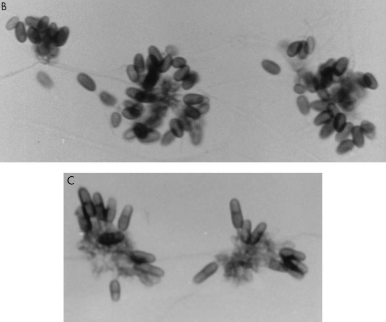
Lactophenol cotton blue mounts of 2-week-old cornmeal agar slide cultures of T. pulvinata incubated at 26 (±1)°C. (A) The three forms of conidia produced by the fungus are (i) brown, oval, and unicellular (upper middle area); (ii) brown, cylindrical, and one-septate (lower middle area); and (iii) hyaline and acicular, with five or six septates (center area). (B) Brown, unicellular conidia. (C) Brown, one-septate conidia.
For identification studies, the isolated fungus was subcultured onto fresh SDA and cornmeal agar media and was incubated at 26 (±1) and 37°C. It grew well on both media and at both temperatures, with moderate growth rates. During the first 3 or 4 days, the colonies appeared creamy and moist. During the next 2 weeks, the centers turned brown and then grey-black as the organism matured (Fig. 2B). Hyphae were hyaline and thin. Some collected in fascicles or coils on which aggregates of hyaline to light-brown, pyriform, sporogenous cells were formed. The fungus produced three forms of conidia: (i) brown, oval, unicellular conidia; (ii) brown, cylindrical, one-septate conidia; and (iii) hyaline, acicular conidia with 5 or 6 septates tapering to a long break (Fig. 3). It is because of these features that the fungus was identified as T. pulvinata (3, 4). The isolates were preserved in our culture collection under the numbers 4289 and 4989.
Examination of the literature did not reveal any previous report of a human infection caused by this fungus. In fact, not only has organism not been implicated in any infection, the published reports on T. pulvinata itself are very limited. It is not familiar to many mycologists. It sporulates readily, but its identification may not be straightforward because of its pleomorphic nature. All species of the genus Triadelphia are pleomorphic, producing at least two forms of conidia from sporogenous cells that agglomerate in a sporodochium-like aggregate (3, 5). No difficulty was encountered in identifying the present isolates because of previous experience with the fungus (2).
It is not clear how the patient acquired his infection. He denied having visited a particular bat site in Saudi Arabia or India, but he occasionally went to farms or moist ecosystem grasses. The case reported herein represents the first human infection caused by T. pulvinata.
Acknowledgments
The contribution to this case by Ibrahim Al-Rabieh from our Dermatology Department is acknowledged.
REFERENCES
- 1.Al-Hedaithy S S A, Leathers C R. Country-wide search in Saudi Arabia for the etiologic agent of histoplasmosis. Proc Saudi Biol Soc. 1987;10:197–207. [Google Scholar]
- 2.Al-Hedaithy S S A, Leathers C R. Presence of Triadelphia pulvinata in Saudi Arabia and its potential pathogenicity. Proc Saudi Biol Soc. 1987;10:209–216. [Google Scholar]
- 3.Constantinescu O, Samson R A. Triadelphia, a pleomorphic genus of Hyphomycetes. Mycotaxon. 1982;15:472–486. [Google Scholar]
- 4.Maggi O, Bartoli A, Rumbelli A. Two new species of Triadelphia from rhizosphere of Loudetia simplex in the Ivory Coast. Trans Br Mycol Soc. 1978;71:148–154. [Google Scholar]
- 5.Shearer C A, Crane J L. Fungi of the Chesapeake Bay and its tributaries. I. Patuxent River. Mycologia. 1971;63:237–260. [Google Scholar]


