
Fig. 1.
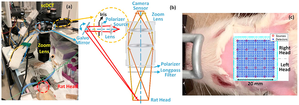
A modified scDCT system for imaging of CBF in rats. (a) Experimental setup for noncontact cerebral imaging of a rat. (b) Optical design of the modified scDCT. An iris was used to limit the size of light spot. (c) Rat head hair was shaved and removed with hair cream to expose a ROI of 20 × 20mm2 on the rat head. 5 × 5 sources (circles) and 21 × 21 detectors (crosses) were evenly distributed within the ROI to acquire boundary flow data for 3D image reconstruction.