Abstract
Background
Clinical outcome after surgery of breast cancer needs more prognostic markers to predict currently. Cluster of differentiation 47 (CD47), due to its overexpression in various tumors and ability to inhibit phagocytosis, has been identified as a new immune checkpoint. Monocarboxylate transporter 1 (MCT1) is a protein involved in the immunomodulatory activities of the tumor microenvironment (TME) by maintaining the pH through aerobic glycolysis.
Methods
We explored the expression of CD47 and MCT1 in breast invasive ductal carcinoma specimens to determine their association with prognosis. A total of 137 breast invasive ductal carcinoma tissues were collected for CD47 and MCT1 immunohistochemical staining.
Results
Statistically analyzed, our study first indicated that in both univariate and multivariate analyses, the coexpression of CD47 and MCT1 was an independent prognostic factor for a poor 10-year overall survival rate (10-OS) and 10-year progression-free survival rate (10-DFS) (P<0.05). In addition, the combined high expression of these two markers also led to worse OS and PFS rates in the TNM (II + III), histologic grade (I + II), HER2 overexpression and basal-like subgroups. High expression of CD47 and MCT1 and combined high expression of CD47 and MCT1 were associated with clinicopathological parameters, such as histological grade, TNM stage, death status, and recurrence status in breast cancer patients. However, in the multivariate survival analysis, high expression of CD47 alone was not an independent prognostic factor for the 10-OS or the 10-DFS (P=0.104; P=0.153), and high expression of MCT1 alone was not an independent predictor for a poor 10-DFS (P=0.177) either.
Conclusions
The coexpression of CD47 and MCT1 can serve as a prognostic biomarker leading to poor survival and an increased risk for recurrence, and this novel information could help guide the development of adjuvant therapy for breast cancer.
Keywords: Cluster of differentiation 47 (CD47), monocarboxylate transporter 1 (MCT1), breast cancer, prognosis
Introduction
Due to the development of adjuvant therapies, the 5-year survival rate of breast cancer patients has increased significantly, and several breast cancer patients survive over 10 years (1). However, recurrence and metastasis are the two biggest in the treatment of breast cancer patients. Interactions between the tumor microenvironment (TME) and cancer cells confer tumor characteristics and represent potential treatment targets. Especially in advanced and specific types of breast cancer, such as basal-like breast cancer which also termed triple-negative breast cancer (TNBC), new prognostic factors and therapeutic strategies can be discovered by detecting new predictive biomarkers.
Immune checkpoint inhibitors targeting programmed cell death protein-1 (PD-1)/programmed cell death ligand 1 (PD-L1) and cytotoxic T lymphocyte-associated antigen-4 (CTLA-4)/B7 have been applied to treat cancer patients (2). Unlike promoting adaptive T cell-mediated immunity as mentioned above, cluster of differentiation 47 (CD47)-mediated therapy to enhance innate immune activity is a new direction for cancer treatment (3). It has been reported that CD47 is widely expressed on the surface of several cancer cells. Signaling regulatory protein α (SIRPα, also termed CD172a or SHPS-1) is another membrane protein of the immunoglobulin superfamily that is particularly overexpressed in macrophages and dendritic cells (DCs). SIRPα expressed on myeloid cells is influenced by CD47 expressed on neighboring cancer cells and leads to the phosphorylation of SIRPα cytoplasmic immunoreceptor tyrosine-based inhibition motifs (ITIM), which results in a decrease in the phagocytosis capacity and clearance function of macrophages (4). The CD47-SIRPα axis indirectly affects the adaptive immune function of cells and participates in T cell activation (5).
Acidic pH value is another characteristic of the TME. High concentrations of lactate contribute significantly to this phenomenon. Lactate transported by monocarboxylate transporters 1 (MCT1) is involved in glucose metabolism in tumor cells. It has been demonstrated that lactate plays a pivotal role in tumor immune surveillance, T cell differentiation and macrophage polarization (6). Macrophages in the TME have been divided into two types based on their different functions. M1 macrophages have tumoricidal effects and can inhibit tumor growth, whereas M2 macrophages participate in promoting tissue repair, suppressing the immune response, and angiogenesis, leading to poor patient prognosis in cancer (7). MCT1 is an important molecule that stimulates the IL-6/IL-6R/Stat3 axis and enhances M2 polarization in breast cancer (8,9). Additionally, utilizing 3-BrPA (a glycolysis inhibitor) to treat glioma suppressed the expression of MCT1 and regulated the progression of malignantly transformed macrophages (10). Type-I interferons (IFNs), such as IFN-α, IFN-β and others, are influenced by lactate and play an important role in elevating host immunity for cancer immune surveillance. Maintaining intracellular and extracellular lactate concentrations of MCT1 directly decreased type-I IFNs and promoted an immunosuppressive TME (6). In TNBC, MCT1 reduced miR-34a and induced the expression of PD-L1 (8). In addition, MCT1 also participates in maintaining the epithelial-mesenchymal transition (EMT) phenotype by promoting the acidic environment, underscoring its importance in the invasion and progression of malignant tumors (8,11).
High expression of CD47 and MCT1 in invasive breast cancer might regulate the characteristics of the TME and affect tumor invasion and metastasis by maintaining the proinflammatory nature and EMT phenotype of tumor cells. In this study, we evaluated the expression of CD47 and MCT1 in 137 breast cancer tissues and showed for the first time that the coexpression of CD47 and MCT1 in breast cancer is potentially a better independent predictor for poor prognosis than the expression of CD47 or MCT1 alone. Therefore, our study provides a new potential immune target for breast cancer treatment.
We present the following article in accordance with the REMARK reporting checklist (available at https://tcr.amegroups.com/article/view/10.21037/tcr-21-1951/rc).
Methods
Tissue samples
This array consisted of 137 tissue samples of primary invasive breast cancer without any treatment before surgery, and all the tested samples were from surgical resection. Exclusion criteria: (I) patients with other malignant tumors; (II) patients with special types of breast cancer (medullary carcinoma, cribriform carcinoma, mucinous colloid breast carcinoma, pleomorphic lobular breast carcinoma, papillary breast carcinoma); (III) patients with severe heart disease (such as dilated cardiomyopathy, after coronary artery bypass graft surgery and valvular surgery). During admission, the following variables were considered: age, gender, definite TNM stage, surgery information, pathological information and imaging data.
The time of surgery for all the patients are between January 2005 and September 2009. Neoadjuvant chemotherapy was not available in our study, and patients with invasive breast cancer received chemotherapy after surgery, the chemotherapy regimens [doxorubicin (DOX)/epidoxorubicin (EPI) + chlorotoxin (CTX) or DOX/EPI + CTX − DTX] and doses were given in accordance with NCCN guidelines. Patients with positive lymph nodes and high risk of recurrence and metastasis were treated with radiotherapy. The criteria for recurrence of the patients based on imaging findings. All the tissue samples were selected from the medical records of the Department of Pathology, Shanghai East Hospital of Tongji University. The average patient age was 55.9 years (range, 29–87 years). The patients’ clinicopathological data were collected including sex, age, tumor node metastasis (TNM) stage (AJCC 6 version), number of positive lymph nodes, histologic grade, estrogen receptor (ER), progesterone receptor (PR), human epidermal growth factor receptor 2 (HER2), recurrence, and survival status.
All the patients followed up for almost 10 years, and the follow-up data was defined as the time between the date of the first diagnosis and the date of death or last follow-up time. Among the dead cases during the 10 years of follow-up, 1 patient died from drug complications and the others died as the result of tumor progression. With respect to the expression of hormone receptor, immunostaining results were considered positive when >5% cells staining positive for both ER and PR status. For HER2 overexpression, the positive status was the current guideline of 3+ staining by immunohistochemistry or fluorescent in situ hybridization (FISH) positive. All tissue samples were selected from the medical records of the Department of Pathology at Shanghai East Hospital of Tongji University.
The study was conducted in accordance with the Declaration of Helsinki (as revised in 2013). The study was approved by the Ethics Committee of Shanghai East Hospital ([2020] Preliminary Examination No. 093) and informed consent was taken from all patients.
Immunohistochemical staining and evaluation
CD47 (A1838, 1:200 dilutions; ABclonal, Wuhan, China) and MCT1 (ab90582, 1:200 dilutions; Abcam, Cambridge, UK) were measured using immunohistochemical staining as described previously (12). Each slide was independently reviewed by two senior pathologists, and any disagreements were resolved by consensus. Pathologists were totally blinded to the clinicopathological data and patient status.
A semiquantitative scoring system was applied to calculate the positive percentage and intensity of tumor cells. The average percentage of positive tumor cells was determined in 5 regions of a given tissue. The magnification was ×400, and the score ranged from 0–1 (0–100%). The staining intensity was scored as negative (−/0), weak positive (+/1), positive (++/2) and strong positive (+++/3). Therefore, each sample value was generated by a weighted score ranging from 0 to 3. Meanwhile, a score ≥0.8 was defined as high expression.
Statistical analysis
All data analysis and image generation were performed using SPSS 22.0 and GraphPad Prism 8.0. Differences were considered significant when the P value was <0.05.
Results
Baseline patient characteristics and immunohistochemical staining for CD47 and MCT1 in breast cancer samples
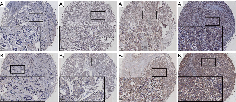
We included 140 individuals diagnosed with breast cancer at Shanghai East Hospital of Tongji University between January 2005 and September 2009. Three patients were lost to follow-up, and several patients had incomplete pathological information (4 with ER, 3 with PR and 4 with HER2). The clinicopathological characteristics of the patients are shown in Table 1. We collected data on TNM stage, and 33 patients were stage I, 60 were stage II, 44 were stage III, and none were stage IV. There were 66 patients who were lymph node negative and 61 who were lymph node positive. Thirty-seven patients experienced recurrence during the follow-up period. The ER, PR, and HER2 status of each patient is shown in Table 1. Immunohistochemical staining for CD47 and MCT1 is shown in Figure 1. Both CD47 and MCT1 were localized to the cytoplasm and cell membrane of breast cancer cells. Considering that membrane expression is mainly for function, all the analyses described were only based on membrane expression.
Table 1. Association between CD47, MCT1 or CD47-CD68 expression and various clinicopathological variables in breast cancer.
| Clinicopathological characteristics | N | CD47 | MCT1 | CD47-MCT1 | ||||||||
|---|---|---|---|---|---|---|---|---|---|---|---|---|
| High expression (%) | χ2 | P | High expression (%) | χ2 | P | High expression (%) | χ2 | P | ||||
| Age (years) | 0.019 | 0.890 | 0.727 | 0.416 | 0.325 | 0.584 | ||||||
| <50 | 43 | 60.5% | 67.4% | 51.2% | ||||||||
| ≥50 | 94 | 61.7% | 74.5% | 56.4% | ||||||||
| Histological grade | 14.270 | ≤0.001* | 12.172 | 0.001* | 18.757 | ≤0.001* | ||||||
| G1–2 | 96 | 51.0% | 63.5% | 42.7% | ||||||||
| G3 | 41 | 85.4% | 92.7% | 82.9% | ||||||||
| TNM stage | 8.135 | 0.017* | 6.459 | 0.044* | 8.632 | 0.013* | ||||||
| I | 33 | 66.7% | 66.7% | 57.6% | ||||||||
| II | 60 | 48.3% | 65.0% | 41.7% | ||||||||
| III | 44 | 75.0% | 86.4% | 70.5% | ||||||||
| ER status | 0.157 | 0.706 | 0.029 | 1.000 | 0.462 | 0.573 | ||||||
| Negative | 41 | 63.4% | 73.2% | 58.5% | ||||||||
| Positive | 92 | 59.8% | 71.7% | 52.2% | ||||||||
| PR status | 0.016 | 1.000 | 0.068 | 0.848 | 0.332 | 0.605 | ||||||
| Negative | 64 | 62.5% | 73.4% | 57.8% | ||||||||
| Positive | 70 | 61.4% | 71.4% | 52.9% | ||||||||
| HER2 status | 0.155 | 0.817 | 0.026 | 1.000 | 0.045 | 1.000 | ||||||
| Negative | 109 | 62.4% | 72.5% | 56.0% | ||||||||
| Positive | 24 | 66.7% | 70.8% | 58.3% | ||||||||
| Lymph node metastasis | 1.252 | 0.278 | 2.295 | 0.165 | 2.444 | 0.153 | ||||||
| No | 66 | 57.6% | 70.2% | 48.5% | ||||||||
| Yes | 61 | 67.2% | 87.5% | 62.3% | ||||||||
| Death | 4.419 | 0.049* | 7.447 | 0.008* | 8.064 | 0.005* | ||||||
| No | 109 | 56.9% | 67.0% | 70.7% | ||||||||
| Yes | 28 | 78.6% | 92.9% | 78.6% | ||||||||
| Recurrence | 4.408 | 0.048* | 0.295 | 0.671 | 4.932 | 0.033* | ||||||
| No | 100 | 56.0% | 71.0% | 49.0% | ||||||||
| Yes | 37 | 75.7% | 75.7% | 70.3% | ||||||||
*, statistically significant. TNM, tumor node metastasis; ER, estrogen receptor; PR, progesterone receptor; HER2, human epidermal growth factor receptor 2; CD47, cluster of differentiation 47; MCT1, monocarboxylate transporter 1.
Figure 1.
Immunohistochemical staining for CD47 and MCT1 in tissues (magnification, ×400; scale bar: 50 µm). (A) CD47 immunoreactivity in invasive ductal carcinoma: (A1) patient 1: score =0 (staining negative) × 100% =0; (A2) patient 2: score =1 (staining weak) ×100% =1; (A3) patient 3: score =2 (staining moderate) ×100% =2; (A4) patient 4: score =3 (staining strong) ×100% =3. (B) MCT1 immunoreactivity in invasive ductal carcinoma: (B1) patient 5: score =0 (staining negative) ×100% =0; (B2) patient 6: score =1 (staining weak) ×100% =1; (B3) patient 7: score =2 (staining moderate) ×100% =2; (B4) patient 8: score =3 (staining strong) ×100% =3. CD47, cluster of differentiation 47; MCT1, monocarboxylate transporter 1.
Expression of CD47 and its prognostic value in breast cancer
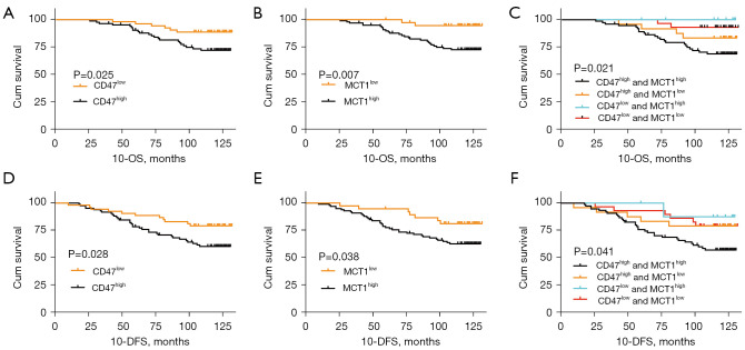
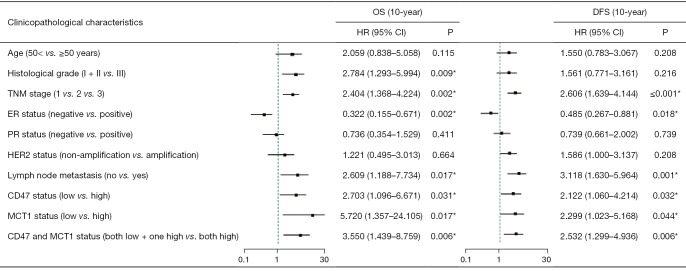
Considering the important role of CD47 in cancer progression, the expression in invasive ductal carcinoma (IDC) tissue was explored by immunohistochemistry. Immunohistochemical staining revealed that 61.3% (n=84) of the IDC samples had high expression of CD47 and 38.7% (n=53) had low expression. Additionally, further analysis showed a significant correlation between high CD47 expression and histological grade (χ2=14.270, P≤0.001), TNM stage (χ2=8.135, P=0.017), death status (χ2=4.419, P=0.049), and recurrence status (χ2=4.408, P=0.048) (Table 1). These results suggest that CD47 may be a prognostic factor for IDC. Furthermore, the Kaplan-Meier analysis and log-rank test demonstrated that the expression of CD47 was associated with shorter 10-year overall survival rate (10-OS, P=0.025) and 10-year disease-free survival rate (10-DFS, P=0.028) (Figure 2). The univariate analysis showed that CD47 was a poor prognostic factor of survival and recurrence in breast cancer patients [10-OS, hazard ratio (HR) =2.703, P=0.031; 10-DFS, HR =2.122, P=0.032] (Figure 3). The results of the multivariate analysis conducted using the Cox proportional hazard model demonstrated that CD47 is not an independent prognostic risk factor for either OS (HR =2.285, P=0.104) or DFS (HR =1.711, P=0.153) (Figure 4).
Figure 2.
Kaplan-Meier survival analysis for 10-OS and 10-DFS of breast cancer patients based on CD47 expression (A,D), MCT1 expression (B,E), and CD47-MCT1 expression (C,F). P values were obtained using the log-rank test. OS, overall survival; DFS, disease-free survival. 10-OS, 10-year overall survival rate; 10-DFS, 10-year progression-free survival rate; CD47, cluster of differentiation 47; MCT1, monocarboxylate transporter 1.
Figure 3.
Univariate analysis of variables associated with overall survival and disease-free survival in patients with breast cancer. *, statistically significant. OS, overall survival; DFS, disease-free survival; TNM, tumor node metastasis; ER, estrogen receptor; PR, progesterone receptor; HER2, human epidermal growth factor receptor 2; CD47, cluster of differentiation 47; MCT1, monocarboxylate transporter 1.
Figure 4.
Multivariate analysis of variables associated with OS and DFS in breast cancer patients with CD47 status. *, statistically significant. OS, overall survival; DFS, disease-free survival; TNM, tumor node metastasis; ER, estrogen receptor; PR, progesterone receptor; HER2, human epidermal growth factor receptor 2.
Expression of MCT1 and its prognostic value in breast cancer
The rate of high MCT1 expression measured by immunohistochemistry in IDC samples was 72.3% (n=99), and the rate of low MCT1 expression was 27.7% (n=38). Moreover, the expression of MCT1 was significantly associated with histological grade (χ2=12.172, P=0.001), advanced TNM stage (χ2=6.459, P=0.044) and death status (χ2=7.447, P=0.008), which was similar to that observed for CD47 (Table 1). Kaplan-Meier analysis and log-rank tests showed that overexpression of MCT1 was strongly associated with reduced 10-year OS (P=0.007) and DFS (P=0.038) (Figure 2). The univariate analysis indicated that MCT1 expression (10-OS, HR =5.720, P=0.017; 10-DFS, HR =2.299, P=0.044), histological grade, TNM stage, ER status, lymph node status and CD47 expression were independent prognostic factors for predicting survival and recurrence rates in IDC patients (Figure 3). According to the multivariate analysis conducted using the Cox proportional hazard model, high expression of MCT1 was only a poor prognostic factor for the 10-OS rate (HR =5.720, P=0.047) and was not an independent predictor of a poor 10-DFS rate (HR =1.779, P=0.177) (Figure 5).
Figure 5.
Multivariate analysis of variables associated with OS and DFS in breast cancer patients with MCT1 status. *, statistically significant. OS, overall survival; DFS, disease-free survival; TNM, tumor node metastasis; ER, estrogen receptor; PR, progesterone receptor; HER2, human epidermal growth factor receptor 2; MCT1, monocarboxylate transporter 1.
Expression of CD47-MCT1 and its prognostic value in breast cancer
CD47 protein has been reported to be highly expressed on cancer cells and to inhibit the phagocytic activity of macrophages, and MCT1 polarizes M1 macrophages to the M2 phenotype. Thus, we hypothesized that the expression of these two proteins promotes the immune escape of cancer cells, and a significant correlation was found between the expression of CD47 and MCT1 (γ=0.733, P≤0.001). The percentage of combined high expression of CD47 and MCT1 (CD47highMCT1high expression) was 54.7% (n=75), percentage of combined low expression of CD47 and MCT1 (CD47lowMCT1low expression) was 21.2% (n=29), percentage of CD47highMCT1low expression was 6.6% (n=9) and percentage of CD47lowMCT1high expression was 17.5% (n=24). Moreover, high expression of CD47 and MCT1 was significantly associated with histological grade (χ2=18.757, P≤0.001), advanced TNM stage (χ2=8.632, P=0.013), death status (χ2=8.064, P=0.005) and recurrence status (χ2=4.932, P=0.033) (Table 1). Kaplan-Meier analysis and log-rank tests suggested that combined high expression of CD47 and MCT1 is associated with the prognosis of breast cancer patients (10-OS, P=0.021; 10-DFS, P=0.041) (Figure 2). According to the univariate analysis, patients with CD47highMCT1high expression had worse 10-OS (HR =3.550, P=0.006) and 10-DFS (HR =2.532, P=0.006) clinical outcomes (Figure 3). The multivariate analysis revealed that CD47highMCT1high expression was also an independent predictive factor for poor survival rates and disease recurrence (10-OS, HR =3.104, P=0.026; 10-DFS, HR =2.176, P=0.033) (Figure 6).
Figure 6.
Multivariate analysis of variables associated with OS and DFS in breast cancer patients with CD47-MCT1 status. *, Statistically significant. OS, overall survival; DFS, disease-free survival; TNM, tumor node metastasis; ER, estrogen receptor; PR, progesterone receptor; HER2, human epidermal growth factor receptor 2; CD47, cluster of differentiation 47; MCT1, monocarboxylate transporter 1.
Coexpression of CD47 and MCT1 and its prognostic value in different subgroups of breast cancer
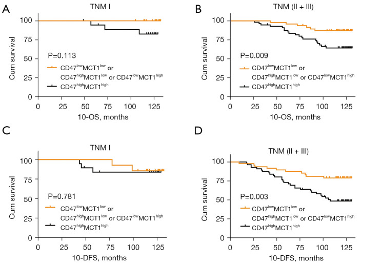
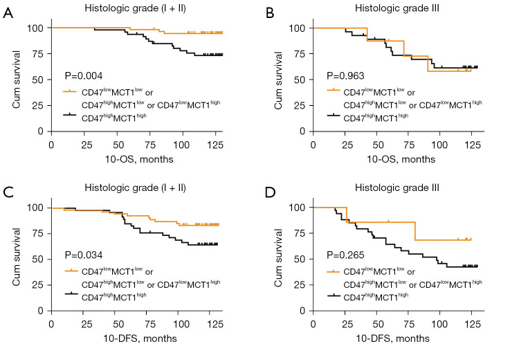
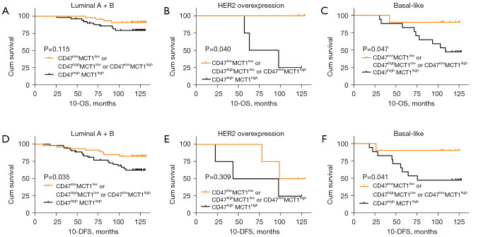
As shown in Table 1, there was a significant difference in the expression of CD47 and MCT1 at different histological grades and TNM stages. Moreover, multivariate analysis by the Cox proportional hazard model with CD47highMCT1high expression suggested that ER status was a protective factor for breast cancer prognosis (10-OS, HR =0.379, P=0.039; 10-DFS, HR =0.479, P=0.047) (Figure 6). To explore the influence of CD47highMCT1high expression in different subgroups of breast cancer, survival curves were plotted by Kaplan-Meier analysis. We detected TNM stage separately by Kaplan-Meier analysis, the result showed no significant difference in single group (TNM I, 10-OS, P=0.113,10-DFS, P=0.781; TNM II, 10-OS, P=0.083,10-DFS, P=0.082; TNM III 10-OS, P=0.229, 10-DFS, P=0.387;) (Figure S1). Considered TNM (I + II) was usually defined as early-stage, CD47highMCT1high expression in different clinical stages, TNM stages were regrouped. In the TNM (I + II) group, CD47highMCT1high expression was associated with poor OS (10-OS, P=0.032; 10-DFS, P=0.134) (Figure S2). To further explored the prognosis of CD47highMCT1high expression in TNM stages, we regroup TNM stages as TNM I vs. TNM (II + III), and the prognosis of patients in TNM (II + III) group was significantly different from that of the other groups (10-OS, P=0.009; 10-DFS, P=0.003) (Figure 7). Similar results were found in the histologic grade (I + II) subgroup (10-OS, P=0.004; 10-DFS, P=0.034) (Figure 8). In regard to basal-like breast cancer, CD47highMCT1high expression correlated with worse 10-OS (P=0.047) and 10-DFS (P=0.041) clinical outcomes. In the HER2 overexpression group (n=8), coexpression of CD47 and MCT1 was associated with poorer OS rates (P=0.040) but had no significant correlation with 10-DFS outcomes (P=0.309) (Figure 9). In the luminal (A+B) subgroup, CD47highMCT1high expression was associated with poor 10-DFS outcomes (P=0.035) but not with 10-OS outcomes (P=0.115).
Figure 7.
Kaplan-Meier survival analysis for different TNM stage breast cancer patients according to CD47-MCT1 expression. (A,C) 10-OS and 10-DFS in TNM I breast cancer patients. (B,D) 10-OS and 10-DFS in TNM (II + III) breast cancer patients. P values were obtained using the log-rank test. TNM, tumor node metastasis; OS, overall survival; DFS, disease-free survival; 10-OS, 10-year overall survival rate; 10-DFS, 10-year progression-free survival rate; CD47, cluster of differentiation 47; MCT1, monocarboxylate transporter 1.
Figure 8.
Kaplan-Meier survival analysis for different histologic stage breast cancer patients according to CD47-MCT1 expression. (A,C) 10-OS and 10-DFS in histologic grade (I + II) breast cancer patients. (B,D) 10-OS and 10-DFS in histologic grade III breast cancer patients. P values were obtained using the log-rank test. OS, overall survival; DFS, disease-free survival; 10-OS, 10-year overall survival rate; 10-DFS, 10-year progression-free survival rate; CD47, cluster of differentiation 47; MCT1, monocarboxylate transporter 1.
Figure 9.
Kaplan-Meier survival analysis for different molecular subtypes of breast patients according to CD47-MCT1 expression. (A,D) 10-OS and 10-DFS in luminal (A+B) breast cancer patients. (B,E) 10-OS and 10-DFS in HER2 overexpression breast cancer patients. (C,F) 10-OS and 10-DFS in basal like breast cancer patients. P values were obtained using the log-rank test. OS, overall survival; DFS, disease-free survival; 10-OS, 10-year overall survival rate; 10-DFS, 10-year progression-free survival rate; HER2, human epidermal growth factor receptor 2; CD47, cluster of differentiation 47; MCT1, monocarboxylate transporter 1.
Discussion
Breast IDC is the leading cause of cancer-related deaths among women (13). Once recurrence and metastases occur, the survival time of breast cancer patients is substantially shortened (14,15). Many patients experience metastasis after radical surgery and drug therapy, and new prognostic factors for breast cancer urgently need to be identified.
Macrophages of the innate immune system phagocytose cancer cells in the TME as one of the first lines of defense against cancer. CD47 has been widely reported to be associated with several cancers, participates in the inhibition of phagocytosis and shortens PFS and OS rates (16-18). There are three main mechanisms by which CD47 is involved in regulating the tumor immune microenvironment. Targeting the CD47-SIRPα axis to remove “do not eat me” signals to reduce immune evasion is one of the main treatment strategies (19). Furthermore, a CD47 inhibitor has been found to promote the capacity of DCs to phagocytize and subsequently present antigens to CD4+ and CD8+ T cells, resulting in an antitumor adaptive immune response (19). Additionally, CD47 activates the innate immune response mediated by antibody-dependent cellular cytotoxicity (ADCC) (20). In TNBC, CD47 has been found to be an independent prognostic factor poor for 5-year DFS outcomes (21). In addition, combined with CD68 expression on macrophages, high expression of CD47 is associated with poor prognosis among breast cancer patients (17). To further estimate the effect of high expression of CD47 on long-term survival outcomes, 137 breast cancer samples were immunohistochemically stained and statistically analyzed to determine the association between CD47 expression levels and 10-OS and 10-DFS rates. In our study, high expression of CD47 significantly reduced 10-OS and 10-DFS rates among breast cancer patients. However, the multivariate analysis indicated that CD47 was not an independent prognostic risk predictor (OS, HR =2.285, P=0.104; DFS, HR =1.711, P=0.153), which is consistent with the findings of other studies. These results suggest that the mechanism by which CD47 promotes breast cancer progression is complex.
According to the Warburg effect, tumor cells that proliferate in hypoxic conditions stimulate glycolysis. MCT proteins are involved in the metabolism of all cells, yet tumor cells or tissues become more dependent on the monocarboxylate transport pathway to obtain energy under hypoxic or ischemic conditions by metabolic symbiosis (19). MCT1 in particular helps tumor cells use lactate as an energy source for glycolysis when oxygen is sufficient. BT474, SKBR3 and MCF cells have been utilized by Hou et al. to demonstrate that upregulation of lactate dehydrogenase-A (LDHA) and MCT1 promotes Taxol resistance and thus mediates breast cancer progression (22). It has also been reported that MCT1 is highly overexpressed in TNBC, and high MCT1 expression level has been found to be associated with a high recurrence rate (HR =2.26, P=0.024) in all breast cancer types (23). In our research, Kaplan-Meier analysis and log-rank tests also showed that overexpression of MCT1 had a strong correlation with poor OS and DFS outcomes in all breast cancer types. However, the multivariate analysis showed that high expression of MCT1 was not an independent predictor of poor DFS outcomes (HR =1.779, P=0.177). We consider that this result is due to multifactorial correction in the Cox multivariate model.
Our findings indicate that combined high expression of CD47 and MCT1 alone is not an independent predictor of poor DFS outcomes, and CD47 expression alone is not an independent predictor of poor OS outcomes. Nevertheless, combined high expression of CD47 and MCT1 served as a significant independent factor for poor OS rates and disease recurrence (10-OS, HR =3.104, P=0.026; 10-DFS, HR =2.176, P=0.033). To understand this phenomenon, conjectural interactions between CD47 and MCT1 were investigated further. The immunosuppressive nature of the local TME was considered first. M2 phenotype macrophages are influenced by microenvironment stimulation and are known to exhibit anti-inflammatory and protumoral effects and are involved in tumor angiogenesis and matrix remodeling (24,25). In the TME, growth factors and chemokines secreted by the stroma and tumor cells recruit peripheral blood monocytes to the tumor site (26). Macrophage colony stimulating factor (MCSF) secreted by cancer cells increases tumor-associated macrophage (TAM) recruitment and induces the M2 phenotype by binding to the tyrosine kinase CSF-receptor 1 (CSF-1R) expressed on monocytes and macrophages (27). M2 macrophages with higher IL-10 and tumor growth factor β (TGF-β) expression levels are usually induced by the IL-6/STAT3 axis to promote cancer metastasis (28). In lung cancer cells, the USP24-p300-NF-κB signaling pathway has been reported to increase IL-6 transcription in M2 macrophages (29). Another important role of macrophages in the TME is lactic acid produced by tumor cells. MCT1 is a protein involved in regulating the release and uptake of lactic acid and changing the pH of the microenvironment (8,30). MCT1 can via hypoxia-inducible factor 1 (HIF-1) maintains the acidity of the TME and is sufficient to induce vascular endothelial growth factor (VEGF) and arginase 1 (ARG1) expressed on TAMs (31,32). VEGF is well known to promote neovascularization and the subsequent growth of tumors (33). ARG1 is known as a marker of M2 macrophages and possibly plays a critical role in tumor proliferation by producing polyamines (26). In the TNBC cell line, MCT1 is overexpressed and secretes PD-L1, IL-6, CCL2 and MCSF to promote the polarization of M2 macrophages (8). The high expression level of MCT1 on cancer cells results in an increase in IL-6 expression through the YY1-EGFR signaling pathway (8). As mentioned above, MCT1 enables tumor cells to survive in hypoxia and maintains the acidic pH of the TME. Continuous hypoxic conditions can induce the production of key monocyte recruitment factors, including CCL2, CCL5, CXCL12, CSF-1, and VEGF, by tumor cells to recruit TAMs into hypoxic regions (34). It has been reported that in hypoxic breast cancer cells, HIF-1 directly activates the transcription of CD47 (35). Genetic or pharmacological inhibition of HIFs blocked the enrichment of CD47+CD73+PD-L1+ TNBC cells (36). Based on these findings, MCT1 likely provides a suitable condition for TAM recruitment and M2 polarization, and the increased expression of CD47 further inhibits phagocytosis of tumor cells in a hypoxic environment. Furthermore, the high expression of PD-L1 in cancer cells downregulates the cytotoxicity of activated T cells. Both mouse and human TAMs express PD-1, and PD-1 interaction with PD-L1 on cancer cells decreases macrophage phagocytic capabilities (37).
It is also important to note that effluent lactic acid in the TME is responsible for the overexpression of hyaluronic acid, TGF-β and CD44, which are cytokines that activate EMT (38,39). TAMs can activate Nrf2 in cancer cells, in turn increasing EMT through paracrine VEGF (40). Cancer cells with an EMT-activated phenotype may exhibit higher expression of CD47 as its proximal promoter directs the binding of SNAI1 and ZEB1 (41). Furthermore, CD47 has been reported to induce EMT by modulating N-cadherin and E-cadherin in ovarian cancer (42). This complex process ultimately leads to an immunosuppressive microenvironment and maintains the EMT phenotype, which may explain why combined high expression of CD47 and CD68 might serve as a prognostic risk factor in breast cancer.
There may be some deficiencies in our study. (I) Although the effect of the interaction between CD47 and MCT1 in breast cancer was speculated, this mechanism needs to be explored further. (II) Both MCT1 and CD47 participate in the maintenance of the stem cell phenotype and EMT, but whether these two markers interact with each other is unknown. In addition to the immune mechanism, the relationship between the two molecules in other pathways also needs to be detected. (III) Our research suggested that CD47highMCT1high expression is an independent factor for poor prognosis. However, we verified this result only at the histological level, thus cytological studies are still needed to confirm it. (IV) In our further analysis, we explored the coexpression of CD47 and MCT1 and its prognostic value in different subgroups of breast cancer. Due to the limited number of patients, there were only 8 samples in the HER2 overexpression subgroup, and the expression of CD47 and MCT1 in this group needs to be verified further. Luminal A and B were not grouped independently because of the lack of Ki-67 status information. CD47highMCT1high expression was associated with poor DFS outcomes in the ER-negative group, suggesting that these two molecules may affect hormone levels to promote the recurrence of breast cancer, which needs further study.
Conclusions
In conclusion, our study provided definitive evidence for the prognostic value of CD47 and MCT1 in breast cancer. In comparison with the expression of CD47 or MCT1 alone, the combined high expression of CD47 and MCT1 may be a better independent factor for poor patient prognosis and disease recurrence, particularly in the TNM (II + III), histologic grade (I + II) and basal-like subgroups. Additionally, CD47 and MCT1 coexpression might be a new immune microenvironment therapeutic target for breast cancer.
Acknowledgments
Funding: This work was financially supported by the Science and Technology Commission of Shanghai Municipality (No. 14DZ2261100, 15DZ1940106), the Joint Project of Health and Family Planning Committee of Pudong New Area (No. PW2017D-10), and the National Natural Science Foundation of China (No. 81860547, 81573008, 81703075).
Ethical Statement: The authors are accountable for all aspects of the work in ensuring that questions related to the accuracy or integrity of any part of the work are appropriately investigated and resolved. The study was conducted in accordance with the Declaration of Helsinki (as revised in 2013). The study was approved by the Ethics Committee of Shanghai East Hospital ([2020] Preliminary Examination No. 093) and informed consent was taken from all patients.
Footnotes
Reporting Checklist: The authors have completed the REMARK reporting checklist. Available at https://tcr.amegroups.com/article/view/10.21037/tcr-21-1951/rc
Data Sharing Statement: Available at https://tcr.amegroups.com/article/view/10.21037/tcr-21-1951/dss
Conflicts of Interest: All authors have completed the ICMJE uniform disclosure form (available at https://tcr.amegroups.com/article/view/10.21037/tcr-21-1951/coif). The authors have no conflicts of interest to declare.
References
- 1.Wu C, Li M, Meng H, et al. Analysis of status and countermeasures of cancer incidence and mortality in China. Sci China Life Sci 2019;62:640-7. 10.1007/s11427-018-9461-5 [DOI] [PubMed] [Google Scholar]
- 2.Emens LA. Breast Cancer Immunotherapy: Facts and Hopes. Clin Cancer Res 2018;24:511-20. 10.1158/1078-0432.CCR-16-3001 [DOI] [PMC free article] [PubMed] [Google Scholar]
- 3.Liu X, Kwon H, Li Z, et al. Is CD47 an innate immune checkpoint for tumor evasion? J Hematol Oncol 2017;10:12. 10.1186/s13045-016-0381-z [DOI] [PMC free article] [PubMed] [Google Scholar]
- 4.Barclay AN, Van den Berg TK. The interaction between signal regulatory protein alpha (SIRPα) and CD47: structure, function, and therapeutic target. Annu Rev Immunol 2014;32:25-50. 10.1146/annurev-immunol-032713-120142 [DOI] [PubMed] [Google Scholar]
- 5.Cham LB, Torrez Dulgeroff LB, Tal MC, et al. Immunotherapeutic Blockade of CD47 Inhibitory Signaling Enhances Innate and Adaptive Immune Responses to Viral Infection. Cell Rep 2020;31:107494. 10.1016/j.celrep.2020.03.058 [DOI] [PMC free article] [PubMed] [Google Scholar]
- 6.Zhang W, Wang G, Xu ZG, et al. Lactate Is a Natural Suppressor of RLR Signaling by Targeting MAVS. Cell 2019;178:176-189.e15. 10.1016/j.cell.2019.05.003 [DOI] [PMC free article] [PubMed] [Google Scholar]
- 7.Klichinsky M, Ruella M, Shestova O, et al. Human chimeric antigen receptor macrophages for cancer immunotherapy. Nat Biotechnol 2020;38:947-53. 10.1038/s41587-020-0462-y [DOI] [PMC free article] [PubMed] [Google Scholar]
- 8.Weng YS, Tseng HY, Chen YA, et al. MCT-1/miR-34a/IL-6/IL-6R signaling axis promotes EMT progression, cancer stemness and M2 macrophage polarization in triple-negative breast cancer. Mol Cancer 2019;18:42. 10.1186/s12943-019-0988-0 [DOI] [PMC free article] [PubMed] [Google Scholar]
- 9.Zhang J, Muri J, Fitzgerald G, et al. Endothelial Lactate Controls Muscle Regeneration from Ischemia by Inducing M2-like Macrophage Polarization. Cell Metab 2020;31:1136-1153.e7. 10.1016/j.cmet.2020.05.004 [DOI] [PMC free article] [PubMed] [Google Scholar]
- 10.Sheng Y, Jiang Q, Dong X, et al. 3-Bromopyruvate inhibits the malignant phenotype of malignantly transformed macrophages and dendritic cells induced by glioma stem cells in the glioma microenvironment via miR-449a/MCT1. Biomed Pharmacother 2020;121:109610. 10.1016/j.biopha.2019.109610 [DOI] [PubMed] [Google Scholar]
- 11.Zhang G, Zhang Y, Dong D, et al. MCT1 regulates aggressive and metabolic phenotypes in bladder cancer. J Cancer 2018;9:2492-501. 10.7150/jca.25257 [DOI] [PMC free article] [PubMed] [Google Scholar]
- 12.Song Z, Feng C, Lu Y, et al. PHGDH is an independent prognosis marker and contributes cell proliferation, migration and invasion in human pancreatic cancer. Gene 2018;642:43-50. 10.1016/j.gene.2017.11.014 [DOI] [PubMed] [Google Scholar]
- 13.Bray F, Ferlay J, Soerjomataram I, et al. Global cancer statistics 2018: GLOBOCAN estimates of incidence and mortality worldwide for 36 cancers in 185 countries. CA Cancer J Clin 2018;68:394-424. 10.3322/caac.21492 [DOI] [PubMed] [Google Scholar]
- 14.Sopik V, Sun P, Narod SA. Predictors of time to death after distant recurrence in breast cancer patients. Breast Cancer Res Treat 2019;173:465-74. 10.1007/s10549-018-5002-9 [DOI] [PubMed] [Google Scholar]
- 15.Tellez-Gabriel M, Knutsen E, Perander M. Current Status of Circulating Tumor Cells, Circulating Tumor DNA, and Exosomes in Breast Cancer Liquid Biopsies. Int J Mol Sci 2020;21:9457. 10.3390/ijms21249457 [DOI] [PMC free article] [PubMed] [Google Scholar]
- 16.Folkes AS, Feng M, Zain JM, et al. Targeting CD47 as a cancer therapeutic strategy: the cutaneous T-cell lymphoma experience. Curr Opin Oncol 2018;30:332-7. 10.1097/CCO.0000000000000468 [DOI] [PMC free article] [PubMed] [Google Scholar]
- 17.Yuan J, He H, Chen C, et al. Combined high expression of CD47 and CD68 is a novel prognostic factor for breast cancer patients. Cancer Cell Int 2019;19:238. 10.1186/s12935-019-0957-0 [DOI] [PMC free article] [PubMed] [Google Scholar]
- 18.Arrieta O, Aviles-Salas A, Orozco-Morales M, et al. Association between CD47 expression, clinical characteristics and prognosis in patients with advanced non-small cell lung cancer. Cancer Med 2020;9:2390-402. 10.1002/cam4.2882 [DOI] [PMC free article] [PubMed] [Google Scholar]
- 19.Liu X, Pu Y, Cron K, et al. CD47 blockade triggers T cell-mediated destruction of immunogenic tumors. Nat Med 2015;21:1209-15. 10.1038/nm.3931 [DOI] [PMC free article] [PubMed] [Google Scholar]
- 20.Zhang X, Chen W, Fan J, et al. Disrupting CD47-SIRPα axis alone or combined with autophagy depletion for the therapy of glioblastoma. Carcinogenesis 2018;39:689-99. 10.1093/carcin/bgy041 [DOI] [PubMed] [Google Scholar]
- 21.Yuan J, Shi X, Chen C, et al. High expression of CD47 in triple negative breast cancer is associated with epithelial-mesenchymal transition and poor prognosis. Oncol Lett 2019;18:3249-55. 10.3892/ol.2019.10618 [DOI] [PMC free article] [PubMed] [Google Scholar]
- 22.Hou L, Zhao Y, Song GQ, et al. Interfering cellular lactate homeostasis overcomes Taxol resistance of breast cancer cells through the microRNA-124-mediated lactate transporter (MCT1) inhibition. Cancer Cell Int 2019;19:193. 10.1186/s12935-019-0904-0 [DOI] [PMC free article] [PubMed] [Google Scholar]
- 23.Johnson JM, Cotzia P, Fratamico R, et al. MCT1 in Invasive Ductal Carcinoma: Monocarboxylate Metabolism and Aggressive Breast Cancer. Front Cell Dev Biol 2017;5:27. 10.3389/fcell.2017.00027 [DOI] [PMC free article] [PubMed] [Google Scholar]
- 24.Shapouri-Moghaddam A, Mohammadian S, Vazini H, et al. Macrophage plasticity, polarization, and function in health and disease. J Cell Physiol 2018;233:6425-40. 10.1002/jcp.26429 [DOI] [PubMed] [Google Scholar]
- 25.Komohara Y, Fujiwara Y, Ohnishi K, et al. Tumor-associated macrophages: Potential therapeutic targets for anti-cancer therapy. Adv Drug Deliv Rev 2016;99:180-5. 10.1016/j.addr.2015.11.009 [DOI] [PubMed] [Google Scholar]
- 26.Van den Bossche J, Baardman J, Otto NA, et al. Mitochondrial Dysfunction Prevents Repolarization of Inflammatory Macrophages. Cell Rep 2016;17:684-96. 10.1016/j.celrep.2016.09.008 [DOI] [PubMed] [Google Scholar]
- 27.Kulkarni A, Chandrasekar V, Natarajan SK, et al. A designer self-assembled supramolecule amplifies macrophage immune responses against aggressive cancer. Nat Biomed Eng 2018;2:589-99. 10.1038/s41551-018-0254-6 [DOI] [PMC free article] [PubMed] [Google Scholar]
- 28.Fu XL, Duan W, Su CY, et al. Interleukin 6 induces M2 macrophage differentiation by STAT3 activation that correlates with gastric cancer progression. Cancer Immunol Immunother 2017;66:1597-608. 10.1007/s00262-017-2052-5 [DOI] [PMC free article] [PubMed] [Google Scholar]
- 29.Wang YC, Wu YS, Hung CY, et al. USP24 induces IL-6 in tumor-associated microenvironment by stabilizing p300 and β-TrCP and promotes cancer malignancy. Nat Commun 2018;9:3996. 10.1038/s41467-018-06178-1 [DOI] [PMC free article] [PubMed] [Google Scholar]
- 30.Benjamin D, Robay D, Hindupur SK, et al. Dual Inhibition of the Lactate Transporters MCT1 and MCT4 Is Synthetic Lethal with Metformin due to NAD+ Depletion in Cancer Cells. Cell Rep 2018;25:3047-3058.e4. 10.1016/j.celrep.2018.11.043 [DOI] [PMC free article] [PubMed] [Google Scholar]
- 31.Sonveaux P, Copetti T, De Saedeleer CJ, et al. Targeting the lactate transporter MCT1 in endothelial cells inhibits lactate-induced HIF-1 activation and tumor angiogenesis. PLoS One 2012;7:e33418. 10.1371/journal.pone.0033418 [DOI] [PMC free article] [PubMed] [Google Scholar]
- 32.Zhang L, Li S. Lactic acid promotes macrophage polarization through MCT-HIF1α signaling in gastric cancer. Exp Cell Res 2020;388:111846. 10.1016/j.yexcr.2020.111846 [DOI] [PubMed] [Google Scholar]
- 33.Viallard C, Larrivée B. Tumor angiogenesis and vascular normalization: alternative therapeutic targets. Angiogenesis 2017;20:409-26. 10.1007/s10456-017-9562-9 [DOI] [PubMed] [Google Scholar]
- 34.DeNardo DG, Ruffell B. Macrophages as regulators of tumour immunity and immunotherapy. Nat Rev Immunol 2019;19:369-82. 10.1038/s41577-019-0127-6 [DOI] [PMC free article] [PubMed] [Google Scholar]
- 35.Zhang H, Lu H, Xiang L, et al. HIF-1 regulates CD47 expression in breast cancer cells to promote evasion of phagocytosis and maintenance of cancer stem cells. Proc Natl Acad Sci U S A 2015;112:E6215-23. 10.1073/pnas.1520032112 [DOI] [PMC free article] [PubMed] [Google Scholar]
- 36.Samanta D, Park Y, Ni X, et al. Chemotherapy induces enrichment of CD47+/CD73+/PDL1+ immune evasive triple-negative breast cancer cells. Proc Natl Acad Sci U S A 2018;115:E1239-48. 10.1073/pnas.1718197115 [DOI] [PMC free article] [PubMed] [Google Scholar]
- 37.Gordon SR, Maute RL, Dulken BW, et al. PD-1 expression by tumour-associated macrophages inhibits phagocytosis and tumour immunity. Nature 2017;545:495-9. 10.1038/nature22396 [DOI] [PMC free article] [PubMed] [Google Scholar]
- 38.Park NR, Cha JH, Jang JW, et al. Synergistic effects of CD44 and TGF-β1 through AKT/GSK-3β/β-catenin signaling during epithelial-mesenchymal transition in liver cancer cells. Biochem Biophys Res Commun 2016;477:568-74. 10.1016/j.bbrc.2016.06.077 [DOI] [PubMed] [Google Scholar]
- 39.Kim BN, Ahn DH, Kang N, et al. TGF-β induced EMT and stemness characteristics are associated with epigenetic regulation in lung cancer. Sci Rep 2020;10:10597. 10.1038/s41598-020-67325-7 [DOI] [PMC free article] [PubMed] [Google Scholar]
- 40.Morrot A, da Fonseca LM, Salustiano EJ, et al. Metabolic Symbiosis and Immunomodulation: How Tumor Cell-Derived Lactate May Disturb Innate and Adaptive Immune Responses. Front Oncol 2018;8:81. 10.3389/fonc.2018.00081 [DOI] [PMC free article] [PubMed] [Google Scholar]
- 41.Noman MZ, Van Moer K, Marani V, et al. CD47 is a direct target of SNAI1 and ZEB1 and its blockade activates the phagocytosis of breast cancer cells undergoing EMT. Oncoimmunology 2018;7:e1345415. 10.1080/2162402X.2017.1345415 [DOI] [PMC free article] [PubMed] [Google Scholar]
- 42.Li Y, Lu S, Xu Y, et al. Overexpression of CD47 predicts poor prognosis and promotes cancer cell invasion in high-grade serous ovarian carcinoma. Am J Transl Res 2017;9:2901-10. [PMC free article] [PubMed] [Google Scholar]


