Abstract
We report a patient who had recovered from pneumonia caused by severe acute respiratory syndrome coronavirus 2 (SARS-CoV-2) presenting with acute cerebral ischemia due to atypical dissection of the left internal carotid artery immediately after an oropharyngeal swab (OPS) for SARS-CoV-2 RT-PCR testing. The symptoms consisting of right-side hemiparesis and aphasia improved after systemic thrombolysis and the patient recovered completely in the further course. We demonstrate findings from imaging (computed tomography, magnetic resonance imaging, neurovascular ultrasound) among other investigations and discuss coronavirus disease 2019 (COVID-19)-related vessel wall vulnerability as well as tissue injury posed by the swab procedure as underlying causes of the dissection. Individuals performing OPSs during the corona pandemic should be aware of this so far undescribed complication.
Keywords: cervical artery dissection, COVID-19, SARS-CoV-2, stroke, swab
Case description
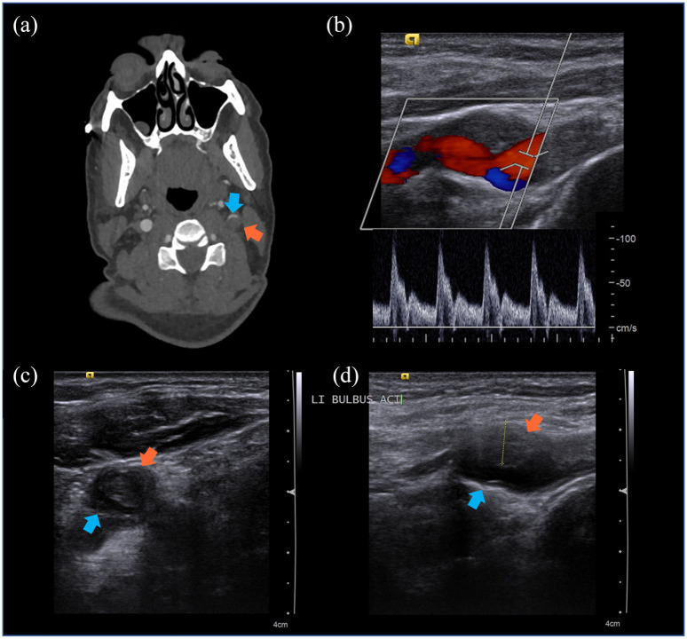
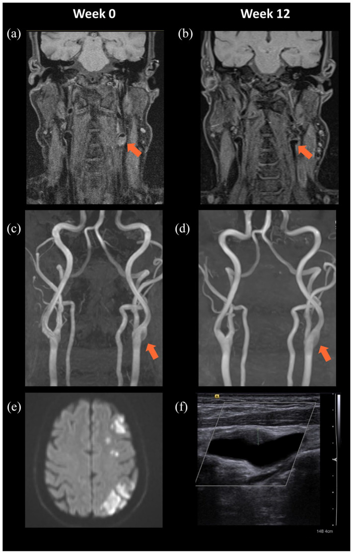
A 52-year-old man who had fully recovered from mild pneumonia due to infection with severe acute respiratory syndrome coronavirus 2 (SARS-CoV-2) in home quarantine 5 days earlier presented to our emergency room with acute severe left middle cerebral artery (MCA) syndrome on 5 May 2020. Just before onset of symptoms, an oropharyngeal swab (OPS) had been collected by a paramedic in a seated position. During the procedure, the patient’s head was hyperextended and he experienced acute neck pain; 30 min later, he was found agitated and unable to communicate by his relatives. Upon presentation to our emergency room 50 min after symptom onset, the patient suffered from high-grade sensorimotor right-side hemiparesis and global aphasia, with an initial National Institutes of Health Stroke Scale (NIHSS)-Score of 17. Immediate computed tomography (CT)-examination of the brain showed no early stroke signs but CT-perfusion revealed large areas of hypoperfusion in left precentral and central areas. Large vessel occlusion was absent in CT-angiography, but irregularities in the left carotid bulb with suspected short-segment stenosis due to atypical dissection were found (Figure 1a). Systemic thrombolysis was administered with 70 mg rtPA at a door-to-needle time of 10 min, 60 min after symptom onset. Symptoms improved quickly subsequently, with mild paraesthesia of the right arm and mild aphasia remaining. Ultrasound examination confirmed a short vessel wall hematoma in the proximal internal carotid artery (ICA) (Figure 1b–d) without signs of arteriosclerosis. The diagnosis of ICA dissection was supported by magnetic resonance imaging (MRI) of the brain and neck with a focal proximal ICA dissection and cerebral infarctions in the left MCA territory (Figure 2). Further diagnostic workup revealed continuous sinus rhythm, unremarkable transoesophageal echocardiography, exclusion of hereditary thrombophilia and normal cerebrospinal fluid findings with negative viral search panel including negative SARS-CoV-2-RT-PCR. There were no signs or symptoms of hereditary tissue disease or migraine; cerebrovascular risk factors (such as smoking, obesity, hypertension) were absent and family history was unremarkable for dissection or stroke. Medical treatment included platelet inhibition with 100 mg/day acetylsalicylic acid and 40 mg/day atorvastatin as lipid lowering therapy. During rehabilitation, symptoms further improved and the patient recovered without sequelae, allowing him to go back to work after 3 months. Follow-up examination in our outpatient clinic including ultrasound and MRI 3 months later confirmed full neurological recovery, albeit with incomplete remission of ICA vessel wall injury (Figure 2d, f).
Figure 1.
(a–d) CT angiography and neurovascular ultrasound at admission. Red arrows: intraarterial mass inside the left proximal ICA, suspicious for hematoma of the vessel wall; blue arrows: residual lumen of the left ICA. (a) CT angiography with short segment stenosis and residual eccentric lumen in the left carotid bulb. (b) Flow signal in close proximity to the wall hematoma. (c) Axial cross-section (B-mode). (d) Sagittal longitudinal section (B-mode). Dotted yellow line: measurement of 6.8 mm.
CT, computed tomography; ICA, internal carotid artery.
Figure 2.
(a–e) Confirmation of left ICA dissection in MRI and follow-up images after 12 weeks. MRI with T1-weighted, fat-saturated images (a–b) and time-of-flight angiography (c–d) showing suspicious ICA dissection in week 0 (a, c) and improvement at follow up in week 12 (b, d). (e) Diffusion-weighted imaging at week 0 shows a diffusion restricted pattern in the territory of the left medial cerebral artery matching haemodynamic infarction. (f) Follow-up neurovascular ultrasound at 12 weeks shows receding vessel wall hematoma (dotted green line: 4.5 mm).
ICA, internal carotid artery; MRI, magnetic resonance imaging.
Discussion
COVID-19 as a predisposing condition for arterial dissections
Ischemic stroke has been described as major cardiovascular complication in patients with coronavirus disease 2019 (COVID-19). 1 Mechanistically, this has been attributed mostly to a higher risk of thrombotic events due to a hypercoagulable state driven by systemic inflammation during the course of COVID-19. 1 Additionally, direct coronavirus-induced endotheliitis has been observed in some patients. 2 It seems plausible that this thrombo-inflammation increases vessel wall vulnerability, which may ultimately lead to dissection even upon mild tissue trauma. 3 In fact, published cases of unusual presentations of arterial dissections in COVID-19 patients include spontaneous bilateral carotid artery dissection and spontaneous coronary artery dissection.4,5 Arterial dissections are not specific for SARS-CoV-2, as also other respiratory infections have been shown to present a risk factor for cervical artery dissections (CAD) in the past.6,7 This risk was shown to be independent from mechanical strain through cough or sneeze, hinting at associated vessel pathology. Interestingly, in our case, vessel wall vulnerability seemed to outlast the acute infection phase as the dissection occurred 5 days after clinical recovery from COVID-19.
Role of tissue trauma following OPS
In the present case, the timing of stroke symptoms is highly suggestive for a causal link between the sampling of the OPS and the occurrence of ICA dissection. It is well established that CAD can appear after only minor trauma or even spontaneously.8,9 Hyperextension of the neck can be considered as such a minor trauma and has been described to be associated with CAD in the past, as it probably leads to compression of the ICA against the transverse processes of cervical vertebrae. 9 Pressure exerted on the posterolateral wall of the oropharynx by the swab could also have added slight force onto the vessel in this case. The patient had been instructed to recline his head to a maximum position, which he described retrospectively as uncomfortable, and he reported that the swab felt locally painful. Remarkably, the site of dissection at the proximal ICA close to the carotid bulb, about 7 cm from the skull base, is untypical, since most ICA dissections occur adjacent to the skull base or 2–3 cm proximal to it.10,11
Conclusion
Since the beginning of the pandemic, the wide application of OPSs or nasopharyngeal swabs for the detection of SARS-CoV-2-RNA have been indispensable for diagnosis and incidence monitoring. 12 Although this procedure can be generally regarded as safe, the present case report underlines the potential risk of rare but severe complications, e.g. dissection of head and neck arteries. Therefore, the manipulation should be performed only by trained personnel and with high caution, particularly in individuals with predisposing risk factors such as existing or currently remitted COVID-19 or a history of arterial dissection. Moreover, swabs testing against SARS-CoV-2 should be restricted to situations reasonable from an infectiological perspective.
Footnotes
Author contributions: LA collected data, wrote the manuscript and created figures. CD and MF supervised and evaluated neuroimaging data. CK supervised diagnosis and treatment. MK collected data and delivered diagnosis and treatment. All authors read, reviewed and approved the final manuscript.
Conflict of interest statement: The authors declare that there is no conflict of interest.
Funding: The authors received no financial support for the research, authorship, and/or publication of this article.
Patient consent: The patient gave written consent to publication of images and medical information.
ORCID iD: Livia Asan  https://orcid.org/0000-0001-5813-3242
Contributor Information
Livia Asan, Department of Neurology and Center for Translational Neuro- and Behavioral Sciences (C-TNBS), University Hospital Essen, Hufelandstr. 55, Essen, 45147, Germany.
Cornelius Deuschl, Institute for Diagnostic and Interventional Radiology and Neuroradiology, University Hospital Essen, Essen, Germany.
Michael Forsting, Institute for Diagnostic and Interventional Radiology and Neuroradiology, University Hospital Essen, Essen, Germany.
Christoph Kleinschnitz, Department of Neurology and Center for Translational Neuro- and Behavioral Sciences (C-TNBS), University Hospital Essen, Essen, Germany.
Martin Köhrmann, Department of Neurology and Center for Translational Neuro- and Behavioral Sciences (C-TNBS), University Hospital Essen, Essen, Germany.
References
- 1. Ellul MA, Benjamin L, Singh, et al. Neurological associations of COVID-19. Lancet Neurol 2020; 19: 767–783. [DOI] [PMC free article] [PubMed] [Google Scholar]
- 2. Varga Z, Flammer AJ, Steiger P, et al. Endothelial cell infection and endotheliitis in COVID-19. Lancet 2020; 395: 1417–1418. [DOI] [PMC free article] [PubMed] [Google Scholar]
- 3. Patel P, Khandelwal P, Gupta G, et al. COVID-19 and cervical artery dissection- a causative association?. J Stroke Cerebrovasc Dis 2020; 29: 105047. [DOI] [PMC free article] [PubMed] [Google Scholar]
- 4. Kumar K, Vogt JC, Divanji PH, et al. Spontaneous coronary artery dissection of the left anterior descending artery in a patient with COVID-19 infection. Catheter Cardiovasc Interv. Epub ahead of print 7 May 2020. DOI: 10.1002/ccd.28960. [DOI] [PMC free article] [PubMed] [Google Scholar]
- 5. Morassi M, Bigni B, Cobelli M, et al. Bilateral carotid artery dissection in a SARS-CoV-2 infected patient: causality or coincidence? J Neurol 2020; 267: 2812–2814. [DOI] [PMC free article] [PubMed] [Google Scholar]
- 6. Grau AJ, Brandt T, Buggle F, et al. Association of cervical artery dissection with recent infection. Arch Neurol 1999; 56: 851–856. [DOI] [PubMed] [Google Scholar]
- 7. Hunter MD, Moon YP, Miller EC, et al. Influenza-like illness is associated with increased short-term risk of cervical artery dissection. J Stroke Cerebrovasc Dis 2021; 30: 105490. [DOI] [PMC free article] [PubMed] [Google Scholar]
- 8. Engelter ST, Grond-Ginsbach C, Metso TM, et al. Cervical artery dissection: trauma and other potential mechanical trigger events. Neurology 2013; 80: 1950–1957. [DOI] [PubMed] [Google Scholar]
- 9. Caso V, Paciaroni M, Bogousslavsky J. Environmental factors and cervical artery dissection. Front Neurol Neurosci 2005; 20: 44–53. [DOI] [PubMed] [Google Scholar]
- 10. Downer J, Nadarajah M, Briggs E, et al. The location of origin of spontaneous extracranial internal carotid artery dissection is adjacent to the skull base. J Med Imaging Radiat Oncol 2014; 58: 408–414. [DOI] [PubMed] [Google Scholar]
- 11. Blum CA, Yaghi S. Cervical artery dissection: a review of the epidemiology, pathophysiology, treatment, and outcome. Arch Neurosci 2015; 2: e26670. [DOI] [PMC free article] [PubMed] [Google Scholar]
- 12. Velavan TP, Meyer CG. COVID-19: a PCR-defined pandemic. Int J Infect Dis. Epub ahead of print 1 December 2020. DOI: 10.1016/j.ijid.2020.11.189. [DOI] [PMC free article] [PubMed] [Google Scholar]


