Abstract
Purpose
To examine the associations of optical coherence tomography angiography (OCTA)–derived retinal capillary flux with systemic determinants of health.
Methods
This is a cross-sectional study of subjects recruited from the African American Eye Disease Study. A commercially available swept-source (SS)-OCTA device was used to image the central 3 × 3 mm macular region. Retinal capillary perfusion was assessed using vessel skeleton density (VSD) and flux. Flux approximates the number of red blood cells moving through vessel segments and is a novel metric, whereas VSD is a previously validated measure commonly used to quantify capillary density. The associations of OCTA derived measures with systemic determinants of health were evaluated using multivariate generalized linear mixed-effects models.
Results
A total of 154 eyes from 83 participants were enrolled. Mean VSD and flux were 0.148 ± 0.009 and 0.156 ± 0.016, respectively. In a model containing age, systolic blood pressure, diabetes status, hematocrit, and presence of retinopathy as covariates, there was a negative correlation between VSD and age (P < 0.001) and retinopathy (P = 0.02), but not with hematocrit (P = 0.85) or other factors. There was a positive correlation between flux and hematocrit (P = 0.02), as well as a negative correlation for flux with age (P < 0.001), systolic blood pressure (P = 0.04), and diabetes status (P = 0.02). A 1% decrease in hematocrit was associated with the same magnitude change in flux as ∼1.24 years of aging. Signal strength was associated with flux (P < 0.001), but not VSD (P = 0.51).
Conclusions
SS-OCTA derived flux provides additional information about retinal perfusion distinct from that obtained with skeleton density-based measures. Flux is appropriate for detecting subclinical changes in perfusion in the absence of clinical retinopathy.
Keywords: angiography, retinal blood flow, flux
The retinal vasculature has been studied extensively as an indicator of systemic vascular diseases. For example, central retinal arteriolar narrowing has been associated with stroke,1 high blood pressure,2,3 and cardiac perfusion4,5 and central retinal venular widening has been noted in anemia6 and diabetes.7 Arteriolar and venular diameters are also significantly correlated with systemic hematologic variables such as hemoglobin and hematocrit levels.8,9 These studies based on fundus photography provide compelling evidence that macroscopic changes in retinal arterioles and venules reflect changes in systemic blood flow and are significant predictors of systemic health and disease. These changes strongly suggest corresponding changes in retinal capillary perfusion, which are much harder to detect in human subjects in a clinically meaningful way.
Optical coherence tomography angiography (OCTA) is a relatively novel, high-resolution, minimally invasive, and Food and Drug Administration–approved imaging modality that allows assessment of retinal perfusion with capillary level precision.10,11 OCTA-based metrics have been used to quantify retinal capillary density in pathology ranging from diabetes to dementia10,12–15 and in quantifying inducible physiological changes in retinal perfusion.16,17 Therefore OCTA-based measures provide an ideal opportunity to further explore and understand the association of systemic vascular variables with retinal capillary perfusion. In this study, we report associations of retinal capillary flux, a novel measure of capillary perfusion, with systemic determinants of health and disease using a state-of-the-art, commercially available swept-source (SS) OCTA system. We compare a previously validated OCTA-derived measure called vessel skeleton density (VSD) with flux.
In vitro18 and in vivo15,18 studies demonstrate that flux approximates the number of red blood cells moving through capillary segments and is a potentially useful measure of retinal perfusion in addition to other OCTA derived metrics. Flux is an approximation of the average number of red blood cells (RBC) passing through capillary segments using the complex-based OCTA technique called optical microangiography (OMAG).18 Flux is independent of flow velocity because of the large amount of time between the A-scans but is dependent on particle concentration.18,19 The relationship of flux with particle concentration has been analytically modeled and validated in vitro using microfluidic phantoms and intralipid solution.18,20,21 These experiments and models suggest that OMAG signal intensity is directly proportional to the concentration of particles moving through an imaging voxel. In vitro experiments have repeated these findings with hematocrit over the range of 32% to 45% and confirmed a positive association with flux and decorrelation signal intensity.22 Because blood flow in capillaries is essentially the same as the movement of RBCs, capillary flux may be more sensitive to changes in retinal perfusion at the capillary level than traditional measures of vessel or skeleton density such as VSD. Specifically, VSD represents the number of perfused capillary segments containing particles with velocities ranging between 0.3 to 3 mm/sec.
This study aims to determine whether retinal capillary flux is a useful tool for the assessment of subclinical changes in commonly used systemic determinants of health. The results of this methodology can provide additional, complementary data to previous studies of large-caliber retinal vascular associations with systemic determinants of health.
Methods
Subjects
A total of 125 participants of the African American Eye Disease Study23 were invited for the study and consented to their participation. The African American Eye Disease Study is a population-based study of 40+-year-old African American residents of Inglewood, California, who underwent a comprehensive ophthalmic examination.23 These participants were enrolled at the Roski Eye Institute of the University of Southern California. The study was approved by and conducted in compliance with the University of Southern California Institutional Review Board. All subjects had the following systemic determinants of health assessed: blood pressure, complete blood count (including white blood cell count, red blood cell count, hemoglobin, hematocrit, and platelet count), and point-of-care hemoglobin A1c and microhematocrit. All subjects completed a medical history questionnaire reporting their sex, cigarette smoking (current smoker or not), diabetes status, hypertensive status, and hypertensive medication use. Subjects were classified as diabetic if they endorsed a history of diabetes mellitus or had a hemoglobin A1c value of ≥6.5%. Subjects were classified as hypertensive if they were prescribed hypertension medication, endorsed a history of hypertension, or presented with a systolic blood pressure >140 mm Hg or diastolic blood pressure >90 mm Hg. Past medical records indicating any type of retinopathy status (including macular edema) were noted. Subjects also underwent an ophthalmic examination that included dilation, refraction, and visual acuity measurements.
Imaging
Swept-source OCTA images were acquired using a commercially available device (PlexElite 9000; Carl Zeiss Meditec, Dublin, CA, USA) with a scan speed of 100 or 200 kHz, respective interscan interval of ∼4 ms or ∼2 ms, central wavelength of 1060 nm, and bandwidth of 100 nm providing an axial resolution of 6 µm and lateral resolution of 20 µm. The device uses an OMAG-based algorithm to measure decorrelation.24 Images were centered on a 3 mm × 3 mm region over the fovea. Each OCTA image consisted of 300 horizontal B-scans with 300 A-scans per B-scans and 1536 pixels per A-scan. A target of three acceptable OCTA volumes was recorded for the eye of each subject. One image from each eye was selected for further analysis, and the signal strength and axial length were noted.
Image Analysis
OCTA images with the following characteristics were selected for analysis from each eye: signal strength >7 (as assessed by the vendor's commercially available native software), centered on the fovea, lacking hyporeflective or hyper-reflective artifacts on en face structural images and with fewer than 10 visibly detectable motion artifacts (defined as horizontal disruptions in continuous vessels of more than 1 pixel width). Analyses were limited to the superficial retinal layer (SRL) to minimize confounding effects of projection artifacts and segmentation errors. Images were also excluded if the segmentation algorithm had errors and either SRL boundary could not be reliably manually segmented by experienced graders. The SRL was defined from the internal limiting membrane to the outer boundary of the inner plexiform layer using the commercially available automated segmentation algorithm.
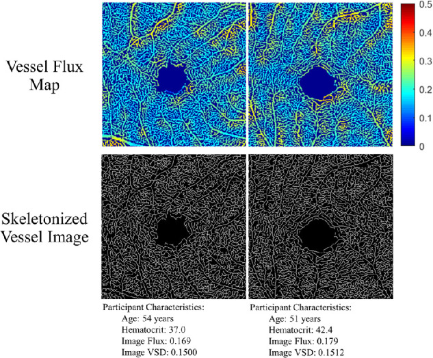
A previously validated, semi-automated, custom MATLAB (MathWorks Inc., Natlick, MA, USA) algorithm was used to calculate vessel skeleton density (VSD10,24,25) and flux.15,18 As described previously, VSD is a measure of overall vessel length within an OCTA image and is computed by reducing all vessel segments to one-pixel width before calculating the ratio of the total length of vessels to the total area of the image.10,24 Flux is calculated by averaging the absolute (non-binarized) decorrelation intensity values from regions of the image occupied by vascular structures.15,18 This is described in more detail in Abdolahi et al.26 Pseudocolor flux and skeletonized vessel maps for two subjects are illustrated in Figure 1.
Figure 1.

Vessel flux and skeletal vessel map from the right eyes of two female participants with OCTA image signal strength of 10 and hematocrit values spanning the normative range from the cohort of subjects in this study. Note that the subject with the higher hematocrit value demonstrates a relatively higher flux value than skeleton density value.
Statistical Analysis
All regression models were analyzed using R (version 3.6.3). VSD and flux were treated as continuous outcomes. The systemic determinants of health that were available and investigated were sex, age, hypertensive status, systolic blood pressure, diastolic blood pressure, diabetes status, hemoglobin a1c, smoking history, hematocrit, hemoglobin, white blood cell count, red blood cell count, platelet count, and the presence of retinopathy. Each of these factors was investigated because of previously shown associations with arteriole or venous retinal diameters8,9,27 or VSD.12 Multivariable analyses were performed for each systemic determinant while adjusting for sex, age, signal strength and correlation between two eyes using mixed-effects linear regression. Determinants of health that were significant (P < 0.05) in either the VSD or flux regression analysis were used to generate a mixed-effects linear regression model of the association of systemic determinants with either VSD or flux. Determinants were modeled as fixed-effects except for the between-eye correlation that was modeled as a random intercept and adjusted using a compound symmetry correlation matrix.28 Semi-partial and total R2 values were calculated to demonstrate how each model element independently and in combination contributed to the variation in OCTA flux and VSD. Semi-partial R2 conceptually is the decrease in the R2 statistic without the variable of interest. The sum of the semi-partial R2 of every determinant need not be equal to the total R2 of the model because every factor may be positively or negatively correlated with one another in varying ways, if at all.29 Least-square means were calculated for selected variables with clinically meaningful cutoffs for continuous variables (age ≥65 or <65, hematocrit ≥40 or <40) to demonstrate the magnitude of their effects on the OCTA parameters. Interactions between two factors were tested by including their product terms in the regression model. Associations were considered significant if P < 0.05. All P values are reported based on two-sided tests.
Results
Characteristics of the Study Population
The study enrolled 85 subjects (170 eyes). OCTA images that met inclusion criteria were available for 83 subjects (27 males and 56 females) (154 eyes). Mean axial length of the cohort was 23.80 mm ± 1.20 mm. Mean signal strength of the OCTA images was 9.32 ± 0.66. The demographic features of this cohort are summarized in Table 1. The imaging characteristics of this cohort are seen in Supplementary Table S1. There were significant differences in flux (P < 0.001), signal strength (P < 0.001), and VSD (P = 0.003) for subjects aged ≥65 versus those younger than 65 years of age. Axial length was the only significantly different (P = 0.04) imaging variable between sexes.
Table 1.
Subject Demographics
| Determinant | All Subjects |
|---|---|
| No. of subjects (no. of eyes) | 83 (154) |
| Age (y) | |
| Mean ± SD | 66.2 ± 8.9 |
| Range | 40–83 |
| Hypertension*, No. of subjects (%) | 63 (75.9%) |
| SBP (mm Hg), mean ± SD | 146.4 ± 24.5 |
| DBP (mm Hg), mean ± SD | 84.8 ± 12.1 |
| Diabetes†, no. of subjects (%) | 22 (26.5%) |
| HbA1c (%), mean ± SD | 6.30 ± 1.57 |
| Current smoker, no. subjects (%) | 10 (12.0%) |
| Hematocrit (%) | |
| Mean ± SD | 39.99 ± 4.36 |
| [Normal Low–Normal High] | [38.80–53.20] |
| Hemoglobin (g/dL) | |
| Mean ± SD | 12.93 ± 1.66 |
| [Normal Low–Normal High] | [13.30–17.70] |
| White blood cell count ×103/µL | |
| Mean ± SD | 6.21 ± 1.99 |
| [Normal Low–Normal High] | [4.00–10.90] |
| RBC count ×106/µL | |
| Mean ± SD | 4.55 ± .48 |
| [Normal Low–Normal High] | [3.80–5.20] |
| Platelet count ×102/µL | |
| Mean ± SD | 248.55 ± 54.5 |
| [Normal Low–Normal High] | [150.00–400.00] |
| Retinopathy‡, no. subjects (%) | 7 (8.4%) |
SD, standard deviation; SBP, systolic blood pressure; DBP, diastolic blood pressure.
Subjects were categorized as hypertensive if they were prescribed hypertension medication, endorsed a history of hypertension, or presented with a systolic blood pressure > 140 and/or diastolic blood pressure > 90.
Subjects were categorized as diabetic if they endorsed a history of diabetes mellitus or had a hemoglobin A1c value of 6.5% or greater.
Subjects were designated as having retinopathy if medical records indicated a history of retinopathy or diabetic macular edema.
Associations Between OCTA Derived Variables and Systemic Determinants
Each systemic determinant was analyzed for significance in its own multivariate regression model with flux or VSD as the dependent variable while accounting for intra-eye correlation and including age, sex, and signal strength as covariates (Table 2). Retinopathy was the only factor significantly correlated (P = 0.03) with VSD where the presence of retinopathy decreased the image VSD. History of diabetes (P = 0.02), systolic blood pressure (P = 0.04), hematocrit (P = 0.02), and hemoglobin (P = 0.01) were significantly correlated with flux whereas the red blood cell count was borderline significant (P = 0.06). Age was significant in all models (P < 0.02), whereas sex was never significant. Although not a systemic determinant, it is important to note that signal strength in the range of 8 to–10 was a significant contributor to flux (P < 0.001) but not VSD in all models evaluated.
Table 2.
Multivariate Analysis of Factors Associated With Flux or VSD
| VSD | Flux | |||
|---|---|---|---|---|
| Laboratory or Historical Characteristic* | Beta† (SE) 10−3 | P Value | Beta† (SE) 10−3 | P Value |
| Hypertension‡ | 0.791 (2.182) | 0.72 | −4.607 (3.287) | 0.17 |
| SBP (mm Hg) | 0.033 (0.038) | 0.39 | −0.120 (0.056) | 0.04 |
| DBP (mm Hg) | −0.042 (0.072) | 0.56 | −0.089 (0.110) | 0.42 |
| Diabetes§ | 1.010 (2.031) | 0.62 | −7.374 (3.000) | 0.02 |
| Ha1c | −0.298 (0.584) | 0.61 | −0.969 (0.888) | 0.28 |
| Smoker|| | −5.135 (2.601) | 0.05 | 0.034 (4.090) | 0.99 |
| Hematocrit (%) | 0.127 (0.208) | 0.54 | 0.749 (0.307) | 0.02 |
| Hemoglobin (g/dL) | 0.211 (0.566) | 0.71 | 2.253 (0.842) | 0.01 |
| White blood cell count ×103/µL | 0.172 (0.451) | 0.70 | −0.254 (0.706) | 0.72 |
| RBC count ×106/µL | 0.736 (1.946) | 0.71 | 5.668 (2.962) | 0.06 |
| Platelet count ×102/µL | 0.018 (0.017) | 0.28 | 0.002 (0.027) | 0.95 |
| Retinopathy¶ | −4.443 (1.954) | 0.03 | −1.717 (2.948) | 0.56 |
SBP, systolic blood pressure; DBP, diastolic blood pressure.
All models adjusted for age, sex and signal strength with the addition of the variable of interest.
Beta values represent the change in the output of interest (VSD or flux) for a single unit increase in the case of continuous variables or by the presence of the characteristic in the case of indicator variables.
Subjects were categorized as hypertensive if they were prescribed hypertension medication, endorsed a history of hypertension, or presented with a systolic blood pressure > 140 and/or diastolic blood pressure > 90.
Subjects were categorized as diabetic if they endorsed a history of diabetes mellitus or had a hemoglobin A1c value of 6.5% or greater.
Smoker was categorized as current smoker versus not.
Subjects were designated as having retinopathy if medical records indicated a history of retinopathy or diabetic macular edema.
We also examined the association of axial length on VSD and flux in this cohort and it was not a significant covariate for either VSD or flux in any of the models we assessed. Therefore it was excluded from the final model.
Multivariable Adjusted Linear-Mixed Effects Model
To estimate the potential contribution of systemic determinants to OCTA-derived measures of VSD and flux, we developed combined models of VSD or flux that included all the significant systemic determinants for either flux or VSD from Table 2 including: age, systolic blood pressure, diabetes status, hematocrit, and the presence of retinopathy. This combined model is described in Table 3.
Table 3.
Combined Mixed Effect Linear Model Parameters for VSD or Flux Models
| VSD | Flux | |||||||
|---|---|---|---|---|---|---|---|---|
| Model Factor | Beta* (SE) 10−3 | P Value | Semi-Partial R2 | Full Model R2 | Beta* (SE) 10−3 | P Value | Semi-Partial R2 | Full Model R2 |
| Age, per year | −0.357 (0.101) | <0.001 | 0.148 | 0.171 | −0.612 (0.170) | <0.001 | 0.161 | 0.353 |
| Systolic blood pressure, per mm Hg | 0.037 (0.038) | 0.34 | 0.013 | −0.131 (0.064) | 0.04 | 0.057 | ||
| Diabetes (yes vs. no)† | 0.166 (1.997) | 0.93 | 0.000 | −7.564 (3.356) | 0.02 | 0.067 | ||
| Hematocrit, per % | −0.037 (0.195) | 0.85 | 0.001 | 0.758 (0.328) | 0.02 | 0.074 | ||
| Retinopathy (yes vs. no) | −4.885 (2.005) | 0.02 | 0.037 | 1.226 (3.233) | 0.71 | 0.001 | ||
SE, standard error.
Beta values represent the change in the output of interest (VSD or flux) for a single unit increase in the case of continuous variables or by the presence of the characteristic in the case of indicator variables.
Subjects were categorized as diabetic if they endorsed a history of diabetes mellitus or had a hemoglobin A1c value of 6.5% or greater.
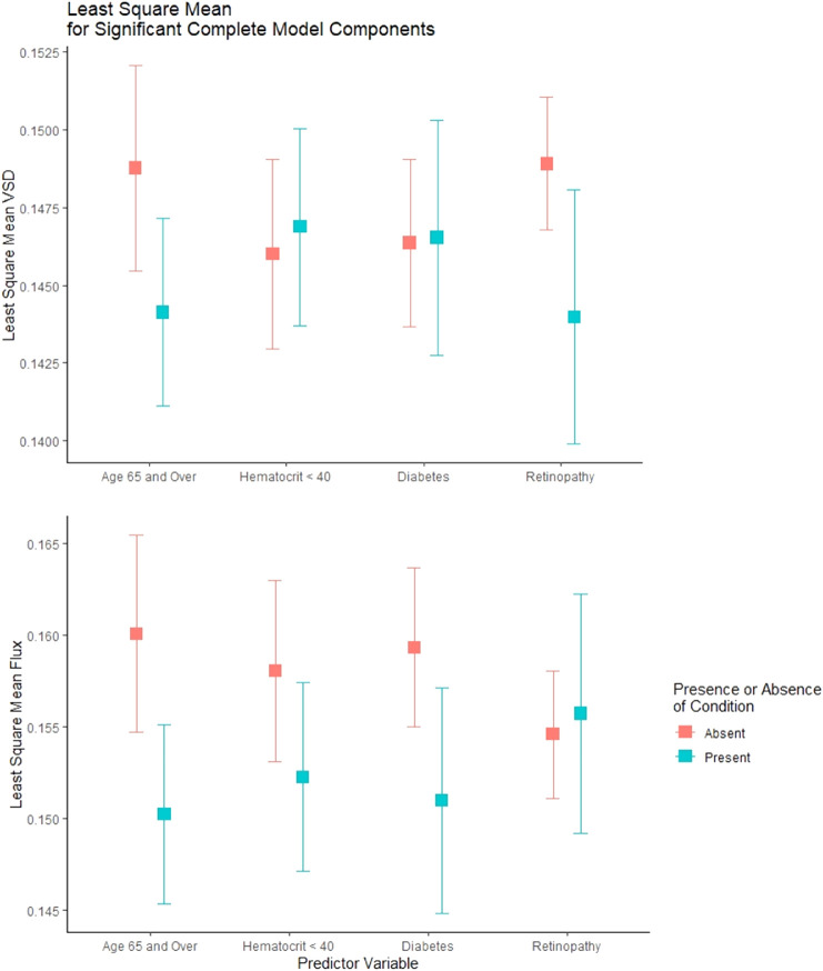
Significant systemic determinants of VSD included age (P < 0.001) and the presence of retinopathy (P = 0.02). All determinants marginally increased the R2 of the model except for diabetes status with the largest contributor being age (R2 = 0.148) for an overall R2 = 0.171. To demonstrate effect sizes of relevant clinical categories on VSD, a revised model with the same determinants was made, but age (≥65 vs. <65) and hematocrit (≥40 vs. <40) were treated as categorical variables and least-square means were estimated. Least square mean VSD and flux values are plotted in Figure 2. Both changes in age category from less than age 65 to age 65 or greater and the presence of retinopathy accounted for a change of about 0.05 in VSD.
Figure 2.
Least square mean estimates for select elements of the complete VSD and flux models.
Significant systemic determinants of flux included age (P < 0.001), diabetes status (P = 0.02), systolic blood pressure (P = 0.04) and hematocrit (P = 0.02). Age (R2 = 0.161) was the largest systemic determinant in the overall model (model R2 = 0.353) whereas hematocrit (R2 = 0.074), diabetes (R2 = 0.067), and systolic blood pressure (R2 = 0.057) were secondary contributors. As with VSD, a revised model with the same determinants was made where age and hematocrit were treated as categorical variables, and least-square means were estimated. Age 65 or older versus age younger than 65 had the greatest effect on the flux estimate (with a difference of 0.01) whereas the hematocrit category (<40% vs. ≥40%) and diabetes status (yes vs. no) were similar in effect size with changes of 0.006 and 0.008, respectively (Fig. 2).
The above-mentioned associations did not change qualitatively when adjusting for signal strength (Supplementary Table S2) or interactions between sex and age.
Discussion
Using a commercially available swept-source OCTA system, we demonstrate that flux is a useful measure of retinal perfusion that is sensitive to well-established systemic determinants of health and disease such as age, systolic blood pressure, hematocrit and diabetes status. This data suggests that flux is a novel measure of subclinical changes in perfusion that may occur very early in retinal vascular disease. For example, it is well understood that retinopathy takes many years to manifest in diabetic patients, but abnormal retinal perfusion and histopathologic capillary damage have been documented much earlier than clinically detectable retinopathy.
It is widely known that impaired regulation of blood flow in non-capillary retinal vessels (arterioles, venules, arteries and veins) is a preclinical finding in subjects with diabetes but with no retinopathy. Our results now suggest that this finding can be extended to impaired capillary flow in diabetic subjects independently of the presence of retinopathy. In fact, flux was not found to be associated with frank retinopathy. This may be due to capillary dropout, which measures such as VSD capture more effectively. However, it is also possible that the small number of retinopathy patients in this study may limit our ability to capture an association between flux and retinopathy. The observation that flux was significantly associated with a diagnosis of diabetes, but not necessarily with hemoglobin a1c, suggests that flux changes may be best used as an early sign of pathology. Flux in conjunction with other OCTA parameters, like VSD, could provide a more complete picture of the vascular health of a patient.
The association of flux with axial length was also considered. Axial length can confound OCTA-based measures via transverse magnification of the imaged area, especially for measures of vascular density that change significantly with radial eccentricity from the fovea. Unlike capillary density–based measures, there is no evidence that capillary blood flow or RBC flux varies as a function of distance from the fovea. Therefore, because flux-based measures are based on pixel intensity rather than pixel number, it may be possible that flux is less likely to be impacted in the same manner as vessel density measures with small variations in the field-of-view. One piece of evidence that supports this idea is that axial length was not found to be associated with flux in this study. In contrast axial length has been shown to be associated with density measures in larger studies.30 In any case, although we have no evidence that axial length was a confounding variable in this particular study, additional studies in larger populations will be necessary to accurately assess the magnitude and significance of axial length on flux as has been done for vessel density measures.30
In this study, vessel skeletal density in the superficial retinal layer was not shown to vary with any of the systemic risk factors that were assessed except for age. Age is a known determinant of vessel skeletal density with a strong association.31,32 We also show that age is a strong determinant for flux. For some factors such as blood pressure derivatives, hemoglobin a1c, and sex, larger sample sizes have found a significant correlation in univariate analysis with vessel skeletal density that we did not.31 However, correlations in vessel skeletal density for each of these factors are inconsistent across the literature and without a clear conclusion.31,33–35 For other parameters such as smoking history, we did not find a correlation consistent with previous studies.31,34,36 This suggests that our data are in agreement with the literature at large and that our significant findings are those with a higher effect size.
Our study also shows the strong dependence of flux on signal strength, a correlation not seen with vessel skeletal density. This is observed even between those with the highest signal strength grades of 9 and 10, which comprises the vast majority of this dataset. This is expected because signal intensity at any single pixel will correlate with the overall signal strength. As seen in Supplementary Table S2, when signal strength was added to the model, it did not change the conclusions of the study. However, the strength of this correlation suggests that only high-quality images should be included in analyses involving flux. As seen in Supplementary Table S1, individuals older than age 65 generally had significantly lower signal strength than those younger than age 65. This implies that controlling for signal strength is especially important for older populations in which imaging quality may be reduced.
We hypothesized that flux is a useful measure of a change in the in vivo RBC content of capillaries similar to what has been shown in in vitro studies.18 This information would not necessarily be captured by VSD (or similar measures of density) especially in the absence of frank capillary loss. The results of our study support this hypothesis in living human subjects by demonstrating a significant correlation between flux and hematocrit, hemoglobin and borderline significant association with RBC count (Table 2).
In summary, this study demonstrates that OCTA-derived flux is a novel and significant indicator of systemic determinants of health and disease. The exploratory nature of this study meant that the sample size was relatively small and the investigation limited in its power. Further validation of these results will require future work, but these data strongly suggest that flux provides information about capillary perfusion that is distinct from more conventional measures of capillary density derived from OCTA. Flux is also less likely to be impacted by magnification errors induced by axial length, but more likely to be sensitive to signal strength.
Supplementary Material
Acknowledgments
Funding by NIH Grants to AHK R01EY030564; NIH Grants to XJ R21EY028721. Unrestricted departmental funding from Research to Prevent Blindness to University of Southern California and Johns Hopkins University.
Carl Zeiss Meditec provided funding in the form of research equipment that was used in this study. Carl Zeiss Meditec was not involved in the conception, data collection, writing, editing or final publication of this manuscript.
Disclosure: S. Kushner-Lenhoff, None; Y. Li, None; Q. Zhang, None; R.K. Wang, Carl Zeiss Meditec (P), Meditec (C), Kowa Inc (P), Insight Photonic Solutions (C); X, Jiang, None; A.H. Kashani, Carl Zeiss Meditec (F, R)
References
- 1. Wong TY, Klein R, Couper DJ, et al.. Retinal microvascular abnormalities and incident stroke: the Atherosclerosis Risk in Communities Study. Lancet. 2001; 358(9288): 1134–1140. [DOI] [PubMed] [Google Scholar]
- 2. Friedenwald H. The Doyne Memorial Lecture: pathological changes in the retinal blood-vessels in arterio-sclerosis and hypertension. Trans Ophthalmol Soc UK. 1930;50452–50531. [Google Scholar]
- 3. Büttner M, Schuster AKG, Vossmerbäumer U, Fischer JE.. Associations of cardiovascular risk factors and retinal vessel dimensions at present and their evolution over time in a healthy working population. Acta Ophthalmologica. 2020; 98(4): e457–e463. [DOI] [PubMed] [Google Scholar]
- 4. Wang Lu, Wong Tien Y, Richey SA, Ronald K, Folsom Aaron R, Michael J-H. Relationship between retinal arteriolar narrowing and myocardial perfusion. Hypertension. 2008; 51(1): 119–126. [DOI] [PubMed] [Google Scholar]
- 5. Cheung N, Bluemke DA, Klein R, et al.. Retinal arteriolar narrowing and left ventricular remodeling: the multi-ethnic study of atherosclerosis. J Am Coll Cardiol. 2007; 50: 48–55. [DOI] [PMC free article] [PubMed] [Google Scholar]
- 6. Aisen ML, Bacon BR, Goodman AM, Chester EM.. Retinal abnormalities associated with anemia. Arch Ophthalmol. 1983; 101: 1049–1052. [DOI] [PubMed] [Google Scholar]
- 7. Klein R, Klein BEK, Moss SE, Wong TY.. Retinal vessel caliber and microvascular and macrovascular disease in type 2 diabetes: XXI: the Wisconsin Epidemiologic Study of Diabetic Retinopathy. Ophthalmology. 2007; 114: 1884–1892. [DOI] [PubMed] [Google Scholar]
- 8. Klein BEK. Complete blood cell count and retinal vessel diameters. Arch Ophthalmol. 2011; 129: 490. [DOI] [PMC free article] [PubMed] [Google Scholar]
- 9. Liew G, Wang JJ, Rochtchina E, Wong TY, Mitchell P.. Complete blood count and retinal vessel calibers. PLoS ONE. 2014; 9(7): e102230. [DOI] [PMC free article] [PubMed] [Google Scholar]
- 10. Kashani AH, Chen CL, Gahm JK, et al.. Optical coherence tomography angiography: a comprehensive review of current methods and clinical applications. Prog Retinal Eye Res. 2017; 60: 66–100. [DOI] [PMC free article] [PubMed] [Google Scholar]
- 11. Matsunaga Douglas, Yi Jack, Puliafito Carmen A, Kashani Amir H.. OCT angiography in healthy human subjects. Ophthalm Surg Lasers Imaging Retina. 2014; 45: 510–515. [DOI] [PubMed] [Google Scholar]
- 12. Kim AY, Chu Z, Shahidzadeh A, Wang RK, Puliafito CA, Kashani AH.. Quantifying microvascular density and morphology in diabetic retinopathy using spectral-domain optical coherence tomography angiography. Invest Ophthalmol Vis Sci. 2016; 57(9): OCT362. [DOI] [PMC free article] [PubMed] [Google Scholar]
- 13. Koulisis N, Kim AY, Chu Z, et al.. Quantitative microvascular analysis of retinal venous occlusions by spectral domain optical coherence tomography angiography. PLoS ONE. 2017; 12(4): e0176404. [DOI] [PMC free article] [PubMed] [Google Scholar]
- 14. Ashimatey BS, D'Orazio LM, Ma SJ, et al.. Lower retinal capillary density in minimal cognitive impairment among older Latinx adults. Alzheimers Dement (Amst). 2020; 12(1): e12071–e12071. [DOI] [PMC free article] [PubMed] [Google Scholar]
- 15. Singer MB, Ringman JM, Chu Z, et al.. Abnormal retinal capillary blood flow in autosomal dominant Alzheimer's disease. Alzheimers Dement (Amst). 2021; 13(1): e12162–e12162. [DOI] [PMC free article] [PubMed] [Google Scholar]
- 16. Ashimatey BS, Green KM, Chu Z, Wang RK, Kashani AH.. Impaired retinal vascular reactivity in diabetic retinopathy as assessed by optical coherence tomography angiography. Invest Ophthalmol Vis Sci. 2019; 60: 2468–2473. [DOI] [PMC free article] [PubMed] [Google Scholar]
- 17. Singer M, Ashimatey BS, Zhou X, Chu Z, Wang R, Kashani AH.. Impaired layer specific retinal vascular reactivity among diabetic subjects. PLOS ONE. 2020; 15(9): e0233871. [DOI] [PMC free article] [PubMed] [Google Scholar]
- 18. Choi WJ, Qin W, Chen CL, et al.. Characterizing relationship between optical microangiography signals and capillary flow using microfluidic channels. Biomed Opt Express. 2016; 7: 2709. [DOI] [PMC free article] [PubMed] [Google Scholar]
- 19. Chen CL, Wang RK.. Optical coherence tomography based angiography. Biomed Opt Express. 2017; 8(2): 1056. [DOI] [PMC free article] [PubMed] [Google Scholar]
- 20. Su JP, Chandwani R, Gao SS, et al.. Calibration of optical coherence tomography angiography with a microfluidic chip. J Biomed Opt. 2016; 21(08): 1. [DOI] [PMC free article] [PubMed] [Google Scholar]
- 21. Spaide RF, Fujimoto JG, Waheed NK, Sadda SR, Staurenghi G.. Optical coherence tomography angiography. Prog Retin Eye Res. 2018; 64: 1–55. [DOI] [PMC free article] [PubMed] [Google Scholar]
- 22. Yang J, Su J, Wang J, et al.. Hematocrit dependence of flow signal in optical coherence tomography angiography. Biomed Opt Express. 2017; 8(2): 776. [DOI] [PMC free article] [PubMed] [Google Scholar]
- 23. McKean-Cowdin R, Fairbrother-Crisp A, Torres M, et al.. The African American Eye Disease Study: design and methods. Ophthalmic Epidemiol. 2018; 25: 306–314. [DOI] [PMC free article] [PubMed] [Google Scholar]
- 24. Reif R, Qin J, An L, Zhi Z, Dziennis S, Wang R.. Quantifying optical microangiography images obtained from a spectral domain optical coherence tomography system. Int J Biomed Imaging. 2012; 2012: 1–11. [DOI] [PMC free article] [PubMed] [Google Scholar]
- 25. Chu Z, Lin J, Gao C, et al.. Quantitative assessment of the retinal microvasculature using optical coherence tomography angiography. J Biomed Opt. 2016; 21(6): 066008. [DOI] [PMC free article] [PubMed] [Google Scholar]
- 26. Abdolahi F, Zhou X, Ashimatey BS, et al.. Optical coherence tomography angiography–derived flux as a measure of physiological changes in retinal capillary blood flow. Trans Vis Sci Tech. 2021; 10(9): 5. [DOI] [PMC free article] [PubMed] [Google Scholar]
- 27. Yanagi M, Misumi M, Kawasaki R, et al.. Is the association between smoking and the retinal venular diameter reversible following smoking cessation? Invest Ophthalmol Vis Sci. 2014; 55(1): 405. [DOI] [PubMed] [Google Scholar]
- 28. shuang Ying G, MG Maguire, Glynn R, Rosner B. Tutorial on biostatistics: linear regression analysis of continuous correlated eye data. Ophthalmic Epidemiol. 2017; 24: 130–140. [DOI] [PMC free article] [PubMed] [Google Scholar]
- 29. Edwards LJ, Muller KE, Wolfinger RD, Qaqish BF, Schabenberger O.. An R2 statistic for fixed effects in the linear mixed model. Stat Med. 2008; 27: 6137–6157. [DOI] [PMC free article] [PubMed] [Google Scholar]
- 30. Richter GM, Lee JC, Khan N, et al.. Ocular and systemic determinants of perifoveal and macular vessel parameters in healthy African Americans [published online ahead of print November 5, 2021]. Br J Ophthalmol, doi: 10.1136/bjophthalmol-2021-319675. [DOI] [PubMed] [Google Scholar]
- 31. You QS, Chan JCH, Ng ALK, et al.. Macular vessel density measured with optical coherence tomography angiography and its associations in a large population-based study. Invest Ophthalmol Vis Sci. 2019; 60: 4830. [DOI] [PubMed] [Google Scholar]
- 32. Wei Y, Jiang H, Shi Y, et al.. Age-related alterations in the retinal microvasculature, microcirculation, and microstructure. Invest Ophthalmol Vis Sci. 2017; 58: 3804. [DOI] [PMC free article] [PubMed] [Google Scholar]
- 33. Zhou W, Yang J, Wang Q, et al.. Systemic stressors and retinal microvascular alterations in people without diabetes: the Kailuan Eye Study. Invest Ophthalmol Vis Sci. 2021; 62(2): 20. [DOI] [PMC free article] [PubMed] [Google Scholar]
- 34. Shaw LT, Khanna S, Chun LY, et al.. Quantitative optical coherence tomography angiography (OCTA) parameters in a black diabetic population and correlations with systemic diseases. Cells. 2021; 10: 551. [DOI] [PMC free article] [PubMed] [Google Scholar]
- 35. Coscas F, Sellam A, Glacet-Bernard A, et al.. Normative data for vascular density in superficial and deep capillary plexuses of healthy adults assessed by optical coherence tomography angiography. Invest Ophthalmol Vis Sci. 2016; 57(9): OCT211. [DOI] [PubMed] [Google Scholar]
- 36. Dogan M, Akdogan M, Gulyesil FF, Sabaner MC, Gobeka HH.. Cigarette smoking reduces deep retinal vascular density. Clin Exp Optom. 2020; 103: 838–842. [DOI] [PubMed] [Google Scholar]
Associated Data
This section collects any data citations, data availability statements, or supplementary materials included in this article.


