Abstract
The purpose of this study was to assess changes in cervical musculature throughout contact-heavy collegiate ice hockey practices during a regular season of NCAA Division III ice hockey teams. In this cross-sectional study, 36 (male n = 13; female n = 23) ice hockey players participated. Data were collected over 3 testing sessions (baseline; pre-practice; post-practice). Neck circumference, neck length, head-neck segment length, isometric strength and electromyography (EMG) activity for flexion and extension were assessed. Assessments were completed approximately 1h before a contact-heavy practice and 15 min after practice. For sternocleidomastoid (SCM) muscles, males had significantly greater peak force and greater time to peak force versus females. For both left and right SCMs, both sexes had significantly greater peak EMG activity pre-practice versus baseline, and right (dominant side) SCM time to peak EMG activity was decreased post-practice compared to pre-practice. There were no significant differences for EMG activity of the upper trapezius musculature, over time or between sexes. Sex differences observed in SCM force and activation patterns of the dominant side SCM may contribute to head stabilization during head impacts. Our study is the first investigation to report changes in cervical muscle strength in men’s and women’s ice hockey players in the practical setting.
Key points.
Concussion prevention strategies, such as cervical muscle strength and activation time, was investigated in the practical setting.
Neck muscle fatigue may not be a contributing factor to a concussive event in contact sports such as ice hockey.
Hand dominance may affect recruitment timing of neck musculature, which can affect the cervical muscle activation response.
Males and females exhibited differences in sternocleidomastoid time to peak force.
Key words: Ice hockey, sternocleidomastoid, sex differences, neck strength
Introduction
Concussion rates in men’s and women’s collegiate ice hockey have been reported at 0.76/1000 athlete exposures (AE) for men and 0.72/1000 AEs for women (Rosene et al., 2017). Accurate diagnosis and complete recovery from SRCs have challenged athletes and health care professionals for decades due to variations in injury, evolving symptoms, and varied approaches to concussion prevention strategies (Eckner et al., 2011; Mansell et al., 2005). Enhanced cervical muscle strength has been proposed as a concussion prevention strategy, yet studies addressing the efficacy of this strategy are typically performed in laboratory-controlled environments (Mansell et al., 2005; Becker et al., 2019; Eckner et al., 2018; Tierney et al., 2005).
Others have proposed cervical stiffness, or anticipatory cervical activation as an aid against concussive events when subjected to head impacts (Collins et al., 2014; Mihalik et al., 2011; Tierney et al., 2005; Viano et al., 2007). Early activation of the cervical musculature may reduce acceleration and velocity of head movement, in turn decreasing concussion risk. As with increased cervical muscle strength, earlier cervical muscle activation may result in increased neck stiffness and its associated connection with the torso. This head/torso connection potentially provides greater head stabilization, thereby reducing head acceleration and velocity in response to a head impact (Reynier et al., 2020).
To understand the utility of enhanced cervical muscle strength and/or early cervical muscle activation as a concussion prevention strategy, studies in the practical setting must be conducted, including consideration of sex differences. Generally, males have greater overall cervical muscle strength compared to females, while females have greater head-neck segment acceleration and displacement compared to males when exposed to an external force to the head (Tierney et al., 2005). Greater head-neck segment acceleration and displacement may result from lower cervical muscular strength, girth, and head mass in females versus males (Tierney et al., 2005). These differences support the need to further investigate enhanced cervical muscle strength as a potential concussion prevention strategy in male and female athletes.
With much of the cervical muscle strength data coming from laboratory-controlled studies (Mansell et al., 2005; Becker et al., 2019), translation into real world applications, for example on the field of play and/or ice, is speculative. The purpose of this investigation was to examine cervical musculature changes throughout contact-heavy collegiate ice hockey practices during a regular season of NCAA Division III men’s and women’s ice hockey teams. We assessed pre- to post-practice measures of isometric neck strength, time to peak torque, electromyography (EMG) activity, and time to peak EMG activity of the sternocleidomastoid (flexion) and upper trapezius (extension) musculature. We hypothesized that following contact heavy practices, cervical muscle strength and EMG measures would decrease.
Methods
Procedures
Thirty-six NCAA Division III collegiate male (N = 13, age= 22.23 ± 1.09 yrs, height = 1.81 ± 0.05 m, mass = 85.89 ± 7.34 kg) and female ice hockey players (N = 23, age = 19.74 ± 1.18yr, height = 1.64 ± 0.06 m, mass = 66.07 ± 9.95kg) participated during the regular ice hockey season. Data were collected at 3 testing time-points (baseline; pre-practice; post-practice) during contact-heavy practices. Contact-heavy practices, defined by the respective coaches, consisted primarily of contact drills. All baseline measures were obtained within 2-weeks prior to the beginning of formal regular season practices. Players were excluded from the study if they had a history of neurological disorders, prior cervical spine injuries, or a diagnosed concussive event within 6-months prior to data collection. Written informed consent was received from each player, and all procedures were approved by the University’s Institutional Review Board. All assessments were completed approximately 1h before a contact-heavy practice and repeated within approximately 15 min following the player’s exit from the ice.
Neck assessments
At the beginning of each testing session, neck assessments were performed for isometric strength and EMG activity, in randomized order for flexion and extension. Measurements of neck circumference, neck length, and head-neck segment length were obtained prior to neck warm-up exercises which consisted of 15s of clockwise neck rotations, 15s of counterclockwise neck rotations, and 2 repetitions each of 15s of neck flexion and 15s of neck extension stretching (Mansell et al., 2005). The skin was then prepared for electrode placement over the right and left upper trapezius between C3 and C5 and right and left sternocleidomastoid muscles. Players were seated with their upper torso secured with a Velcro strap to minimize additional body movement. Each player was fitted with headgear snug to the head attached with a chin strap, with the top of the headgear serving as the attachment for the resistance cable during the trials. The attachment sites for the resistance cable were the top of the head gear and a secure, fixed attachment on the wall, with the cable maintaining a position parallel to the ground.
Once secured, players performed 3 maximal voluntary isometric contractions for both flexion and extension in random order. For each movement, the arms were crossed at the chest with feet off the ground. The 3 trials consisted of an isometric contraction held for 3s using maximum effort, with a 30s break between each trial.
Head-neck segment mass was calculated using body mass multiplied by gender specific head-neck segment to total body mass percentage (8.26% - male, 8.20% - female) (Tierney et al., 2005). Neck circumference was measured just below the thyroid cartilage. Neck length was measured from the 7th cervical vertebra to the base of the occiput, and head-neck segment length was measured from the 7th cervical vertebra to the top of the head with the head in neutral.
Electromyography
Electromyographic (EMG) activity of the left and right sternocleidomastoid and left and right upper trapezius muscles were assessed with the Noraxon Telemyo System (Noraxon U.S.A., Inc. Scottsdale, AZ). These muscles were specifically chosen for assessment due to their involvement in head-neck kinematics (Tierney et al., 2005). Following skin preparation (slight abrasion, and cleaning with 70% isopropyl alcohol), self-adhesive HEX Dual snap electrodes for surface EMG (Noraxon U.S.A., Inc. Scottsdale, AZ) were placed at the center of the muscle belly, parallel to muscle fiber direction. A force sensor (DTS Force Sensor) was connected to an Interface cable force transducer (MFG, Scottsdale, AZ), which sent a signal to a 4 EMG DTS Desk Receiver (Noraxon U.S.A., Inc. Scottsdale, AZ). The DTS Desk Receiver had a low pass filter (1000 Hz) which further amplified the signal from the force transducer and EMG surface electrodes. The analog signal was stored in the MyoResearch Software, version 3.10.64 (Noraxon U.S.A., Inc. Scottsdale, AZ). The raw digital signal was sampled at a rate of 3000 Hz, rectified, and smoothed using a root mean square algorithm over a 400-ms moving window.
Data collected from each session was used to determine peak force (N), time to peak force (sec), peak EMG muscle activity (mV) and time to peak EMG muscle activity (sec) for both right and left sternocleidomastoid and upper trapezius muscles. For analysis, EMG activity data was smoothed at a 1ms smoothing rate. Peak force was defined as the highest force recorded during each of three trials. Time to peak force was measured from the onset of force application to the peak force. Peak EMG activity was defined as the maximum recorded EMG muscle activity. Time to peak EMG activity was determined from the point at which EMG activity first began recording to the peak EMG activity. For all neck muscle force and EMG data, the mean of 3 trials was used for analysis.
Statistical Analysis
Analyses included differences between the sexes for EMG data for pre- and post-practice measurements. Independent sample t-tests compared demographic differences between sexes, which included comparisons for age, height, and weight, as well as neck girth, neck length, head-neck segment length, and head-neck mass. For the examination of EMG data, 2X3 repeated measures ANOVAs were utilized, and Fisher’s LSD post-hoc tests were implemented as necessary. All statistical analyses were performed using SPSS software Version 26. The level of significance was set at p ≤ 0.05.
Results
Demographic and Anthropometric Data
Demographic, and neck anthropometric data are presented in Table 1. As expected, males were significantly greater in all demographic and anthropometric measures (p < 0.05).
Table 1.
Head and neck anthropometrics between groups. Data are means ± SD.
| Variable |
*Males (n = 13) |
Females (n = 23) |
P value |
|---|---|---|---|
| Neck Girth | 38.11 ± 1.01 | 31.96 ± 1.86 | 0.000 |
| Neck Length | 11.82 ± 1.25 | 9.98 ± 1.29 | 0.000 |
| Head/Neck Segment | 25.25 ± 2.04 | 23.51 ± 1.35 | 0.004 |
| Head/Neck Mass | 7.09 ± 0.61 | 5.42 ± 0.82 | 0.000 |
*Males were significantly greater in all demographic and anthropometric measures.
Electromyography Data
Electromyography assessments were conducted for differences among baseline, pre- and post-practice, between sexes, left and right sternocleidomastoid (SCM), and the left and right upper trapezius. For the SCM, males (M) had significantly greater peak force outputs (N) compared to females (F) (M: baseline = 86.74 ± 8.48N, pre-practice = 91.56 ± 37.89N, post-practice = 94.51 ± 34.87N; F: baseline = 62.88 ± 14.31N, pre-practice = 63.47 ± 15.06N, post-practice = 65.74 ± 13.87N) (p = 0.002) (Figure 1). There was a significantly greater SCM overall time to peak force for males compared to females (M = 2.16 ± 0.62 sec; F = 1.71 ± 0.59 sec) (p = 0.001) (Figure 2).
Figure 1.
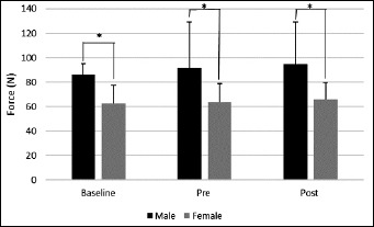
Mean of baseline, pre- and post-practice conditions for sternocleidomastoid (SCM) peak force. Females had significantly less force output compared to males in each of the three conditions (baseline, pre- and post-practice). *p = 0.002
Figure 2.
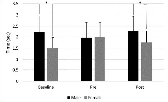
Mean of baseline, pre- and post-practice conditions for sternocleidomastoid (SCM) time to peak force between males and females. Females had significantly less time to peak force compared to males at baseline and post-practice. *p = 0.001
We then examined SCM EMG activity by side, left and right. For the left SCM, males and females had greater peak EMG activity pre-practice when compared to baseline (M: baseline = 234.3 ± 121.1 mV, pre-practice = 331.15 ± 158.9 mV; F: baseline = 246.3 ± 111.9 mV, pre-practice = 264.0 ± 120.8 mV), (p = 0.015), and at post-practice versus baseline (M: baseline = 234.3 ± 121.1mV, post-practice = 316.3 ± 143.9mV; F: baseline = 246.3 ± 111.9mV, post-practice = 289.6 ± 110.7mV) (p = 0.001) (Figure 3). For the right SCM, both males and females had greater right SCM EMG activity pre-practice compared to baseline (M: baseline = 261.7 ± 157.0 mV, pre-practice = 363.72 ± 206.6mV; F: baseline = 250.1 ± 100.7mV, pre-practice = 262.6 ± 88.9 mV) (p = 0.046), and post-practice compared to baseline (M: baseline = 261.7 ± 157.0 mV, post-practice = 382.92 ± 233.7 mV; F: baseline = 250.1 ± 100.7 mV, post-practice = 288.3 ± 72.1 mV) (p = 0.008) (Figure 4). For both males and females, right SCM time to peak EMG activity was decreased post-practice when compared to pre-practice (M: pre-practice = 2.20 ± 0.73 sec, post-practice = 1.94 ± 0.48 sec; F: pre-practice = 2.05 ± 0.55 sec, post-practice = 1.52 ± 0.48 sec) (p = 0.002) (Figure 5). There were no other significant differences for SCM EMG measures. For EMG activity of the upper trapezius musculature, there were no significant differences over time or between sexes (p > 0.05).
Figure 3.
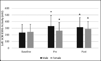
Both males and females had greater left SCM EMG activity at pre-practice (*) compared to baseline and from post-practice (^) compared to baseline. *p = 0.001. No significant differences were observed between males and females.
Figure 4.
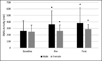
Both males and females had significantly greater right SCM EMG activity pre-practice (*) compared to baseline and post-practice (^) compared to baseline. *p = 0.008. No significant differences were observed between males and females.
Figure 5.
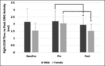
In both males and females, right SCM time to peak EMG activity was significantly decreased post-practice when compared to pre-practice. *p = 0.002. No significant differences were observed between males and females or at baseline for either sex.
Discussion
The potential importance of cervical muscle strength and stiffness as a protective mechanism against concussive events and muscle fatigue has been previously reported (Erkmen et al., 2009; Fox et al., 2008; Kerr et al., 2021; Toninato et al., 2018; Wilkins et al., 2004), from controlled laboratory settings. Data identifying changes in cervical muscle strength and stiffness in practical, non-laboratory settings are not available. The importance of understanding the association of sports activity with changes in cervical muscle strength and stiffness during play and/or practice settings will allow clinicians to devise cervical strengthening programs potentially reducing the severity of head impact injury.
Significant differences in SCM peak force activity were observed between sexes at baseline, pre- and post-practice with males having greater SCM peak force versus females. This result was not surprising as males have been reported to have greater peak force output in neck musculature versus females (Tierney et al., 2005). Males in general have greater neck muscle mass compared to females which leads to greater force production potential. In the present study, males had 17.55% greater neck girth compared to females.
Time to peak force was greater in males versus females for both baseline and post-practice. Tierney et al. (2005) reported females initiated SCM activity earlier than males when an external force was applied to the head. Earlier SCM activity was attributed to greater head-neck peak angular acceleration and displacement in females versus males, in an attempt to minimize head acceleration and displacement compared to males. Wilcox et al. (2015) reported no differences in peak linear acceleration between male and female collegiate ice hockey players. These data demonstrate the potential importance of the neck musculature in reducing acceleration forces upon impact in ice hockey.
Differences between the sexes have potential implications when designing concussion prevention programs should cervical musculature be associated with program design. The current results and those of Tierney et al. (2005) demonstrate differences in firing rates of the cervical musculature between sexes when the head is exposed to an external force. These differences in firing rates between the sexes, along with the potential importance of cervical strength in concussion prevention warrant further investigation to better develop concussion prevention programs that focus on the cervical musculature in both men and women.
Examination of the right and left SCM peak EMG activity resulted in greater peak EMG activity for each side pre- and post-practice versus baseline, yet no differences between pre- and post-practice. The lack of difference between pre- and post-practice EMG activity suggests that similar motor unit activation occurred in each condition. What is of interest however, is the time to peak EMG activity in the right versus left SCM. Right SCM time to peak EMG activity was decreased post-practice versus pre-practice, yet there was no change in time to peak EMG activity for the left SCM. The time to right SCM peak EMG activity suggests a lower activation threshold post-practice (Adam et al., 1998), possibly due to greater neuronal excitability in the right SCM which may relate to the handedness or shot preference of players. In this study the majority of players were right handed shots (R = 24; L = 12). These data show potential differences in activation patterns between dominant and non-dominant sides of cervical musculature, which may influence the consequences of head impacts.
The right SCM is responsible for contralateral head rotation and right lateral flexion. In the sport of ice hockey, the handedness of the player can impact the activity of right versus left SCM. For the right handed player, the right or dominant SCM is potentially more active versus the left SCM. Players look down and to the right as they control the puck on their stick, since most control is utilized with the forehand (curved part of the stick blade). In addition, forehand passing and shooting require the head to rotate left on the follow-through. Greater reliance on the dominant side SCM potentially enhances neurological excitability thereby leading to a reduced activation time as practices and/or games progress. This has implications for head stabilization when receiving a head impact. There is the potential for head rotation in response to an external force, which has been postulated as a mechanism for concussive events (Antonoff et al., 2021; Post and Hoshizaki, 2012; Zhang et al., 2006). More research is required to understand the potential consequences of the differences in rate of activation between dominant side and non-dominant side SCM resulting from practice and/or game play in ice hockey.
Clinical implications
Concussive events continue to be of concern in contact and collision sports. Among the potential concussion prevention paradigms, increased cervical muscle strength and/or pre-activation are widely believed to contribute to reductions in concussive events. Attention to the differences between the sexes relative to cervical muscle firing rates will enable clinicians to design more effective cervical strengthening programs for the purpose of concussion prevention. Focus on improving firing rate in men is recommended while in women, a focus on increasing overall cervical muscle strength will be of benefit.
Conclusion
Our study is the first to explore the consequences of an ice hockey contact-heavy practice on cervical muscle strength between the sexes. Enhanced cervical muscle strength continues to be postulated as a preventative strategy for concussion reduction in sports, yet there is a paucity of data examining the effectiveness of this strategy in the sport of ice hockey. Of note were the sex differences in activation time of the SCM and the differences in time to peak activation of the dominant side SCM versus non-dominant. These findings begin to provide guidance to improve cervical strengthening programs that are sex specific. Cervical muscle strengthening programs should begin to focus on improving time to peak activation of the SCM in males to create a more rapid head/neck stabilization when reacting to an external load. In addition, these programs should address the propensity of the dominant SCM to reach peak activation in less time versus the non-dominant SCM. Continued examination of activation patterns of the SCM in ice hockey may aid in more robust concussion prevention programs enhancing ice hockey player health and safety.
Acknowledgements
We thank the players, coaches, and athletic trainers of the men’s and women’s University of New England ice hockey teams for their participation and cooperation with this study. This work was supported in part by a grant from the University of New England Office of Research and Scholarship. The experiments comply with the current laws of the country in which they were performed. The authors have no conflict of interest to declare. The datasets generated during and/or analyzed during the current study are not publicly available, but are available from the corresponding author who was an organizer of the study.
Biographies
Caitlin A. GALLO
Employment
Department of Exercise and Sport Performance at the University of New England.
Degree
BS
Research interests
Concussions
E-mail: cgallo2@une.edu
Gabrielle N. DESROCHERS
Employment
Department of Exercise and Sport Performance, University of New England
Degree
BS
E-mail: gdesrochers@une.edu
Garett J. MORRIS
Employment
Department of Exercise and Sport Performance, University of New England
Degree
BS
E-mail: gmorris2@une.edu
Chad D. RUMNEY
Employment
Department of Exercise and Sport Performance, University of New England
Degree
BS
E-mail: crumney@une.edu
Sydney J. SANDELL
Employment
Department of Exercise and Sport Performance, University of New England
Degree
BS
E-mail: ssandell@une.edu
Jane K. MCDEVITT
Employment
Ass. Prof.,Temple University Department of Health and Rehabilitation Sciences
Degree
PhD
Research interests
Concussion mechanisms
E-mail: tua84996@temple.edu
Dianne LANGFORD
Employment
Ass. Dean for Research Prof., Department of Neuroscience, Lewis Katz School of Medicine at Temple University
Degree
PhD
Research interests
Concussion mechanisms and prevention
E-mail: tdl@temple.edu
John M. ROSENE
Employment
Clinical Prof., Department of Exercise and Sport Performance University of New England
Degree
DPE
Research interests
Concussion mechanisms and prevention
E-mail: jrosene@une.edu
References
- Adam A., De Luca C.J., Erim Z. (1998) Hand dominance and motor unit firing behavior. Journal of Neurophysiology 80, 1373-1382. https://doi.org/10.1152/jn.1998.80.3.1373 10.1152/jn.1998.80.3.1373 [DOI] [PubMed] [Google Scholar]
- Antonoff D., Goss J., Langevin T., Renodin C., Spahr L., McDevitt J., Langford D., Rosene J. (2021) Unexpected findings from a pilot study on vision training as a potential intervention to reduce sub-concussive head impacts during a collegiate ice hockey season. Journal of Neurotrauma 38, 1783-1790. https://doi.org/10.1089/neu.2020.7397 10.1089/neu.2020.7397 [DOI] [PubMed] [Google Scholar]
- Becker S., Berger J., Backfisch M., Ludwig O., Kelm J., Frohlich M. (2019) Effects of a 6-Week Strength Training of the Neck Flexors and Extensors on the Head Acceleration during Headers in Soccer. Journal of Sports Science and Medicine 18, 729-737. https://pubmed.ncbi.nlm.nih.gov/31827358/ [PMC free article] [PubMed] [Google Scholar]
- Collins E.L., Fletcher E.N., Fields S.K., Kluchurosky L., Rohrkemper M.K., Comstock R. D., Cantu R.C. (2014) Neck strength: a protective factor reducing risk for concussion in high school sports. Journal of Primary Prevention 35, 309-319. https://doi.org/10.1007/s10935-014-0355-2 10.1007/s10935-014-0355-2 [DOI] [PubMed] [Google Scholar]
- Covassin T., Weiss L., Powell J., Womack C. (2007) Effects of a maximal exercise test on neurocognitive function. British Journal of Sports Medicine 41, 370-374. https://doi.org/10.1136/bjsm.2006.032334 10.1136/bjsm.2006.032334 [DOI] [PMC free article] [PubMed] [Google Scholar]
- Derave W., De Clercq D., Bouckaert J., Pannier J.L. (1998) The influence of exercise and dehydration on postural stability. Ergonomics 41, 782-789. https://doi.org/10.1080/001401398186630 10.1080/001401398186630 [DOI] [PubMed] [Google Scholar]
- Eckner J.T., Lipps D.B., Kim H., Richardson J.K., Ashton-Miller J.A. (2011) Can a clinical test of reaction time predict a functional head-protective response? Medicine Science in Sports and Exercise 43, 382-387. https://doi.org/10.1249/MSS.0b013e3181f1cc51 10.1249/MSS.0b013e3181f1cc51 [DOI] [PMC free article] [PubMed] [Google Scholar]
- Eckner J.T., Goshtasbi A., Curtis K., Kapshai A., Myyra E., Franco L.M., Favre M., Jacobson J.A. (2018) Feasibility and Effect of Cervical Resistance Training on Head Kinematics in Youth Athletes: A Pilot Study. American Journal of Physical Medicine and Rehabilitation 97, 292-297. https://doi.org/10.1097/PHM.0000000000000843 10.1097/PHM.0000000000000843 [DOI] [PMC free article] [PubMed] [Google Scholar]
- Erkmen N.T.H., Kaplan T., Sanioglu A. (2009) The effect of fatiguing exercise on balance performance as measured by the balance error scoring system. Isokinetics and Exercise Science 17, 121-127. https://doi.org/10.3233/IES-2009-0343 10.3233/IES-2009-0343 [DOI] [Google Scholar]
- Finnoff J.T., Peterson V.J., Hollman J.H., Smith J. (2009) Intrarater and interrater reliability of the Balance Error Scoring System (BESS). Physical Medicine and Rehabilitation: The Journal of Injury, Function and Rehabilitation 1, 50-54. https://doi.org/10.1016/j.pmrj.2008.06.002 10.1016/j.pmrj.2008.06.002 [DOI] [PubMed] [Google Scholar]
- Fox Z.G., Mihalik J.P., Blackburn J.T., Battaglini C.L., Guskiewicz K.M. (2008) Return of postural control to baseline after anaerobic and aerobic exercise protocols. Journal of Athletic Training 43, 456-463. https://doi.org/10.4085/1062-6050-43.5.456 10.4085/1062-6050-43.5.456 [DOI] [PMC free article] [PubMed] [Google Scholar]
- Guskiewicz K.M., Ross S.E., Marshall S.W. (2001) Postural Stability and Neuropsychological Deficits After Concussion in Collegiate Athletes. Journal of Athletic Training 36, 263-273. [PMC free article] [PubMed] [Google Scholar]
- Kawata K., Rubin H.R., Lee J.H., Sim T., Takahagi M., Szwanki V., Darvish K., Assari S., Henderer J.D., Tierney R. (2016) Association of football subconcussive head impacts with ocular near point of convergence. JAMA Ophthalmology 134, 763-769. https://doi.org/10.1001/jamaophthalmol.2016.1085 10.1001/jamaophthalmol.2016.1085 [DOI] [PubMed] [Google Scholar]
- Kawata K., Tierney R., Phillips J., Jeka J.J. (2016) Effect of Repetitive Sub-concussive Head Impacts on Ocular Near Point of Convergence. International Journal of Sports Medicine 37, 405-410. https://doi.org/10.1055/s-0035-1569290 10.1055/s-0035-1569290 [DOI] [PubMed] [Google Scholar]
- Kerr Z.Y., Pierpoint L.A., Rosene J.M. (2021) Epidemiology of Concussions in High School Boys' Ice Hockey, 2008/09 to 2016/17 School Years. Clinical Journal of Sports Medicine 31, 21-28. [DOI] [PubMed] [Google Scholar]
- Koscs M.K.T., Swanik C.B., Edwards D.G. (2009) Effects of exertional exercise on the standardized assessment of concussion (SAC) score. Athletic Training and Sports Health Care 1, 1-24. https://doi.org/10.3928/19425864-20090101-01 10.3928/19425864-20090101-01 [DOI] [Google Scholar]
- Lepers R., Bigard A.X., Diard J.P., Gouteyron J.F., Guezennec C.Y. (1997) Posture control after prolonged exercise. European Journal of Applied Physiology and Occupational Physiology 76, 55-61. https://doi.org/10.1007/s004210050212 10.1007/s004210050212 [DOI] [PubMed] [Google Scholar]
- Lindsey J.C.K., Mansell J.L., Phillips J., Tierney R.T. (2017) Effect of fatigue on ocular motor assessments. Athletic Training and Sports Health Care 9, 177-183. https://doi.org/10.3928/19425864-20170420-03 10.3928/19425864-20170420-03 [DOI] [Google Scholar]
- Mansell J., Tierney R.T., Sitler M.R., Swanik K.A., Stearne D. (2005) Resistance training and head-neck segment dynamic stabilization in male and female collegiate soccer players. Journal of Athletic Training 40, 310-319. [PMC free article] [PubMed] [Google Scholar]
- Mihalik J.P., Guskiewicz K.M., Marshall S.W., Greenwald R.M., Blackburn J.T., Cantu R.C. (2011) Does cervical muscle strength in youth ice hockey players affect head impact biomechanics? Clinical Journal of Sports Medicine 21, 416-421. https://doi.org/10.1097/JSM.0B013E31822C8A5C 10.1097/JSM.0B013E31822C8A5C [DOI] [PubMed] [Google Scholar]
- Mucha A., Collins M.W., Elbin R.J., Furman J.M., Troutman-Enseki C., DeWolf R.M., Marchetti G., Kontos A.P. (2014) A Brief Vestibular/Ocular Motor Screening (VOMS) assessment to evaluate concussions: preliminary findings. American Journal of Sports Medcine 42, 2479-2486. https://doi.org/10.1177/0363546514543775 10.1177/0363546514543775 [DOI] [PMC free article] [PubMed] [Google Scholar]
- Post A., Hoshizaki T.B. (2012) Mechanisms of brain impact injuries and their prediction: A review. Trauma 14, 327-349. https://doi.org/10.1177/1460408612446573 10.1177/1460408612446573 [DOI] [Google Scholar]
- Reynier K.A., Alshareef A., Sanchez E.J., Shedd D.F., Walton S.R., Erdman N.K., Newman B.T., Giudice J.S., Higgins M.J., Funk J.R., Broshek D.K., Druzgal T.J., Resch J.E., Panzer M.B. (2020) The effect of muscle activation on head kinematic during non-injurious head impacts in human subjects. Journal of Biomedical Engineering 48, 2751-2762. https://doi.org/10.1007/s10439-020-02609-7 10.1007/s10439-020-02609-7 [DOI] [PubMed] [Google Scholar]
- Riemann B.L., Guskiewicz K.M. (2000) Effects of mild head injury on postural stability as measured through clinical balance testing. Journal of Athletic Training 35, 19-25. [PMC free article] [PubMed] [Google Scholar]
- Rosene J.M., Raksnis B, Silva B, Woefel T, Visich P.S., Dompier T.P., Kerr Z.Y. (2017) Comparison of Concussion Rates Between NCAA Division I and Division III Men’s and Women’s Ice Hockey Players. American Journal of Sports Medicine 45, 2622-2629. https://doi.org/10.1177/0363546517710005 10.1177/0363546517710005 [DOI] [PubMed] [Google Scholar]
- Schneiders A.G., Sullivan S.J., Handcock P., Gray A., McCrory P.R. (2012) Sports concussion assessment: the effect of exercise on dynamic and static balance. Scandanavian Journal of Medicine and Science in Sports 22, 85-90. https://doi.org/10.1111/j.1600-0838.2010.01141.x 10.1111/j.1600-0838.2010.01141.x [DOI] [PubMed] [Google Scholar]
- Susco T.M., Valovich McLeod T.C., Gansneder B.M., Shultz S.J. (2004) Balance Recovers Within 20 Minutes After Exertion as Measured by the Balance Error Scoring System. Journal of Athletic Training 39, 241-246. [PMC free article] [PubMed] [Google Scholar]
- Tierney R.T., Sitler M.R., Swanik C.B., Swanik K.A., Higgins M., Torg J. (2005) Gender differences in head-neck segment dynamic stabilization during head acceleration. Medicine and Science in Sports and Exercise 37, 272-279. https://doi.org/10.1249/01.MSS.0000152734.47516.AA 10.1249/01.MSS.0000152734.47516.AA [DOI] [PubMed] [Google Scholar]
- Viano D.C., Casson I.R., Pellman E.J. (2007) Concussion in professional football: biomechanics of the struck player: part 14. Neurosurgery 61, 313-327. https://doi.org/10.1227/01.NEU.0000279969.02685.D0 10.1227/01.NEU.0000279969.02685.D0 [DOI] [PubMed] [Google Scholar]
- Toninato J., Casey H., Uppal M., Abdallah T., Bergman T., Eckner J., Samadani U. (2018) Traumatic brain injury reduction in athletes by neck strengthening (TRAIN). Contemporary Clinical Trials Communications 11, 102-106. https://doi.org/10.1016/j.conctc.2018.06.007 10.1016/j.conctc.2018.06.007 [DOI] [PMC free article] [PubMed] [Google Scholar]
- Wilcox B.J., Beckwith J.G., Greenwald R.M., Raukar N.P., Chu J.J., McAllister T.W., Flashman L.A., Maerlender A.C. (2015) Biomechanics of head impacts associated with diagnosed concussion in female collegiate ice hockey players. Journal of Biomechanics 48, 2201-2204. https://doi.org/10.1016/j.jbiomech.2015.04.005 10.1016/j.jbiomech.2015.04.005 [DOI] [PMC free article] [PubMed] [Google Scholar]
- Wilkins J.C., Valovich McLeod T.C., Perrin D.H., Gansneder B.M. (2004) Performance on the Balance Error Scoring System Decreases After Fatigue. Journal of Athletic Training 39, 156-161. [PMC free article] [PubMed] [Google Scholar]
- Zhang J., Yoganandan N., Pintar F.A., Gennarelli T.A. (2006) Role of translational and rotational accelerations on brain strain in lateral head impact. Biomedical Sciences Instrumentation 42, 501-506. [PubMed] [Google Scholar]


